. 2021 May 24;11(30):18685-18692.
doi: 10.1039/d1ra01296g. eCollection 2021 May 19.

Synergy of molecularly mobile polyrotaxane surfaces with endothelial cell co-culture for mesenchymal stem cell mineralization

Affiliations

Synergy of molecularly mobile polyrotaxane surfaces with endothelial cell co-culture for mesenchymal stem cell mineralization

Hiroki Masuda et al. RSC Adv. .

Abstract

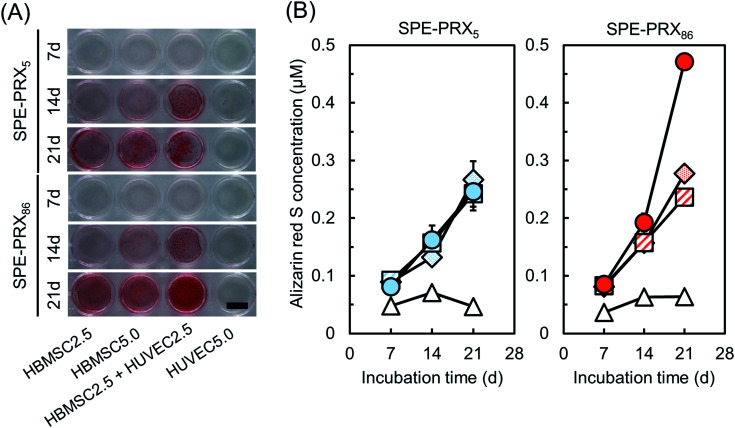
Stem cell-based bone tissue engineering is a promising strategy for the treatment of bone defects. Since regeneration of bone tissue takes a long time, promoting osteogenesis of stem cells is desired for earlier recovery from dysfunctions caused by bone defects. Here, we combined endothelial cell co-culture using the molecularly mobile sulfonated polyrotaxane (PRX) surfaces to enhance the mineralization of human bone marrow derived mesenchymal stem cells (HBMSCs). Sulfonated PRXs are composed of sulfopropyl ether-modified α-cyclodextrins (α-CDs) threaded on a polyethylene glycol chain. The molecular mobility of PRX, α-CDs moving along the polymer, can be modulated by the number of α-CDs. When osteoblastic differentiation was induced in HBMSCs and human umbilical vein endothelial cells (HUVECs), co-culture groups on sulfonated PRX surfaces with low molecular mobility showed the highest mineralization, which is about two times as high as co-culture groups on sulfonated PRX surfaces with high molecular mobility. Nuclear accumulation of yes-associated proteins in HBMSCs and cell-cell communication via cytokines or cadherin may play an important role in synergistically induced mineralization of HBMSCs.


Conflict of interest statement

There are no conflicts to declare.

Figures

Fig. 1
Fig. 1. (A) Chemical structure of SPE-PRX. (B) Preparation of SPE-PRX surfaces and mineralization of HBMSCs co-cultured with HUVECs on SPE-PRX surfaces. For induction of osteoblastic differentiation, HBMSCs and HUVECs were seeded on SPE-PRX5 or SPE-PRX86 surfaces at the following cell densities: HBMSCs at 2.5 × 10⁴ cells per cm2 (HBMSC2.5), HBMSCs at 5.0 × 10⁴ cells per cm2 (HBMSC5.0), HBMSCs and HUVECs (1 : 1) at total density of 5.0 × 10⁴ cells per cm2 (HBMSC2.5 + HUVEC2.5), and HUVECs at 5.0 × 10⁴ cells per cm2 (HUVEC5.0).
Fig. 2
Fig. 2. Box plots of the adhesion area (A) and aspect ratio (B) of HBMSCs on SPE-PRX5 and SPE-PRX86 surfaces. The top and bottom of the boxes correspond to the first and third quartiles. The line in the middle corresponds to the median, the squares represent the mean, and the whiskers represent the maximum and minimum values of data sets. (C) Growth curves of HBMSCs on SPE-PRX5 (blue) and SPE-PRX86 (orange) surfaces. Data are presented as mean ± S.D., n = 4.
Fig. 3
Fig. 3. (A) Fluorescent images of YAP localization in HBMSCs on SPE-PRX5 and SPE-PRX86 surfaces after 2 d of culture. Scale bar: 50 μm. (B) The proportion of YAP localized in nucleus (filled bars), both nucleus and cytoplasm (hatched bars), or cytoplasm (open bars) in HBMSCs.
Fig. 4
Fig. 4. Gene expression levels of BMP-2, VEGF, N-cadherin, and COLI in HBMSCs and HUVECs on SPE-PRX5 or SPE-PRX86 surfaces after 7 d of culture. Data are presented as mean ± S.D., n = 4. Statistical analyses were conducted via one-way analysis of variance and post hoc analysis using Tukey's range test for multiple comparisons. *p < 0.05 indicates significance.
Fig. 5
Fig. 5. ALP staining images of HBMSCs and HUVECs on SPE-PRX5 and SPE-PRX86 surfaces after 3, 7, and 14 d of incubation in mixed differentiation medium. Whole well images are shown. Scale bar: 10 mm.
Fig. 6
Fig. 6. (A) Alizarin red S staining images of HBMSCs and HUVECs on SPE-PRX5 and SPE-PRX86 surfaces after 7, 14, and 21 d of incubation in mixed differentiation medium. Whole well images are shown. Scale bar: 10 mm. (B) Time courses of alizarin red S concentration of HBMSC2.5 (diamond), HBMSC5.0 (square), HBMSC2.5 + HUVEC2.5 (circle), and HUVEC5.0 (triangle) on SPE-PRX5 (blue) surfaces and SPE-PRX86 (red) surfaces on day 7, 14, and 21. Data are presented as mean ± S.D., n = 4.
Fig. 7
Fig. 7. Promoting mineralization of HBMSCs on SPE-PRX surfaces by synergistic effects of translocating YAP to the nucleus by molecular mobility of SPE-PRX and enhanced cell interaction with HUVECs.

References

    1. Myeroff C. Archdeacon M. J. Bone Jt. Surg., Am. Vol. 2011;93:2227. doi: 10.2106/JBJS.J.01513. - DOI - PubMed
    1. Schwartz Z. Mellonig J. T. Carnes, Jr D. L. De La Fontaine J. Cocharan D. L. Dean D. D. Boyan B. D. J. Periodontol. 1996;67:918. doi: 10.1902/jop.1996.67.9.918. - DOI - PubMed
    1. Sopyan I. Mel M. Ramesh S. Khalid K. A. Sci. Technol. Adv. Mater. 2007;8:116. doi: 10.1016/j.stam.2006.11.017. - DOI
    1. Vo T. N. Kasper F. K. Mikos A. G. Adv. Drug Delivery Rev. 2012;64:1292. doi: 10.1016/j.addr.2012.01.016. - DOI - PMC - PubMed
    1. Marolt D. Knezevic M. Vunjak-Novakovic G. Stem Cell Res. Ther. 2010;1:10. doi: 10.1186/scrt10. - DOI - PMC - PubMed