

Biomechanical Effect of Coronal Alignment and Ligament Laxity in Total Knee Arthroplasty: A Simulation Study
- PMID: 35480980
- PMCID: PMC9035799
- DOI: 10.3389/fbioe.2022.851495
Biomechanical Effect of Coronal Alignment and Ligament Laxity in Total Knee Arthroplasty: A Simulation Study
Abstract
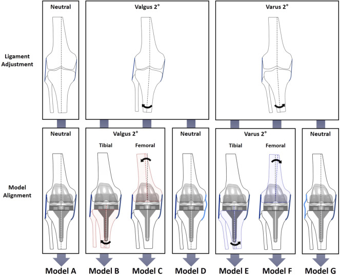
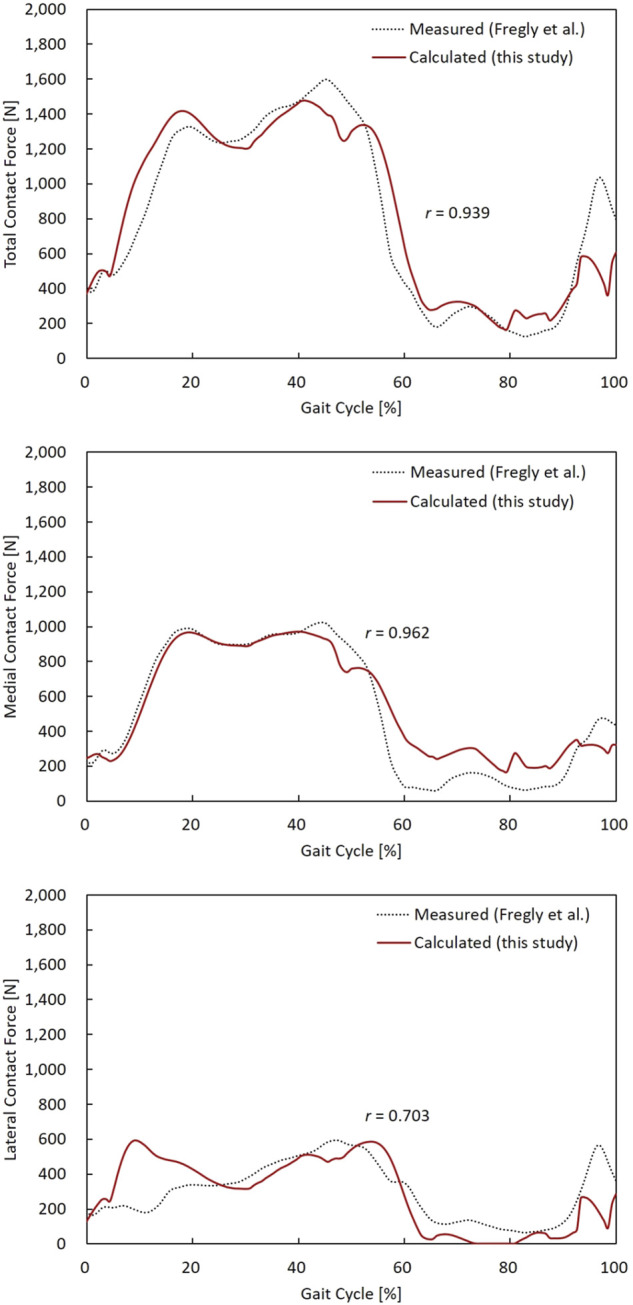
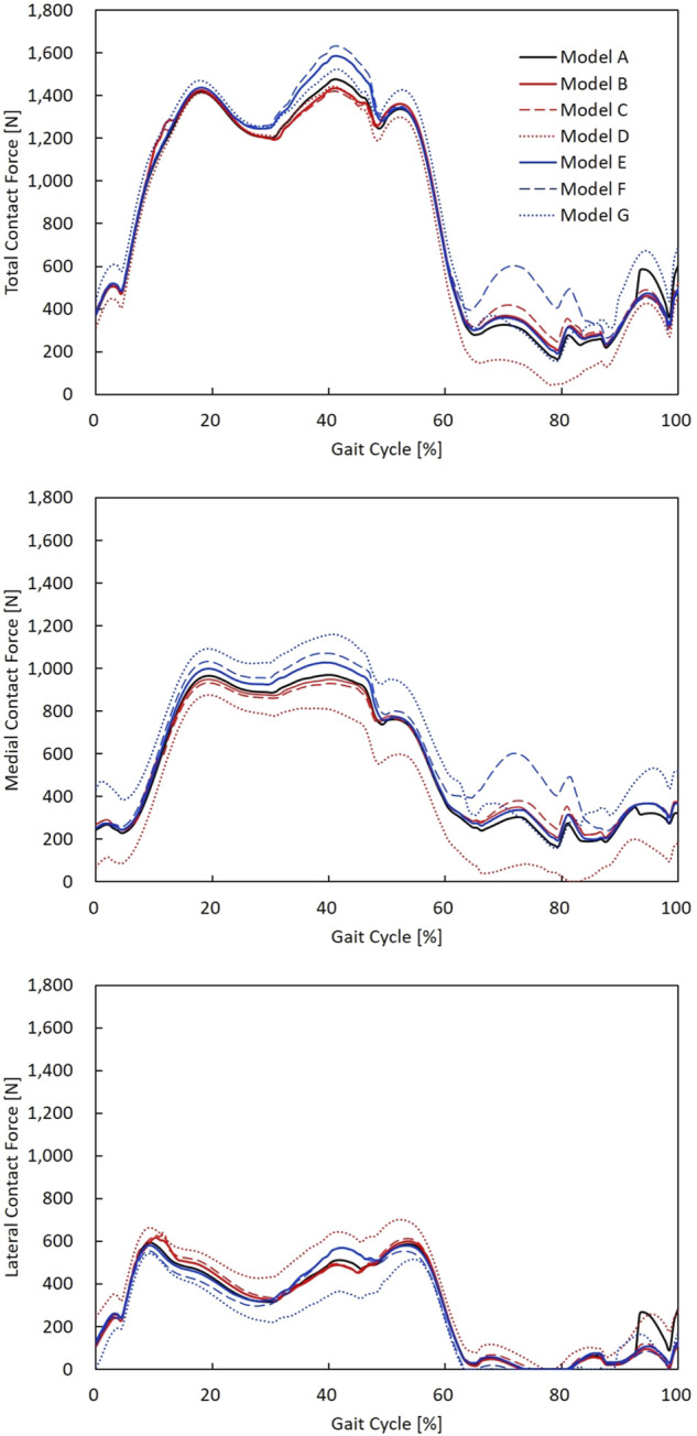
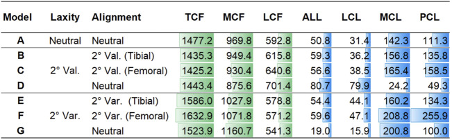
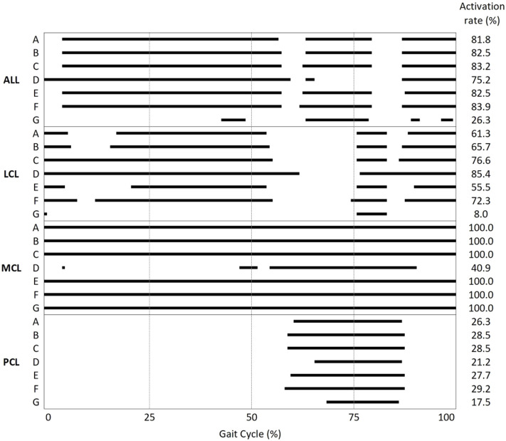
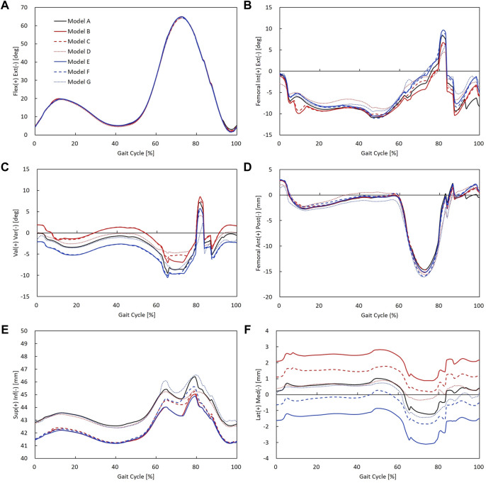
The purposes of this study were to develop a cruciate-retaining total knee arthroplasty musculoskeletal model, which enables the adjustment of ligament length and implant alignment; validate the model; and evaluate the effects of varus/valgus alignment adjustment and unbalanced medial/lateral ligament laxity during gait. A cruciate-retaining total knee arthroplasty musculoskeletal model was constructed and validated against the in vivo contact forces. This model was transformed to 2° varus/valgus alignment of femoral or tibial replacement models and 2° medial/lateral laxity models. The contact forces and ligament tensions of the adjusted models were calculated. The contact forces in the model showed good agreement with the in vivo contact forces. Valgus replacement alignment with balanced ligament models showed a lower contact force at the medial compartment than at the neutral alignment model, whereas the varus replacement alignment with balanced ligament models showed a greater contact force at the medial compartment and medial/posterior cruciate ligament tension. The medial laxity with neutral alignment model showed a similar contact force with decreased medial ligament tension compared to the balanced neutral alignment model, whereas the lateral laxity with the neutral alignment model showed a greater contact force and decreased lateral ligament tension. The cruciate-retaining total knee arthroplasty model was validated using in vivo contact forces (r = 0.939) Two degrees of valgus alignment adjustment with balanced ligament or neutral alignment with 2° of medial laxity can be safe without increasing contact force or ligament tension compared to neutral alignment with a balanced extension gap. However, 2° of varus alignment adjustment with balanced ligament or neutral alignment with 2° of lateral laxity may be unfavorable due to the overloading of the joints and knee ligaments.
Keywords: collateral ligament tension; contact force; coronal alignment; knee arthroplasty; model.
Copyright © 2022 Ro, Ro, Kang, Han and Shin.
Conflict of interest statement
JR and YK were employed by Corentec. DR was employed by CONNECTEVE Co., Ltd. The remaining authors declare that the research was conducted in the absence of any commercial or financial relationships that could be construed as a potential conflict of interest.
Figures
Similar articles
-
Preservation of femoral and tibial coronal alignment to improve biomechanical effects of medial unicompartment knee arthroplasty: Computational study.Biomed Mater Eng. 2018;29(5):651-664. doi: 10.3233/BME-181015. Biomed Mater Eng. 2018. PMID: 30400078
-
Limited effect of anatomical insert geometry on in vitro laxity in balanced anatomic posterior cruciate ligament retaining total knee arthroplasty.Knee Surg Sports Traumatol Arthrosc. 2022 Apr;30(4):1273-1281. doi: 10.1007/s00167-021-06564-1. Epub 2021 Apr 15. Knee Surg Sports Traumatol Arthrosc. 2022. PMID: 33860338
-
Femoral component alignment in unicompartmental knee arthroplasty leads to biomechanical change in contact stress and collateral ligament force in knee joint.Arch Orthop Trauma Surg. 2018 Apr;138(4):563-572. doi: 10.1007/s00402-018-2884-2. Epub 2018 Jan 22. Arch Orthop Trauma Surg. 2018. PMID: 29356941
-
[Adjusted mechanical alignment: operative technique-Tips and tricks].Orthopade. 2020 Jul;49(7):562-569. doi: 10.1007/s00132-020-03929-1. Orthopade. 2020. PMID: 32494903 Review. German.
-
Kinematic alignment in total knee arthroplasty.Oper Orthop Traumatol. 2021 Dec;33(6):525-537. doi: 10.1007/s00064-021-00729-4. Epub 2021 Aug 19. Oper Orthop Traumatol. 2021. PMID: 34414467 Review. English.
Cited by
-
Comparison of navigation systems for total knee arthroplasty: A systematic review and meta-analysis.Front Surg. 2023 Jan 17;10:1112147. doi: 10.3389/fsurg.2023.1112147. eCollection 2023. Front Surg. 2023. PMID: 36733891 Free PMC article. Review.
-
Effect of tibiofemoral alignment on simulated knee contact forces during gait in mechanically and kinematically aligned total knee arthroplasty patients.Sci Rep. 2024 Nov 11;14(1):27510. doi: 10.1038/s41598-024-78618-6. Sci Rep. 2024. PMID: 39528651 Free PMC article.
-
Learning curve of robot-assisted total knee arthroplasty and its effects on implant position in asian patients: a prospective study.BMC Musculoskelet Disord. 2023 Apr 27;24(1):332. doi: 10.1186/s12891-023-06422-w. BMC Musculoskelet Disord. 2023. PMID: 37106353 Free PMC article.
-
Contributions of External, Muscle, and Ligament Forces to Tibiofemoral Contact Loads in Patients with Knee Osteoarthritis and Healthy Individuals.Bioengineering (Basel). 2025 May 31;12(6):600. doi: 10.3390/bioengineering12060600. Bioengineering (Basel). 2025. PMID: 40564417 Free PMC article.
-
Effect of subscapularis repair on joint contact forces based on degree of posterior-superior rotator cuff tear severity in reverse shoulder arthroplasty.Front Bioeng Biotechnol. 2023 Dec 7;11:1229646. doi: 10.3389/fbioe.2023.1229646. eCollection 2023. Front Bioeng Biotechnol. 2023. PMID: 38130822 Free PMC article.
References
-
- Andersen M. S., Rasmussen J. (2011). “Total Knee Replacement Musculoskeletal Model Using a Novel Simulation Method for Non-conforming Joints,” in Proceedings of the International Society of Biomechanics Conference2011 Jul 03-07, Brussels, Belgium (International Society of Biomechanics, ISB; ).
LinkOut - more resources
Full Text Sources
Miscellaneous