. 2022:2:24.
doi: 10.1038/s43586-022-00104-y. Epub 2022 Apr 14.

Nanodelivery of nucleic acids

Affiliations

Nanodelivery of nucleic acids

Bárbara B Mendes et al. Nat Rev Methods Primers. 2022.

Abstract

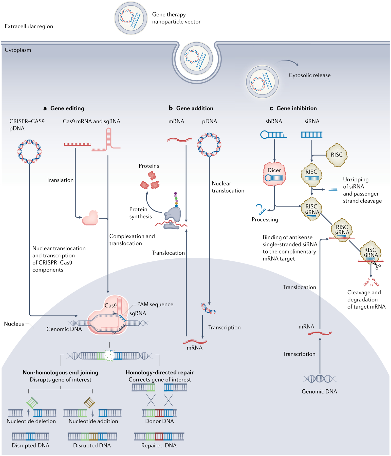
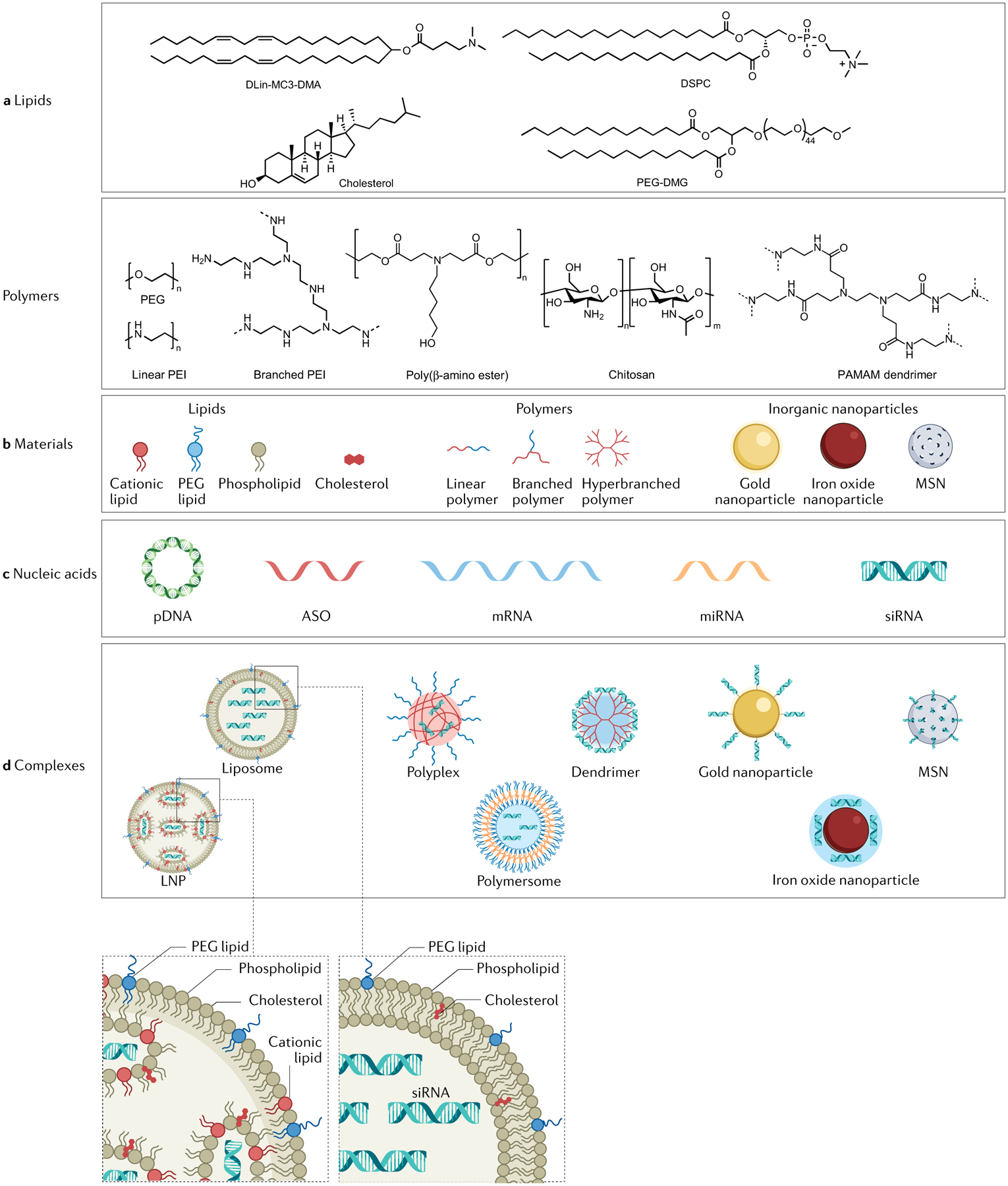
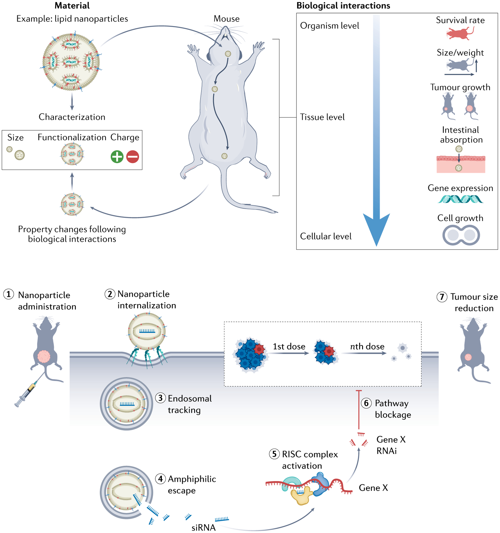
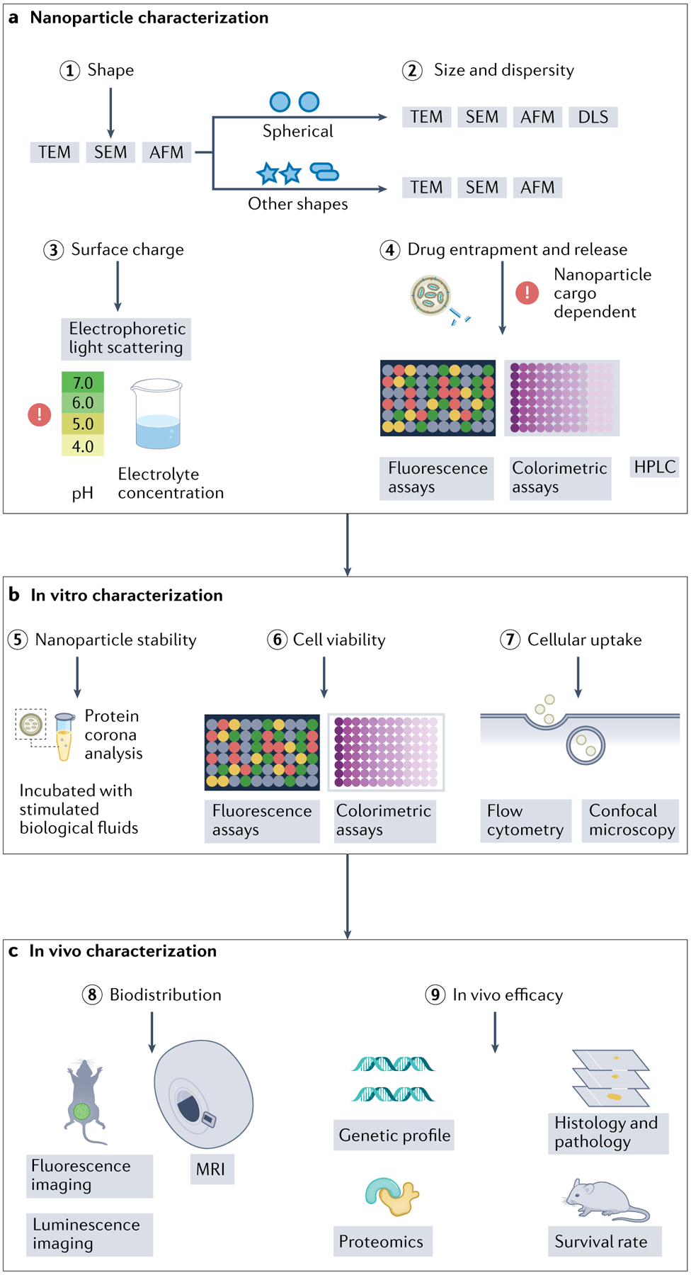
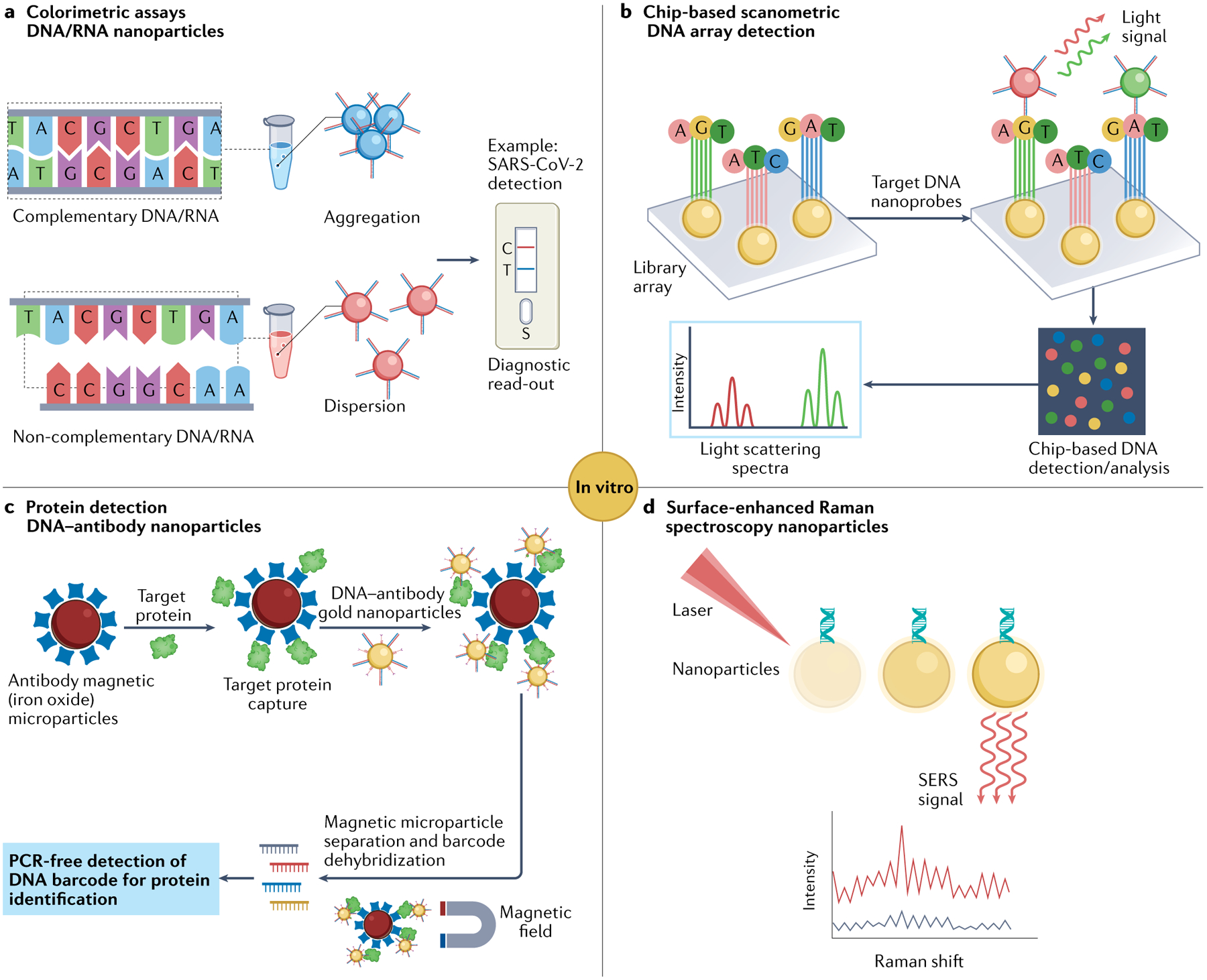
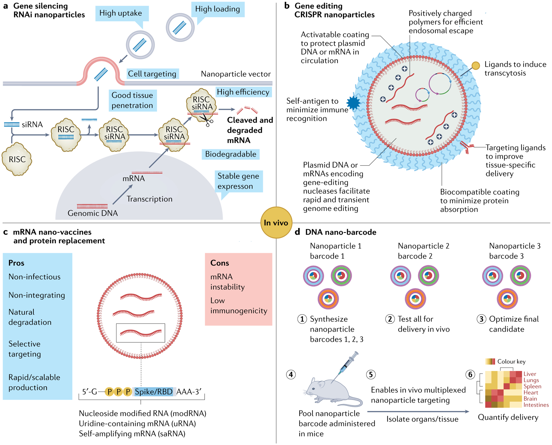
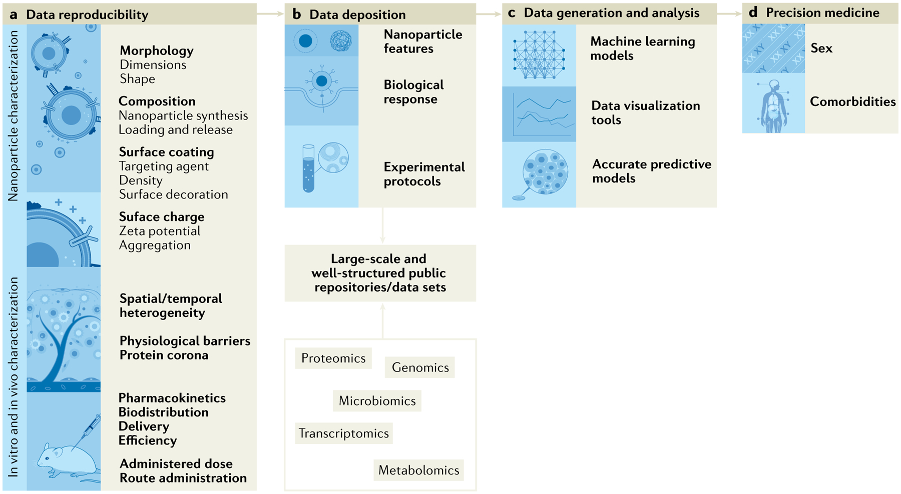
There is growing need for a safe, efficient, specific and non-pathogenic means for delivery of gene therapy materials. Nanomaterials for nucleic acid delivery offer an unprecedented opportunity to overcome these drawbacks; owing to their tunability with diverse physico-chemical properties, they can readily be functionalized with any type of biomolecules/moieties for selective targeting. Nucleic acid therapeutics such as antisense DNA, mRNA, small interfering RNA (siRNA) or microRNA (miRNA) have been widely explored to modulate DNA or RNA expression Strikingly, gene therapies combined with nanoscale delivery systems have broadened the therapeutic and biomedical applications of these molecules, such as bioanalysis, gene silencing, protein replacement and vaccines. Here, we overview how to design smart nucleic acid delivery methods, which provide functionality and efficacy in the layout of molecular diagnostics and therapeutic systems. It is crucial to outline some of the general design considerations of nucleic acid delivery nanoparticles, their extraordinary properties and the structure-function relationships of these nanomaterials with biological systems and diseased cells and tissues.


Conflict of interest statement

Competing interests J. Conde is a co-founder and shareholder of TargTex S.A. The other authors declare no competing interests.

Figures

Fig. 1 |
Fig. 1 |. Schematic pathways of three gene therapy avenues.
a | Gene editing. CRISPR–Cas9 coding DNA plasmid (pDNA) or Cas9 mRNA and single guide RNA (sgRNA) that contains a targeting sequence are delivered to the cell and enter the cytoplasm. pDNA translocates to the nucleus followed by transcription to Cas9 mRNA and sgRNA. Following translocation to the cytosol, Cas9 mRNA is translated to Cas9 protein and complexes sgRNA. Thereafter, Cas9–sgRNA complex translocates to the nucleus, where it binds the genomic DNA target sequence containing a proto-spacer adjacent motif (PAM) sequence at its 3’ end. Specific binding causes double-stranded DNA breakage resulting in non-homologous end joining or homology-directed repair, leading to mutations or changes within the gene sequence, respectively. b | Gene addition. After entering the cytoplasm, pDNA carrying the desired gene sequence translocates to the nucleus and undergoes transcription to mRNA. Next, transcripted or external mRNA is translated by the ribosome, promoting desired protein synthesis. c | Gene inhibition. After entering the cytoplasm, short hairpin RNA (shRNA) is detected and processed by Dicer protein to generate small interfering RNA (siRNA) later loaded onto RNA-induced silencing complex (RISC). Subsequently, double-stranded siRNA is unzipped, and passenger strand is cleaved. Antisense strand guides RISC towards target mRNA and aligns with it, thus leading to mRNA cleavage and degradation.
Fig. 2 |
Fig. 2 |. Scheme for the main nanoparticle design used in gene delivery.
a | Chemical structures of typically used lipids on lipid nanoparticle (LNP) formulations and representative formulation materials for polymeric nanoparticles: cationic lipid (dilinoleylmethyl-4-dimethylaminobutyrate (DLin-MC3-DMA)), phospholipid (1,2-distearoyl-sn-glycero-3-phosphocholine (DSPC)), cholesterol, PEG lipid (1,2-dimyristoyl-sn-glycerol, methoxypolyethylene glycol and polyethylene glycol-dimyristoyl glycerol (PEG-DMG)), PEG, linear and branched polyethylenimine (PEI), poly(β-amino ester), chitosan and polyamidoamine (PAMAM) dendrimer. b | Needed molecular structures for construction of lipid-based nanoparticles (liposomes and LNPs), polymeric nanoparticles (polyplexes, polymersomes and dendrimers) and commonly used inorganic nanoparticles for gene delivery (including gold nanoparticles, iron oxide nanoparticles and mesoporous silica nanoparticles (MSNs)). c | Molecular structures of typical nucleic acids for gene delivery using lipid-based, polymeric and inorganic nanoparticles. d | Structures of commonly used nanoparticle complexes containing nucleic acids. ASO, antisense oligonucleotide; miRNA, microRNA; pDNA, plasmid DNA; PEG, polyethylene glycol; siRNA, small interfering RNA.
Fig. 3 |
Fig. 3 |. Experimental workflow of nucleic acid-loaded LNP delivery in cancer therapies.
After physico-chemical characterization, nucleic acid-loaded lipid nanoparticle (LNP) formulations are administered in vivo. LNP intrinsic features will determine their biological behaviour at organism, tissue and cellular levels. Upon successful LNP administration (1), internalization (2) and siRNA release/escape (3–4), gene silencing (5–6) and subsequent tumour size reduction (7) are achieved. RISC, RNA-induced silencing complex ; RNAi, RNA interference; siRNA, small interfering RNA.
Fig. 4 |
Fig. 4 |. Overview of the common nucleic acid delivery system characterization methods.
a | Upon nanoparticle synthesis, physical and chemical features are determined, namely morphology (step 1), size (step 2), surface charge (step 3), and nucleic acid loading and profile release (step 4). b | Nanoparticle physico-chemical properties fine-tune their in vitro biological response, which ranges from characterization of the produced protein corona (step 5) to study of nanoparticle cellular uptake mechanisms (steps 6,7). c | Although study of in vivo nanoparticle behaviour will depend on target tissue and/or disease (step 8), some of the most commonly used laboratory procedures are recommended to evaluate nanoparticle efficacy and toxicity in vivo, such as animal survival rate, proteomics, genetic profile, histology and pathology (step 9). AFM, atomic force microscopy; DLS, dynamic light scattering; HPLC, high-performance liquid chromatography; SEM, scanning electron microscopy; TEM, transmission electron microscopy.
Fig. 5 |
Fig. 5 |. Nanoparticle-based nucleic acid platforms for diagnosis applications.
a | Antisense oligonucleotide (ASO)-capped gold nanoparticles aggregate in the presence of target RNA sequence of the virus, leading to change in surface plasmon resonance of gold nanoparticles and, thus, observed colour. b | Chip-based scanometric DNA array detection using oligonucleotide-modified gold nanoparticles and silver enhancing for amplification of light scattering signal. c | Ultra-sensitive protein detection using antibody-functionalized magnetic microparticle and DNA–antibody co-functionalized nanoparticle for signal amplification. d | Surface-enhanced Raman spectroscopy (SERS).
Fig. 6 |
Fig. 6 |. Nanoparticle-based nucleic acid delivery systems for therapeutical applications.
a | Small interfering RNA (siRNA) nanoparticle-mediated gene silencing. b | Design and assembly of nucleic acid-based CRISPR nanoparticles.c | Pros and cons of mRNA lipid nanoparticles (LNPs) for vaccination and protein replacement therapy. For example, Pfizer/BioNTech vaccine or individual LNP encapsulated mRNA vaccines (nucleoside modified RNA, one uridine-containing mRNA, one self-amplifying RNA (saRNA)) encoding spike protein or receptor-binding domain (RBD). d | Specific DNA sequences can be incorporated in many different nanoparticles (1) as DNA barcodes to assess their in vivo delivery profiles (2–3) at once in a single experiment. DNA barcode system enables multiplexed nanoparticle-targeted studies in vivo (4–6). RISC, RNA-induced silencing complex; RNAi, RNA interference.
Fig. 7 |
Fig. 7 |. Key limitations of nucleic acid-based nanoparticle clinical translation and potential strategies to tackle their limitations.
a | Scientific community efforts focus on the importance of standardized nanoparticles and bio–nano interface characterization, and their impact on data reproducibility. b | Development of large-scale and well-structured repositories is challenging owing to the broad procedures, terms, abbreviations and acronyms of the scientific field. c | Machine learning will allow generation of predictable models for in silico nanoparticle design taking into account physico-chemical properties (size, zeta potential) and their in vitro and in vivo response,. d | Nanoparticle design is envisioned to focus on precision medicine therapies, instead of one-size-fits-all strategies.

References

    1. Alvarez EM et al. The global burden of adolescent and young adult cancer in 2019: a systematic analysis for the Global Burden of Disease Study 2019. Lancet Oncol . 23, 27–52 (2022). - PMC - PubMed
    1. Global Burden of Disease 2019 Cancer Collaboration. Cancer incidence, mortality, years of life lost, years lived with disability, and disability-adjusted life years for 29 cancer groups from 2010 to 2019: a systematic analysis for the Global Burden of Disease Study 2019. JAMA Oncol . 10.1001/jamaoncol.2021.6987 (2021). - DOI - PMC - PubMed
    1. Vos T et al. Global burden of 369 diseases and injuries in 204 countries and territories, 1990–2019: a systematic analysis for the Global Burden of Disease Study 2019. Lancet 396, 1204–1222 (2020). - PMC - PubMed
    1. Roth AG et al. Global burden of cardiovascular diseases and risk factors, 1990–2019. J. Am. Coll. Cardiol . 76, 2982–3021 (2020). - PMC - PubMed
    1. Forouzanfar MH et al. Global, regional, and national comparative risk assessment of 79 behavioural, environmental and occupational, and metabolic risks or clusters of risks, 1990–2015: a systematic analysis for the Global Burden of Disease Study 2015. Lancet 388, 1659–1724 (2016). - PMC - PubMed

LinkOut - more resources