. 2022 Apr 23;11(1):2067944.
doi: 10.1080/2162402X.2022.2067944. eCollection 2022.

Characterization of plasma circulating small extracellular vesicles in patients with metastatic solid tumors and newly diagnosed brain metastasis

Affiliations

Characterization of plasma circulating small extracellular vesicles in patients with metastatic solid tumors and newly diagnosed brain metastasis

Alberto Carretero-González et al. Oncoimmunology. .

Abstract

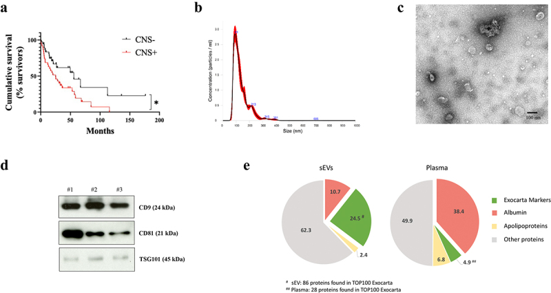
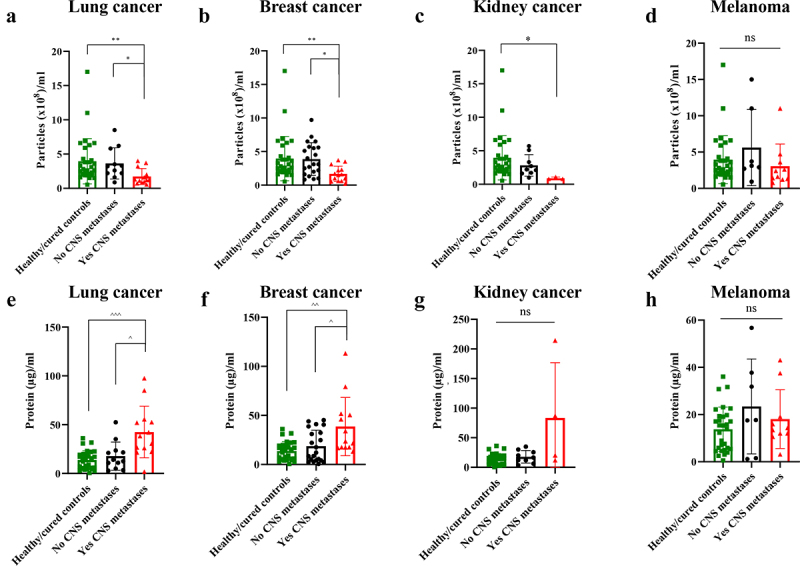
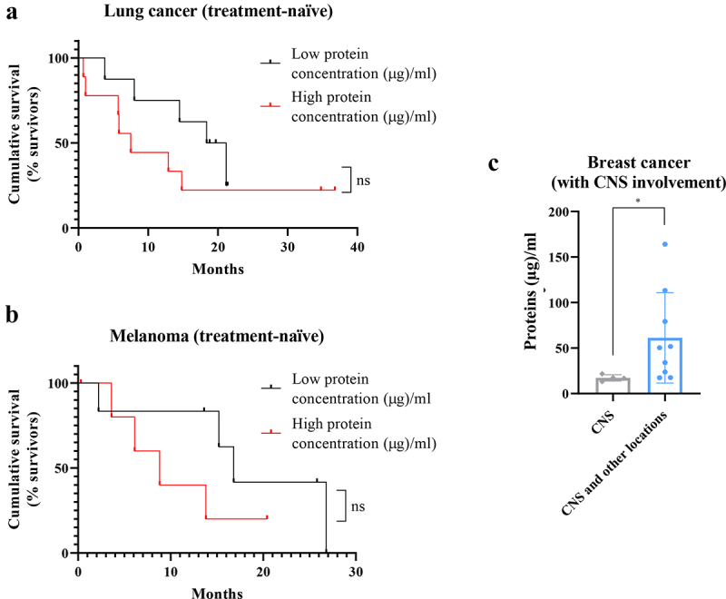
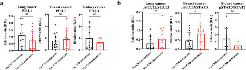
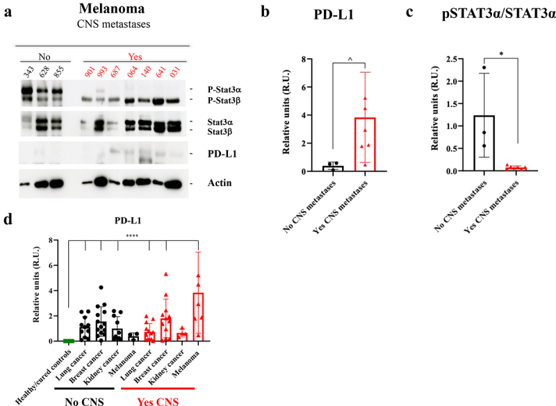
Nearly 40% of the advanced cancer patients will present brain metastases during the course of their disease, with a 2-year life expectancy of less than 10%. Immune system impairment, including the modulation of both STAT3 and PD-L1, is one of the hallmarks of brain metastases. Liquid biopsy could offer several advantages in brain metastases management, such as the possibility of noninvasive dynamic monitoring. Extracellular vesicles (EVs) have been recently proposed as novel biomarkers especially useful in liquid biopsy due to their secretion in biofluids and their role in cell communication during tumor progression. The main aim of this work was to characterize the size and protein cargo of plasma circulating EVs in patients with solid tumors and their correlation with newly diagnosed brain metastases, in addition to their association with other relevant clinical variables. We analyzed circulating EVs in the plasma of 123 patients: 42 patients with brain metastases, 50 without brain metastases and 31 healthy controls. Patients with newly diagnosed brain metastases had a lower number of circulating EVs in the plasma and a higher protein concentration in small EVs (sEVs) compared to patients without brain metastases and healthy controls. Interestingly, melanoma patients with brain metastases presented decreased STAT3 activation and increased PD-L1 levels in circulating sEVs compared to patients without central nervous system metastases. Decreased STAT3 activation and increased PD-L1 in plasma circulating sEVs identify melanoma patients with brain metastasis.

Keywords: PD-L1; STAT3; Small extracellular vesicles; brain metastasis; exosomes.


Conflict of interest statement

No potential conflict of interest was reported by the author(s).

Figures

Figure 1.
Figure 1.
Overall survival analysis of patients included in the study and characterization of plasma-circulating sEVs. a. Survival analysis and graphical representation using Kaplan-Meier curves showing the cumulative survival probability in the study population according to the absence (CNS-) or presence (CNS+) of central nervous system (CNS) metastases. * p value 0.02. Differences were assessed using the Log-Rank test. b. Representative image of the particle content (x108) by NTA analysis of a plasma sample from a melanoma patient. c. Representative electron microscopy imaging of sEVs from the same patient´s plasma. d. Representative Western blot of the analysis of exosome markers CD9, CD81, TSG101 and in sEVs isolated from the plasma of three different melanoma patients. Ponceau staining was used as loading control (see Supplementary Figure 4A). e. Proteomic analysis of sEVs and plasma paired samples derived from melanoma patients. Venn diagrams showing an enrichment of Exocarta markers (green) and a reduction albumin and apoliproteins (red) in sEVs (left diagram) compared to proteins detected in plasma samples (right diagram).
Figure 2.
Figure 2.
Quantitative analysis of plasma-circulating sEVs from patients included in the study according to their central nervous system (CNS) metastases status (including healthy/cured controls). a. Particles/ml in lung cancer. b. Particles/ml in breast cancer. c. Particles/ml in kidney cancer. d. Particles/ml in melanoma. e. Proteins/ml in lung cancer. f. Proteins/ml in breast cancer. g. Proteins/ml in kidney cancer. h. Proteins/ml in melanoma. * p value < .05. ** p value < .01. ^ p value < .05. ^^ p value 0.003. ^^^ p value 0.0003. ns: not significant.
Figure 3.
Figure 3.
Complementary studies for total protein concentration in patients plasma-circulating sEVs showing a correlation between a high protein amount and a worse prognosis. a. Survival analysis showing the cumulative survival probability in patients with previously untreated metastatic lung cancer according to the protein concentration in plasma-circulating sEVs (taking the median value of the group as reference). b. Survival analysis showing the cumulative survival probability in patients with previously untreated metastatic melanoma according to the protein concentration in plasma-circulating sEVs (taking the median value of the group as reference). Differences were assessed using the Log-Rank test. c. Analysis of protein concentration in circulating sEVs regarding the type of progression experienced in patients with metastatic breast cancer and known central nervous system (CNS) involvement: at CNS only or at CNS and other locations. * p value 0.02. ns: not significant.
Figure 4.
Figure 4.
Analysis of PD-L1 and pSTAT3/STAT3 expression by Western blot in plasma-circulating sEVs from patients with lung, breast or kidney cancer. a. Quantification of PD-L1 expression levels and statistical analysis of samples obtained. The data obtained for PD-L1 in densitometry were normalized to Ponceau values (see Supplementary Figure 4). b. Quantification of pSTAT3/STAT3 expression levels and statistical analysis of samples obtained. * p value 0.03. ns: not significant.
Figure 5.
Figure 5.
Study of pSTAT3/STAT3 and PD-L1 expression by Western blot in plasma-circulating sEVs from patients with melanoma. a. Western blot image of the expression of different proteins (indicated on the right) in sEVs from melanoma patients according to the absence (No) or presence (Yes) of central nervous system (CNS) metastases. Ponceau staining was used as loading control (see Supplementary Figure 4 F). b. Quantification of PD-L1 expression levels and statistical analysis of samples considered in A. The data obtained for PD-L1 in densitometry were normalized to Ponceau values (see Supplementary Figure 4). c. Quantification of pSTAT3α/STAT3α band expression levels and statistical analysis of samples considered in A. d. Quantification of PD-L1 expression levels and statistical analysis of the whole population of this study, including the healthy/cured controls group. The data obtained for PD-L1 in densitometry were normalized to Ponceau values (see Supplementary Figure 4). No CNS: Absence of CNS metastases. Yes CNS: Presence of CNS metastases. ^ p value 0.03. * p value 0.02. **** p value < .0001.

References

    1. Gavrilovic IT, Posner JB.. Brain metastases: epidemiology and pathophysiology. J Neurooncol. 2005;75(1):5–11. doi:10.1007/s11060-004-8093-6. - DOI - PubMed
    1. Tabouret E, Chinot O, Metellus P, Tallet A, Viens P, Gonçalves A. Recent trends in epidemiology of brain metastases: an overview. Anticancer Res. 2012;32(11):4655–4662. - PubMed
    1. Hall WA, Djalilian HR, Nussbaum ES, Cho KH. Long-term survival with metastatic cancer to the brain. Med Oncol Northwood Lond Engl. 2000;17(4):279–286. doi:10.1007/BF02782192. - DOI - PubMed
    1. Brastianos PK, Carter SL, Santagata S, Cahill DP, Taylor-Weiner A, Jones RT, Van Allen EM, Lawrence MS, Horowitz PM, Cibulskis K, et al. Genomic characterization of brain metastases reveals branched evolution and potential therapeutic targets. Cancer Discov. 2015;5(11):1164–1177. doi:10.1158/2159-8290.CD-15-0369. - DOI - PMC - PubMed
    1. Fischer GM, Jalali A, Kircher DA, Lee W-C, McQuade JL, Haydu LE, Joon AY, Reuben A, de Macedo MP, Carapeto FCL, et al. Molecular profiling reveals unique immune and metabolic features of melanoma brain metastases. Cancer Discov. 2019;9(5):628–645. doi:10.1158/2159-8290.CD-18-1489. - DOI - PMC - PubMed

Publication types