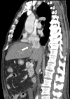
Hepatocellular Carcinoma With Right Atrium Metastases
- PMID: 35481312
- PMCID: PMC9033640
- DOI: 10.7759/cureus.23416
Hepatocellular Carcinoma With Right Atrium Metastases
Abstract
Hepatocellular carcinoma is common in patients with cirrhosis regardless of the etiology. Its presentation is uncommon in patients without known cirrhosis. It can spread commonly to the lungs, abdominal lymph nodes and bone but cardiac metastases are rare. The screening and early diagnosis impact the treatment feasibility and prognosis. The most common etiologies of cirrhosis and hepatocellular carcinoma are alcohol consumption and viral hepatitis B and C, however, non-alcoholic fatty liver disease (NAFLD) is becoming a more pronounced known risk factor for steatosis, advanced liver fibrosis, cirrhosis, and thus hepatocellular carcinoma due to the rise of metabolic syndrome prevalence. Although a known risk factor, there are no current recommendations for cancer surveillance in patients with NAFLD. The aim of this paper is to raise awareness of this rising complication by describing a rare initial presentation of hepatocellular carcinoma.
Keywords: cardiac metastases; cardiac tumours; chronic liver disease; cirrhosis; hepatocellular carcinoma.
Copyright © 2022, Teixeira et al.
Conflict of interest statement
The authors have declared that no competing interests exist.
Figures


References
-
- Hepatocellular carcinoma: a review. Balogh J, Victor D II, Asham EH, et al. https://www.ncbi.nlm.nih.gov/pmc/articles/PMC5063561/pdf/jhc-3-041.pdf. J Hepatocell Carcinoma. 2016;3:41–53. - PMC - PubMed
-
- EASL clinical practice guidelines: management of hepatocellular carcinoma. European Association for the Study of the Liver. J Hepatol. 2018;69:182–236. - PubMed
-
- Sensitivity of commonly available screening tests in detecting hepatocellular carcinoma in cirrhotic patients undergoing liver transplantation. Gambarin-Gelwan M, Wolf DC, Shapiro R, Schwartz ME, Min AD. Am J Gastroenterol. 2000;95:1535–1538. - PubMed
-
- Diagnosis, staging, and management of hepatocellular carcinoma: 2018 practice guidance by the American Association for the Study of Liver Diseases. Marrero JA, Kulik LM, Sirlin CB, et al. Hepatology. 2018;68:723–750. - PubMed
-
- Cardiac metastasis of hepatocellular carcinoma in a young non-cirrhotic patient, to the left ventricle. Tastekin E, Usta U, Ege T, Kazindir G, Kutlu AK. Ann Hepatol. 2012;11:392–394. - PubMed
Publication types
LinkOut - more resources
Full Text Sources
