

Targeted Delivery of DNA Topoisomerase Inhibitor SN38 to Intracranial Tumors of Glioblastoma Using Sub-5 Ultrafine Iron Oxide Nanoparticles
- PMID: 35481625
- PMCID: PMC9308697
- DOI: 10.1002/adhm.202102816
Targeted Delivery of DNA Topoisomerase Inhibitor SN38 to Intracranial Tumors of Glioblastoma Using Sub-5 Ultrafine Iron Oxide Nanoparticles
Abstract
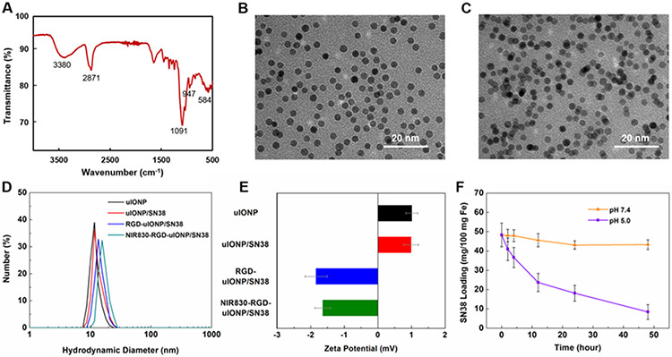
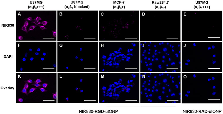
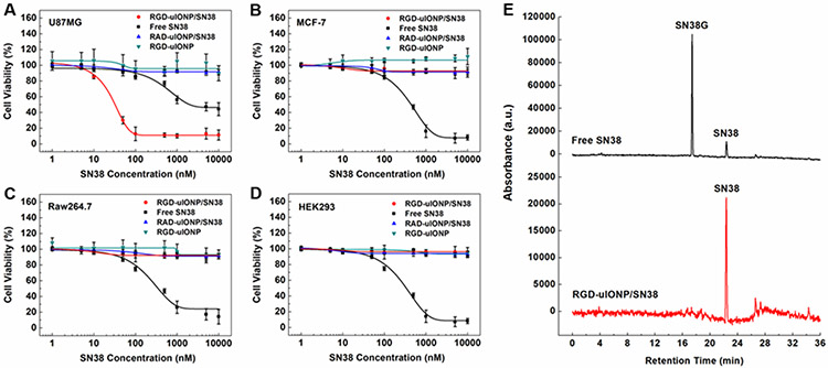
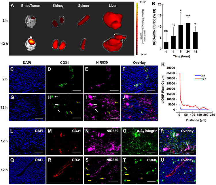
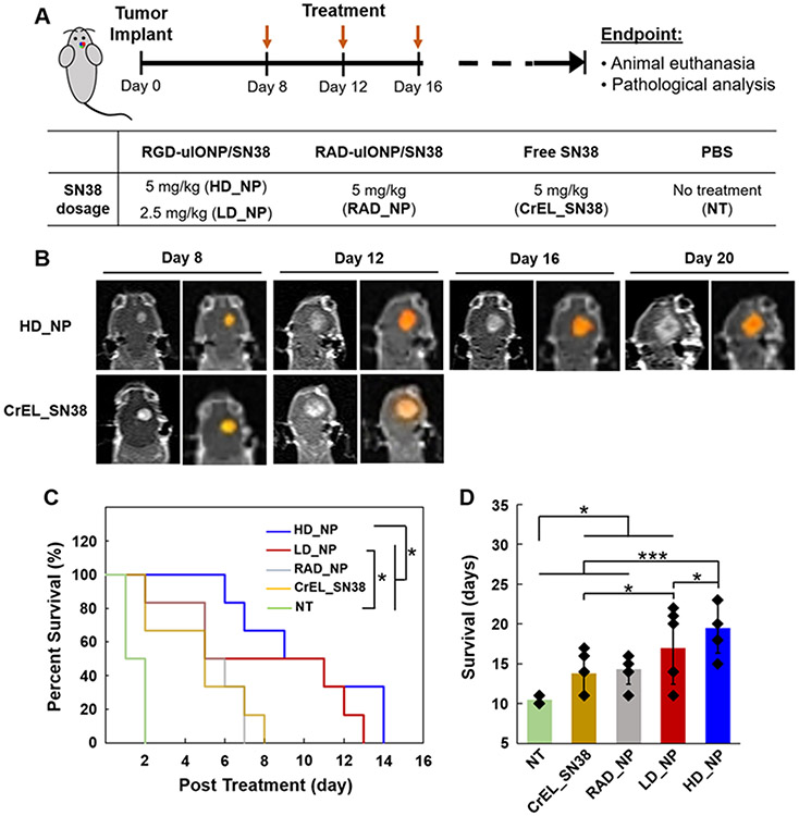
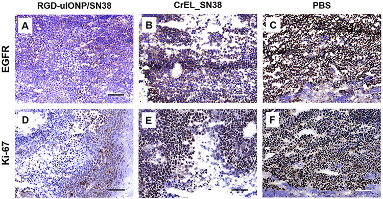
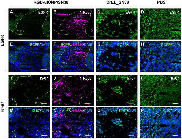
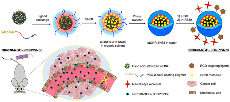
Effectively delivering therapeutics for treating brain tumors is hindered by the physical and biological barriers in the brain. Even with the compromised blood-brain barrier and highly angiogenic blood-tumor barrier seen in glioblastoma (GBM), most drugs, including nanomaterial-based formulations, hardly reach intracranial tumors. This work investigates sub-5 nm ultrafine iron oxide nanoparticles (uIONP) with 3.5 nm core diameter as a carrier for delivering DNA topoisomerase inhibitor 7-ethyl-10-hydroxyl camptothecin (SN38) to treat GBM. Given a higher surface-to-volume ratio, uIONP shows one- or three-folds higher SN38 loading efficiency (48.3 ± 6.1%, mg/mg Fe) than those with core sizes of 10 or 20 nm. SN38 encapsulated in the coating polymer exhibits pH sensitive release with <10% over 48 h at pH 7.4, but 86% at pH 5, thus being protected from converting to inactive glucuronide by UDP-glucuronosyltransferase 1A1. Conjugating αv β3 -integrin-targeted cyclo(Arg-Gly-Asp-D-Phe-Cys) (RGD) as ligands, RGD-uIONP/SN38 demonstrates targeted cytotoxicity to αv β3 -integrin-overexpressed U87MG GBM cells with a half-maximal inhibitory concentration (IC50 ) of 30.9 ± 2.2 nm. The efficacy study using an orthotopic mouse model of GBM reveals tumor-specific delivery of 11.5% injected RGD-uIONP/SN38 (10 mg Fe kg-1 ), significantly prolonging the survival in mice by 41%, comparing to those treated with SN38 alone (p < 0.001).
Keywords: SN38; brain tumors; drug delivery; glioblastoma; iron oxide nanoparticles; theranostics; tumor integrin.
© 2022 Wiley-VCH GmbH.
Conflict of interest statement
Conflict of interest
The authors declare no conflict of interest.
Figures
Similar articles
-
Actively targeted delivery of SN38 by ultrafine iron oxide nanoparticle for treating pancreatic cancer.Invest New Drugs. 2022 Jun;40(3):546-555. doi: 10.1007/s10637-022-01231-9. Epub 2022 Mar 15. Invest New Drugs. 2022. PMID: 35290548
-
A subtype specific probe for targeted magnetic resonance imaging of M2 tumor-associated macrophages in brain tumors.Acta Biomater. 2025 Mar 1;194:336-351. doi: 10.1016/j.actbio.2025.01.003. Epub 2025 Jan 11. Acta Biomater. 2025. PMID: 39805525
-
Doxorubicin-loaded iron oxide nanoparticles for glioblastoma therapy: a combinational approach for enhanced delivery of nanoparticles.Sci Rep. 2020 Jul 9;10(1):11292. doi: 10.1038/s41598-020-68017-y. Sci Rep. 2020. PMID: 32647151 Free PMC article.
-
Tumor-targeted nanotherapeutics: overcoming treatment barriers for glioblastoma.Wiley Interdiscip Rev Nanomed Nanobiotechnol. 2017 Jul;9(4):10.1002/wnan.1439. doi: 10.1002/wnan.1439. Epub 2016 Nov 4. Wiley Interdiscip Rev Nanomed Nanobiotechnol. 2017. PMID: 27813323 Free PMC article. Review.
-
Breaching barriers in glioblastoma. Part II: Targeted drug delivery and lipid nanoparticles.Int J Pharm. 2017 Oct 5;531(1):389-410. doi: 10.1016/j.ijpharm.2017.07.049. Epub 2017 Aug 9. Int J Pharm. 2017. PMID: 28801108 Review.
Cited by
-
Development of a Targeted SN-38-Conjugate for the Treatment of Glioblastoma.ACS Omega. 2024 Jan 4;9(2):2615-2628. doi: 10.1021/acsomega.3c07486. eCollection 2024 Jan 16. ACS Omega. 2024. PMID: 38250376 Free PMC article.
-
Shape-dependent cellular uptake of iron oxide nanorods: mechanisms of endocytosis and implications on cell labeling and cellular delivery.Nanoscale. 2024 Nov 28;16(46):21398-21415. doi: 10.1039/d4nr02408g. Nanoscale. 2024. PMID: 39329423 Free PMC article.
-
Research Progress of SN38 Drug Delivery System in Cancer Treatment.Int J Nanomedicine. 2024 Jan 26;19:945-964. doi: 10.2147/IJN.S435407. eCollection 2024. Int J Nanomedicine. 2024. PMID: 38293612 Free PMC article. Review.
-
Tumor integrin targeted theranostic iron oxide nanoparticles for delivery of caffeic acid phenethyl ester: preparation, characterization, and anti-myeloma activities.Front Pharmacol. 2024 Mar 6;15:1325196. doi: 10.3389/fphar.2024.1325196. eCollection 2024. Front Pharmacol. 2024. PMID: 38510655 Free PMC article.
-
Recent advances in nanomaterial-based brain-targeted delivery systems for glioblastoma therapy.Nanomedicine (Lond). 2025 Jun;20(12):1495-1511. doi: 10.1080/17435889.2025.2503694. Epub 2025 May 12. Nanomedicine (Lond). 2025. PMID: 40353316 Review.
References
-
- Hobbs SK, Monsky WL, Yuan F, Roberts WG, Griffith L, Torchilin VP, Jain RK, Proceedings of the National Academy of Sciences of the United States of America 1998, 95, 4607; - PMC - PubMed
- Monsky WL, Carreira CM, Tsuzuki Y, Gohongi T, Fukumura D, Jain RK, Clinical Cancer Research 2002, 8, 1008; - PubMed
- Pitz MW, Desai A, Grossman SA, Blakeley JO, Journal of Neuo-oncology 2011, 104, 629. - PMC - PubMed
-
- Banks WA, Nature Reviews Drug Discovery 2016, 15, 275. - PubMed
Publication types
MeSH terms
Substances
Grants and funding
LinkOut - more resources
Full Text Sources
Medical
