. 2022 May;31(5):e4301.
doi: 10.1002/pro.4301.

Functionalization of a symmetric protein scaffold: Redundant folding nuclei and alternative oligomeric folding pathways

Affiliations

Functionalization of a symmetric protein scaffold: Redundant folding nuclei and alternative oligomeric folding pathways

Connie A Tenorio et al. Protein Sci. 2022 May.

Abstract

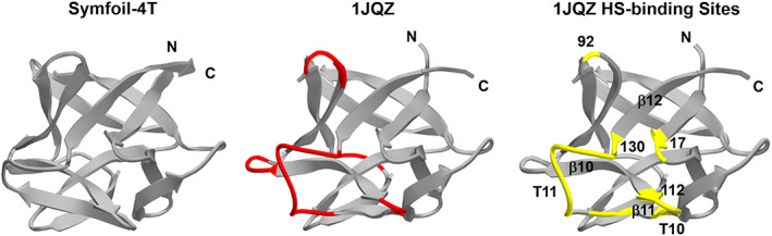
Successful de novo protein design ideally targets specific folding kinetics, stability thermodynamics, and biochemical functionality, and the simultaneous achievement of all these criteria in a single step design is challenging. Protein design is potentially simplified by separating the problem into two steps: (a) an initial design of a protein "scaffold" having appropriate folding kinetics and stability thermodynamics, followed by (b) appropriate functional mutation-possibly involving insertion of a peptide functional "cassette." This stepwise approach can also separate the orthogonal effects of the "stability/function" and "foldability/function" tradeoffs commonly observed in protein design. If the scaffold is a protein architecture having an exact rotational symmetry, then there is the potential for redundant folding nuclei and multiple equivalent sites of functionalization; thereby enabling broader functional adaptation. We describe such a "scaffold" and functional "cassette" design strategy applied to a β-trefoil threefold symmetric architecture and a heparin ligand functionality. The results support the availability of redundant folding nuclei within this symmetric architecture, and also identify a minimal peptide cassette conferring heparin affinity. The results also identify an energy barrier of destabilization that switches the protein folding pathway from monomeric to trimeric, thereby identifying another potential advantage of symmetric protein architecture in de novo design.

Keywords: de novo design; heparin affinity; oligomerization; protein folding; protein stability.


Conflict of interest statement

Michael Blaber is a cofounder and has equity ownership in Trefoil Therapeutics Inc.

Figures

FIGURE 1
FIGURE 1
Conserved and variable tertiary structure in β‐trefoil proteins. “Ribbon” diagrams of representative β‐trefoil proteins clostridium neurotoxin (1EPW), inositol 1,4,5P3 receptor (1N4K), and C. cinereal protease inhibitor (3VWC). The conserved fundamental β‐trefoil structural region is indicated by gray shading, and the variable regions are colored. Also shown is the structure of the Symfoil‐4T de novo designed symmetric protein (3O4B). The variable regions of the different β‐trefoil proteins describe surface loops conferring functionality; in contrast, Symfoil‐4T is an essential β‐trefoil “scaffold” devoid of function. The C3 axis of rotational symmetry, characteristic of the β‐trefoil architecture, is aligned vertically in this view. Also indicated are the locations of the N‐ and C‐termini, which are in proximity to each other and define a “discontinuous surface turn” in the overall symmetric architecture
FIGURE 2
FIGURE 2
Size exclusion chromatography (SEC) of HSM1, 2, and 3 proteins in phosphate buffer. The chromatograms of individual HSM1, 2, and 3 proteins resolved on Superdex 200 SEC are provided; also shown are mass standards (see text for details) and the indicated mass is kDa
FIGURE 3
FIGURE 3
Size exclusion chromatography (SEC) chromatograms of half and quarter HSM1 proteins. Mutant proteins were resolved on Superdex 200 SEC in phosphate buffer. HSM1 and Symfoil‐4T reference chromatograms are also shown. Mass standards are omitted for clarity, but are identical to those provided in Figure 2
FIGURE 4
FIGURE 4
Analytical ultracentrifugation (AUC) data and derived sedimentation coefficients for Symfoil‐4T and heparin sulfate mutants (HSM) proteins. AUC analysis of HSM proteins was performed in phosphate buffer pH 7.0, 293 K, and with protein concentrations of 10–16 μM
FIGURE 5
FIGURE 5
Isothermal equilibrium denaturation (IED) analysis of HSM1, 2, and 3 mutants and Symfoil‐4T. IED analysis of heparin sulfate mutants proteins was performed in phosphate buffer pH 7.0, 298 K, and with protein concentrations of 5.0 μM
FIGURE 6
FIGURE 6
Isothermal equilibrium denaturation (IED) of half and quarter cassette HSM1 proteins. IED analysis of heparin sulfate mutants proteins was performed in phosphate buffer pH 7.0, 298 K, and with protein concentrations of 5.0 μM
FIGURE 7
FIGURE 7
Analytical heparin Sepharose chromatography of heparin sulfate mutants proteins and HS peptide cassette. Upper panel: HSM1, 2, and 3 proteins. The chromatogram covers the [NaCl] gradient subsequent to loading and washing with phosphate buffer. The Symfoil‐4T protein does not bind and elutes in the column wash. The [NaCl] gradient used to elute bound proteins is indicated by the dotted line. Also included with the set of HS mutants is the 24‐mer HS cassette peptide, and the fibroblast growth factor‐1 (FGF‐1) protein. Lower panel: Half and quarter HS cassette mutant proteins. Only the LH cassette and LQ2 cassette mutants bind to Heparin Sepharose; the RH, LQ1, RQ1, and RQ2 mutants are present in the column load flow through. Also included for reference in this figure are the elution profiles of the 24‐mer HS cassette peptide and the HSM1 protein
FIGURE 8
FIGURE 8
Design of a heparin sulfate (HS)‐binding 24‐mer peptide “cassette” derived from bovine and human FGF‐1 and human FGF‐2. The sequences of human/bovine FGF‐1 and human FGF‐2 in the region 108–131 (using the numbering scheme of the 140 amino acid form of human FGF‐1) which form the majority of the HS ligand binding interactions are shown color coded by protein. Boxed positions indicate conserved amino acid positions between all three proteins. Blue positions in the designed HS binding cassette indicate conserved hydrophobic core residues in the Symfoil‐4T scaffold protein, and were therefore retained. The magenta Ala117 positions is an optimized mutation designed to eliminate the reactive Cys thiol at this position. Half‐ and quarter‐cassette designs are also indicated. The origin of residues in the HS binding cassette based upon a consensus or composite of sequences is indicated by color code
FIGURE 9
FIGURE 9
24‐mer heparin sulfate (HS)‐binding “cassette” and sites of insertion in the Symfoil‐4T β‐trefoil “scaffold.” The designed HS‐binding cassette 24‐mer peptide is indicated in the shaded red box. The equivalent structural location in FGF‐1 is indicated in top panel (“Heparin binding mutant #1”). The center and lower panel indicate two equivalent positions related by the threefold rotational symmetry intrinsic to the Symfoil‐4T protein, generating the HSM2 and HSM3 mutants, respectively
FIGURE 10
FIGURE 10
Structurally heterogenous surface loops and heparin sulfate (HS)‐binding positions in FGF‐1/FGF‐2. The structurally heterogenous surface loops in FGF‐1 (1JQZ) compared to Symfoil‐4T are indicated in red (center image). The positions associated with HS‐binding interactions in human/bovine FGF‐1 and FGF‐2 (Table S1) are shown in yellow (right image). Most of the surface loops unique to FGF‐1/FGF‐2 compared to the fundamental β‐trefoil architecture represented by Symfoil‐4T are associated with HS‐binding functionality
FIGURE 11
FIGURE 11
Relationship between oligomeric state and stability for heparin sulfate (HS) mutants. A transition from monomeric to trimeric oligomerization for the set of HS mutants occurs upon a destabilization greater than ~25 kJ/mol

Similar articles

Cited by

References

    1. Sawyer N, Speltz EB, Regan L. NextGen protein design. Biochem Soc Trans. 2013;41:1131–1136. - PMC - PubMed
    1. Chevalier A, Silva D‐A, Rocklin GJ, et al. Massively parallel de novo protein design for targeted therapeutics. Nature. 2017;550:74–79. - PMC - PubMed
    1. Gwyther REA, Jones DD, Worthy HL. Better together: Building protein oligomers naturally and by design. Biochem Soc Trans. 2019;47:1773–1780. - PMC - PubMed
    1. Zhou W, Šmidlehner T, Jerala R. Synthetic biology principles for the design of protein with novel structures and functions. FEBS Lett. 2020;594:2199–2212. - PubMed
    1. Zhang Y, Chen Y, Wang C, et al. ProDCoNN: Protein design using a convolutional neural network. Proteins. 2020;88:819–829. - PMC - PubMed

Publication types

LinkOut - more resources