. 2022 Jul;24(7):1567-1582.
doi: 10.1016/j.gim.2022.03.014. Epub 2022 Apr 28.

A novel DPH5-related diphthamide-deficiency syndrome causing embryonic lethality or profound neurodevelopmental disorder

Collaborators, Affiliations

A novel DPH5-related diphthamide-deficiency syndrome causing embryonic lethality or profound neurodevelopmental disorder

Suma P Shankar et al. Genet Med. 2022 Jul.

Erratum in

Abstract

Purpose: Diphthamide is a post-translationally modified histidine essential for messenger RNA translation and ribosomal protein synthesis. We present evidence for DPH5 as a novel cause of embryonic lethality and profound neurodevelopmental delays (NDDs).

Methods: Molecular testing was performed using exome or genome sequencing. A targeted Dph5 knockin mouse (C57BL/6Ncrl-Dph5em1Mbp/Mmucd) was created for a DPH5 p.His260Arg homozygous variant identified in 1 family. Adenosine diphosphate-ribosylation assays in DPH5-knockout human and yeast cells and in silico modeling were performed for the identified DPH5 potential pathogenic variants.

Results: DPH5 variants p.His260Arg (homozygous), p.Asn110Ser and p.Arg207Ter (heterozygous), and p.Asn174LysfsTer10 (homozygous) were identified in 3 unrelated families with distinct overlapping craniofacial features, profound NDDs, multisystem abnormalities, and miscarriages. Dph5 p.His260Arg homozygous knockin was embryonically lethal with only 1 subviable mouse exhibiting impaired growth, craniofacial dysmorphology, and multisystem dysfunction recapitulating the human phenotype. Adenosine diphosphate-ribosylation assays showed absent to decreased function in DPH5-knockout human and yeast cells. In silico modeling of the variants showed altered DPH5 structure and disruption of its interaction with eEF2.

Conclusion: We provide strong clinical, biochemical, and functional evidence for DPH5 as a novel cause of embryonic lethality or profound NDDs with multisystem involvement and expand diphthamide-deficiency syndromes and ribosomopathies.

Keywords: Nonverbal neurodevelopment delays; Novel gene discovery; Precision animal modeling; Precision genomics; Translational genetics.


Conflict of interest statement

Conflicts of Interest K.G.M. is an employee of GeneDx, Inc. K.M. and U.B. are members of and employed by Roche Pharma Research & Early Development. Roche is interested in targeted therapies and diagnostics. All other authors declare no conflicts of interest.

Figures
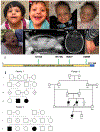
Figure 1
Figure 1. Clinical features of individuals with DPH5 variants.
A., B. Siblings from family 1. C., D. Siblings from family 2. E-G. Proband from family 3. Craniofacial appearance, including sparse hair and eyebrows, broad forehead with frontal bossing, epicanthal folds, broad nasal bridge, upturned nasal tip, and triangular chin, seen in all individuals; down turned corners of the mouth seen in individuals (A) and (E). B. Poor dentition. F. Chest and abdominal X-ray with pneumoperitoneum owing to perforated bowel. G. Axial view of head computed tomography showing bilateral minimal tentorial subdural hemorrhage and enlarged cisterna magna. H. Brachydactyly of toes for individual (B) from family 1. I. Schematic of DPH5 protein showing that all variants identified in the 3 families fall within the conserved domain diphthine methyl ester synthase. J. Family pedigrees of 3 families harboring DPH5 variants. Pedigree 1 is for family 1 with 2 affected siblings born to a consanguineous family from Syria. Pedigree 2 is for family 2 with 2 affected siblings of European ancestry from Boston. Pedigree 3 is for family 3 from Saudi Arabia showing additional affected sibling and an affected first cousin.
Figure 2
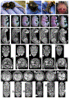
Figure 2. Images of prenatal and postnatal homozygous Dph5 mice.
A-C. Homozygous female Dph5_pH260R mouse aged at 21 days presenting with decreased body size, dysmorphic head morphology, depigmented patch on abdomen, and polydactyly of left hind foot. D. Postmortem photo at age 24 days with observed hemorrhage around the lambdoid skull sutures. E., F. Photomicrographs of embryonic day (ED) 18.5(1) Wild type (WT) and (2-7) homozygous Dph5 embryos with their corresponding micro computed tomography (μCT) images (sagittal sections) showing: (4, 7) facial clefts, (7) exencephaly, (5,6) microphthalmia, (7) anophthalmia, (4-6) shortened frontonasal prominence, and (5,6) vascular hemorrhage with edema. G., H. μCT images (transverse sections) of ED 18.5 (1) WT and (2, 4, 6, 7) homozygous Dph5 embryos through (7) head and (8) heart showing facial cleft (arrow i), microphthalmia (arrow ii), anophthalmia with exencephaly (arrow iii), situs inversus of the dorsal aorta (arrow iv) (and arch aorta, not shown), and ventricular septal defects (arrow v). Arrows in(H, 1) WT show normal position of dorsal aorta and closed heart interventricular septum. I., J. μCT images of visceral cavity (transverse section), and frontal sections of (1) WT and (2-6) homozygous embryos showing diaphragmatic hernia (arrow vi), edema in pleural cavity, lymphatic sacs, brain vesicles and trunk (arrow vii), hypoplastic/absent pineal gland (arrow viii), and hypoplastic stomach (arrow ix).
Figure 3
Figure 3. Homology models of wild type (WT), p.His260Arg, p.Asn110Ser, p Arg207Ter, and p.Asn174LysfsTer10 variants of human DPH5 and interaction with eEF2.
A. Human WT DPH5 dimer (gray/golden) in complex with eEF2 model (green). B. Zoomed image of (A), p.His260 interacts favorably with p.Arg711 and p.Lys582 of eEF2. In yeast DPH5, p.His260 corresponds to p.His257, interacting in the same way with eEF2.16. C., D. p.His260Arg homology modeling fold is identical to the WT. p.Arg260 will be positioned at the protein interface interacting with eEF2. In the S cervisae system, the corresponding residue interacts with p.Arg711 and p.Lys582 of eEF2; the p.His260Arg variant will introduce repulsion owing to its positive charge and weakened protein–protein interaction. C. Sideview (eEF2 interacting from the right; not shown). D. Rotated (90°) zoom-in facing the interaction region binding to eEF2. E. p.Asn110 is located at the DPH5 monomer–monomer interface region. p.Asn110Ser variant may weaken the dimer stability and thus lead to malfunctioning DPH5-eEF2 interaction. It is also possible that exchange of SAM (as eEF2 is methylated 4 times by DPH5) is impaired by the variant. F., G. p.Arg207Ter truncation variant. F. This truncation will remove a large part of the region interacting with eEF2, including p.His260 (dark blue showing the truncated parts of monomer 1), as well as SAM binding (light blue showing the truncated part of monomer 2). It can thus be expected that DPH5–eEF2 interaction as well as DPH5 functionality is severely disrupted. G. DPH5 truncated at p.Arg207, showing the exposed SAM and the much smaller area of interaction toward eEF2. H., I. The p.Asn174LysfsTer10 introduces a stop codon after p.Leu182 in the p.Asn174LysfsTer10 variant, leading to premature truncation and reduced area of interaction with eEF2. I. The interaction between the p.Asn174LysfsTer10 variant DPH5 and eEF2 is significantly reduced, and SAM is largely solvent exposed. ACP, 3-amino-3carboxypropyl; SAM, S-adenosyl methionine.
Figure 4
Figure 4. XXX.
A. ADPR assays indicate loss of function of DPH5 p.Arg207Ter and p.Asn174LysfsTer10. Radioimmunoprecipitation assay extracts of MCF7 DPH5-knockout (KO) cells transfected with expression plasmids encoding DPH5 WT, p.Asn174LysfsTer10, p.Asn110Ser, p.Arg207Ter, and p.His260Arg were incubated with DT and biotinylated nicotinamide adenine dinucleotide (Bio-NAD) to assess the presence or absence of diphthamide. Successful ADPR with Bio-NAD as substrate generates biotinylated eEF2, which can be detected in sodium dodecyl sulfate–polyacrylamide gel electrophoresis blots probed with streptavidin-horseradish peroxidase and peroxidase substrate. Presence of diphthamide (generated by recombinant DPH5 in the DPH5-KO background) therefore produces a band at approximately 100 kDa and is seen with WT, p.Asn110Ser, and p.His260Arg. Reactions without DT and extracts of nontransfected or mock-transfected cells serve as controls. B-D. Assays diagnostic for yeast diphthamide modification capacity in vivo. B. ADPR assay with total protein extracts of yeast BY4741 strains WT (DPH5), dph5Δ, p.Asn111Ser, and p.His257Arg. Extracts were incubated with DT and Bio-NAD to assess the presence or absence of diphthamide. Presence of diphthamide results in successful ADPR and produces a band at approximately 100 kDa as seen with WT and p.Asn111Ser and p.His257Arg variants. Reactions without DT serve as controls. C. Phenotypic spot assay investigating growth inhibition by the diphthamide-dependent DT. Cells carrying the galactose inducible DT expression vector pSU8 were 10-fold serially diluted and cultivated on medium containing the indicated concentrations (%[weight/volume]) of raf and/or gal at 30 °C for 3 days. Cells with the 2 variants p.Asn111Ser and p.His257Arg exhibited higher tolerance to DT than WT but much less than the cells with dph5Δ. D. Assay displaying sensitivity to diphthamide-indicative translation inhibitor drug hygromycin B was examined by cultivation on medium containing the indicated doses of hygromycin B at 30 °C for 2 days. Cells with the p.Asn111Ser and p.His257Arg variants were more sensitive to hygromycin than WT cells but less sensitive than cells with dph5Δ. ADPR, adenosine diphosphate-ribosylation; DT, diphtheria toxin; gal, galatose; raf, raffinose; WT, wild type.

Similar articles

Cited by

References

    1. Gilissen C, Hehir-Kwa JY, Thung DT, et al. Genome sequencing identifies major causes of severe intellectual disability. Nature. 2014;511(7509):344–347. 10.1038/nature13394. - DOI - PubMed
    1. Ismail FY, Shapiro BK. What are neurodevelopmental disorders? Curr Opin Neurol. 2019;32(4):611–616. 10.1097/WCO.0000000000000710. - DOI - PubMed
    1. Lee H, Nelson SF. The frontiers of sequencing in undiagnosed neurodevelopmental diseases. Curr Opin Genet Dev. 2020;65:76–83. 10.1016/j.gde.2020.05.003. - DOI - PMC - PubMed
    1. Vissers LE, Gilissen C, Veltman JA. Genetic studies in intellectual disability and related disorders. Nat Rev Genet. 2016;17(1):9–18. 10.1038/nrg3999. - DOI - PubMed
    1. Soden SE, Saunders CJ, Willig LK, et al. Effectiveness of exome and genome sequencing guided by acuity of illness for diagnosis of neurodevelopmental disorders. Sci Transl Med. 2014;6(265):265ra168. 10.1126/scitranslmed.3010076. - DOI - PMC - PubMed

Publication types

MeSH terms