

Assessment of malignancy and PSMA expression of uncertain bone foci in [18F]PSMA-1007 PET/CT for prostate cancer-a single-centre experience of PET-guided biopsies
- PMID: 35482114
- PMCID: PMC9399054
- DOI: 10.1007/s00259-022-05745-5
Assessment of malignancy and PSMA expression of uncertain bone foci in [18F]PSMA-1007 PET/CT for prostate cancer-a single-centre experience of PET-guided biopsies
Abstract
Purpose: Uncertain focal bone uptake (UBU) with intensive radiopharmaceutical avidity are frequently observed in patients undergoing [18F]PSMA-1007 PET/CT for the detection of prostate cancer (PC). Such foci can pose diagnostic conundrums and risk incorrect staging. The aim of this short communication is to share the results of PET-guided biopsies of such foci.
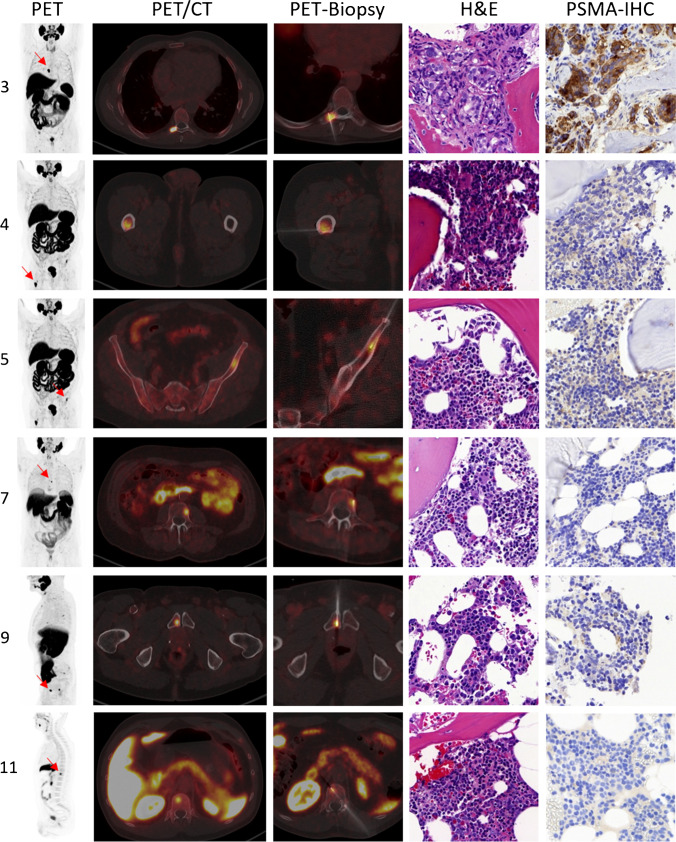
Methods: A retrospective analysis revealed 10 patients who were referred to our department for PET-guided biopsy of UBU visible in a previous [18F]PSMA-1007 PET/CT. [18F]-PSMA-1007 PET-guided biopsy was conducted for 11 PSMA-avid bone foci in these 10 patients. The biopsy materials were analysed for tissue typing, and immunohistochemistry (IHC) was performed for prostate-specific-membrane-antigen (PSMA) expression. The scans were analysed by two experienced physicians in a consensus read for clinical characteristics and radiopharmaceutical uptake of foci.
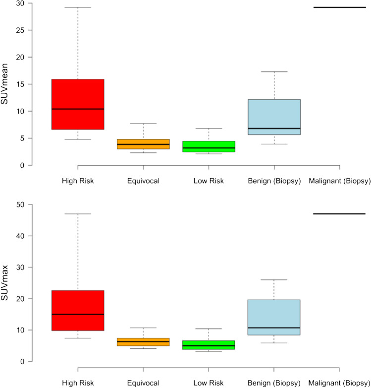
Results: One out of 11 (9.1%) of the foci biopsied was confirmed as bone metastasis of PC with intense PSMA-expression, while 10/11 (90.9%) foci were revealed to be unremarkable bone tissue without evidence of PSMA expression at IHC. Amongst all bone foci assessed by biopsy, eight were visually classified as being at high risk of malignancy in the PET/CT (SUVmean 12.0 ± 8.1; SUVmax 18.8 ± 13.1), three as equivocal (SUVmean 4.6 ± 2.1; SUVmax 7.2 ± 3.0) and zero as low risk. No UBU had any CT correlate.
Conclusions: This cohort biopsy revealed that a small but relevant number of UBU are true metastases. For those confirmed as benign, no PSMA expression at IHC was observed, suggesting a non-PSMA mediated cause for intensive [18F]PSMA-1007 uptake of which the reason remains unclear. Readers must interpret such foci with caution in order to reduce the risk of erroneous staging and subsequent treatment. PET-guided biopsy, particularly in the absence of morphological changes in the CT, can be a useful method to clarify such foci.
Keywords: Biopsy; PET-guided; PET/CT; PSMA; Prostate cancer; Prostate-specific membrane antigen; [18F]-PSMA-1007.
© 2022. The Author(s).
Conflict of interest statement
The authors declare no competing interests.
Figures
References
-
- Afshar-Oromieh A, Malcher A, Eder M, Eisenhut M, Linhart HG, Hadaschik BA, et al. PET imaging with a [68Ga]gallium-labelled PSMA ligand for the diagnosis of prostate cancer: biodistribution in humans and first evaluation of tumour lesions. Eur J Nucl Med Mol Imaging. 2013;40:486–495. doi: 10.1007/s00259-012-2298-2. - DOI - PubMed
-
- Grunig H, Maurer A, Thali Y, Kovacs Z, Strobel K, Burger IA, et al. Focal unspecific bone uptake on [(18)F]-PSMA-1007 PET: a multicenter retrospective evaluation of the distribution, frequency, and quantitative parameters of a potential pitfall in prostate cancer imaging. Eur J Nucl Med Mol Imaging. 2021 doi: 10.1007/s00259-021-05424-x. - DOI - PMC - PubMed
MeSH terms
Substances
LinkOut - more resources
Full Text Sources
Other Literature Sources
Medical
Miscellaneous
