

Preliminary Research: Application of Non-Invasive Measure of Cytochrome c Oxidase Redox States and Mitochondrial Function in a Porcine Model of Carbon Monoxide Poisoning
- PMID: 35482181
- PMCID: PMC9198167
- DOI: 10.1007/s13181-022-00892-5
Preliminary Research: Application of Non-Invasive Measure of Cytochrome c Oxidase Redox States and Mitochondrial Function in a Porcine Model of Carbon Monoxide Poisoning
Erratum in
-
Correction to: Preliminary Research: Application of Non-Invasive Measure of Cytochrome c Oxidase Redox States and Mitochondrial Function in a Porcine Model of Carbon Monoxide Poisoning.J Med Toxicol. 2023 Jan;19(1):53. doi: 10.1007/s13181-022-00913-3. J Med Toxicol. 2023. PMID: 36508082 Free PMC article. No abstract available.
Abstract
Introduction: Carbon monoxide (CO) is a colorless and odorless gas that is a leading cause of environmental poisoning in the USA with substantial mortality and morbidity. The mechanism of CO poisoning is complex and includes hypoxia, inflammation, and leukocyte sequestration in brain microvessel segments leading to increased reactive oxygen species. Another important pathway is the effects of CO on the mitochondria, specifically at cytochrome c oxidase, also known as Complex IV (CIV). The purpose of this ongoing study is the preliminary development of a porcine model of CO poisoning for investigation of alterations in brain mitochondrial physiology.
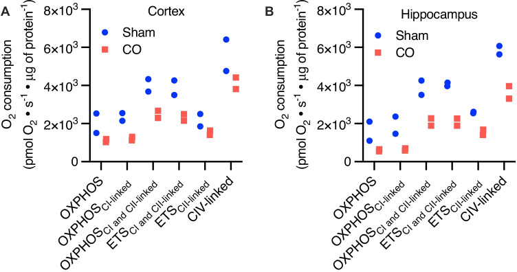
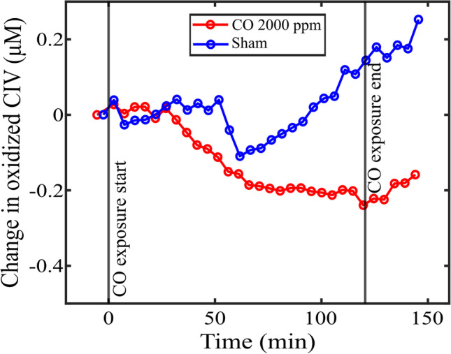
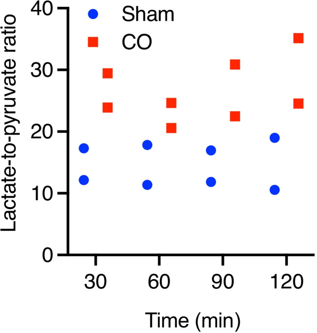
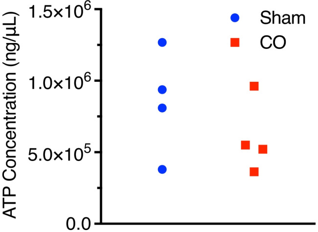
Methods: Four pigs (10 kg) were divided into two groups: Sham (n = 2) and CO (n = 2). Administration of a dose of CO at 2000 ppm to the CO group over 120 minutes followed by 30 minutes of re-oxygenation at room air. The control group received room air for 150 minutes. Non-invasive optical monitoring was used to measure CIV redox states. Cerebral microdialysis was performed to obtain semi real-time measurements of cerebral metabolic status. At the end of the exposure, fresh brain tissue (cortical and hippocampal) was immediately harvested to measure mitochondrial respiration. Snap frozen cortical tissue was also used for ATP concentrations and western blotting.
Results: While a preliminary ongoing study, animals in the CO group showed possible early decreases in brain mitochondrial respiration, citrate synthase density, CIV redox changes measured with optics, and an increase in the lactate-to-pyruvate ratio.
Conclusions: There is a possible observable phenotype highlighting the important role of mitochondrial function in the injury of CO poisoning.
Keywords: Basic science; Biomarker; Carbon monoxide; Mitochondria; Optics.
© 2022. American College of Medical Toxicology.
Conflict of interest statement
None
Figures
References
-
- Pepe G, Castelli M, Nazerian P, Vanni S, Del Panta M, Gambassi F, et al. Delayed neuropsychological sequelae after carbon monoxide poisoning: predictive risk factors in the Emergency Department. A retrospective study. Scand J Trauma Resusc Emerg Med. 2011;19:16. doi: 10.1186/1757-7241-19-16. - DOI - PMC - PubMed
Publication types
MeSH terms
Substances
Grants and funding
- R01NS113945/NS/NINDS NIH HHS/United States
- R03HL154232/HL/NHLBI NIH HHS/United States
- R03 HL154232/HL/NHLBI NIH HHS/United States
- P30 ES013508/ES/NIEHS NIH HHS/United States
- R56 HL158696/HL/NHLBI NIH HHS/United States
- K08 HL136858/HL/NHLBI NIH HHS/United States
- R01 NS113945/NS/NINDS NIH HHS/United States
- R21 ES031243/ES/NIEHS NIH HHS/United States
- R56HL158696/HL/NHLBI NIH HHS/United States
- R01 HL141386/HL/NHLBI NIH HHS/United States
- R21ES031243/ES/NIEHS NIH HHS/United States
- R01HL141386/HL/NHLBI NIH HHS/United States
- K08HL136858/HL/NHLBI NIH HHS/United States
LinkOut - more resources
Full Text Sources
Medical
