

Isolating Anti-amyloid Antibodies from Yeast-Displayed Libraries
- PMID: 35482203
- PMCID: PMC9351425
- DOI: 10.1007/978-1-0716-2285-8_22
Isolating Anti-amyloid Antibodies from Yeast-Displayed Libraries
Abstract
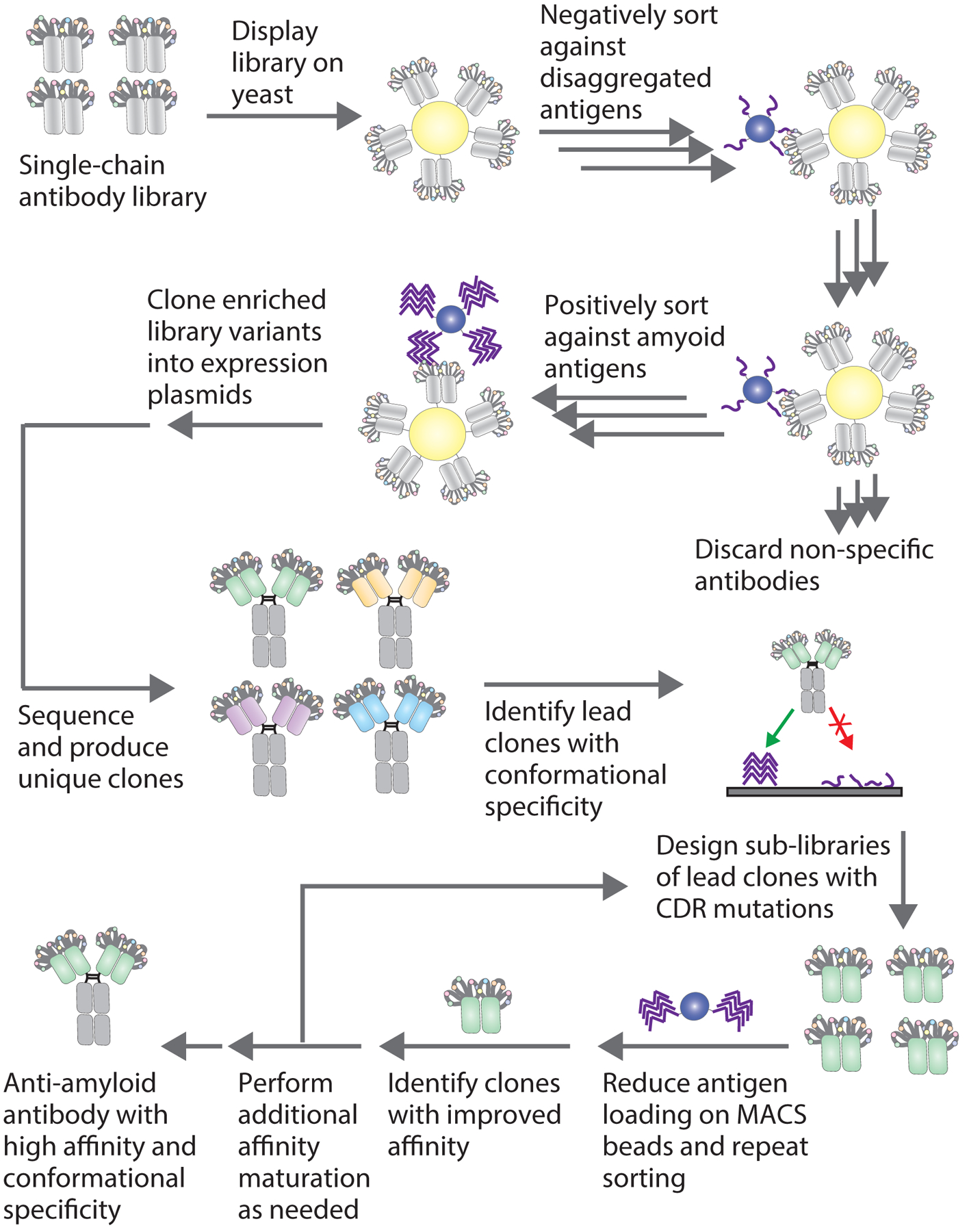
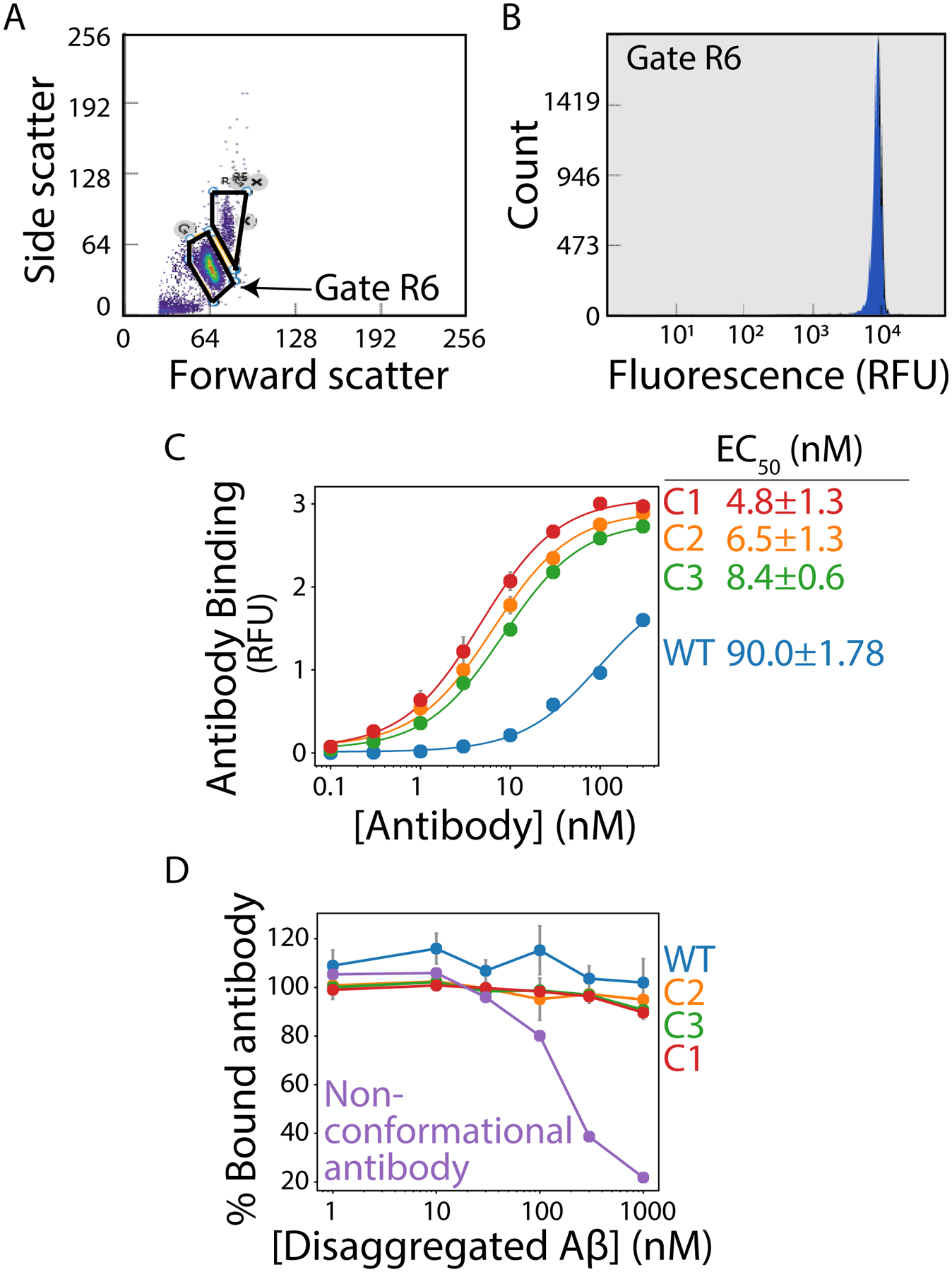
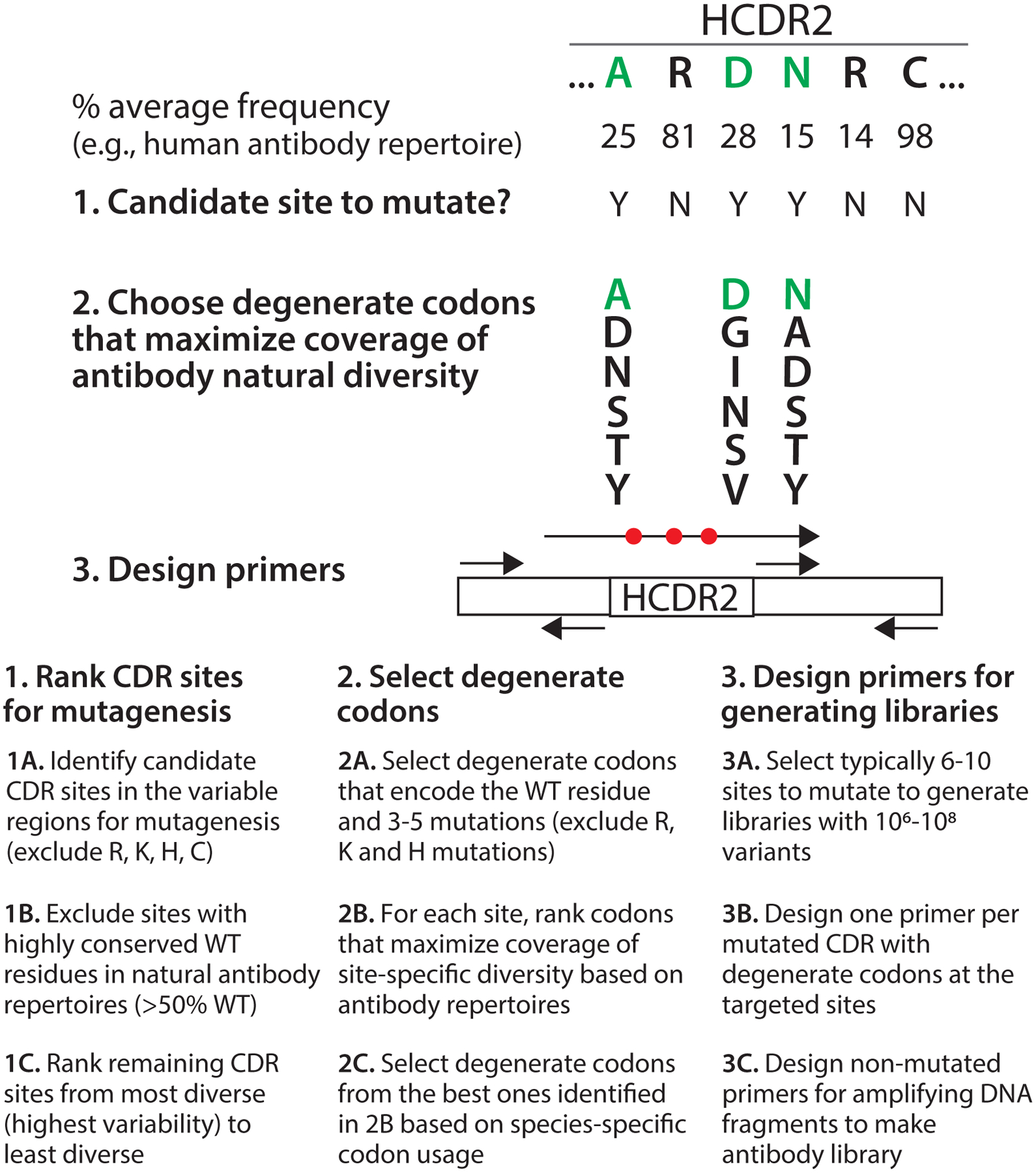
Conformational antibodies specific for amyloid-forming peptides and proteins are important for a range of biomedical applications, including detecting, inhibiting, and potentially treating protein aggregation disorders ranging from Alzheimer's to Parkinson's diseases. Generation of anti-amyloid antibodies is greatly complicated by the complex, heterogeneous and insoluble nature of amyloid antigens. Here we describe systematic methods for isolating and affinity maturing anti-amyloid antibodies using yeast surface display. Magnetic-activated cell sorting is used to sort single-chain antibody libraries positively for binding to amyloid antigens and negatively against the corresponding disaggregated antigens to remove antibodies that bind in a conformation-independent manner. Isolated lead antibody clones with conformational specificity are affinity matured via targeted CDR mutagenesis and magnetic-activated cell sorting.
Keywords: Aggregate; Aggregation; Alzheimer’s; Conformation; Conformational; Directed evolution; Fiber; Fibril; Oligomer; Parkinson’s; Yeast surface display.
© 2022. The Author(s), under exclusive license to Springer Science+Business Media, LLC, part of Springer Nature.
Conflict of interest statement
CONFLICTS OF INTEREST
There are no conflicts of interest.
Figures
Similar articles
-
Rational affinity maturation of anti-amyloid antibodies with high conformational and sequence specificity.J Biol Chem. 2021 Jan-Jun;296:100508. doi: 10.1016/j.jbc.2021.100508. Epub 2021 Mar 4. J Biol Chem. 2021. PMID: 33675750 Free PMC article.
-
Nature-inspired design and evolution of anti-amyloid antibodies.J Biol Chem. 2019 May 24;294(21):8438-8451. doi: 10.1074/jbc.RA118.004731. Epub 2019 Mar 27. J Biol Chem. 2019. PMID: 30918024 Free PMC article.
-
Sensitive detection of glucagon aggregation using amyloid fibril-specific antibodies.Biotechnol Bioeng. 2019 Aug;116(8):1868-1877. doi: 10.1002/bit.26994. Epub 2019 May 8. Biotechnol Bioeng. 2019. PMID: 30982957
-
Simulation Studies of Amyloidogenic Polypeptides and Their Aggregates.Chem Rev. 2019 Jun 26;119(12):6956-6993. doi: 10.1021/acs.chemrev.8b00731. Epub 2019 Apr 11. Chem Rev. 2019. PMID: 30973229 Review.
-
Impact of Amyloid Polymorphism on Prion-Chaperone Interactions in Yeast.Viruses. 2019 Apr 16;11(4):349. doi: 10.3390/v11040349. Viruses. 2019. PMID: 30995727 Free PMC article. Review.
Cited by
-
Development of a pan-tau multivalent nanobody that binds tau aggregation motifs and recognizes pathological tau aggregates.Biotechnol Prog. 2024 Sep-Oct;40(5):e3463. doi: 10.1002/btpr.3463. Epub 2024 Apr 3. Biotechnol Prog. 2024. PMID: 38568030 Free PMC article.
-
Quantitative flow cytometric selection of tau conformational nanobodies specific for pathological aggregates.Front Immunol. 2023 Aug 9;14:1164080. doi: 10.3389/fimmu.2023.1164080. eCollection 2023. Front Immunol. 2023. PMID: 37622125 Free PMC article.
-
Optimization of therapeutic antibodies for reduced self-association and non-specific binding via interpretable machine learning.Nat Biomed Eng. 2024 Jan;8(1):45-56. doi: 10.1038/s41551-023-01074-6. Epub 2023 Sep 4. Nat Biomed Eng. 2024. PMID: 37666923 Free PMC article.
-
Facile generation of drug-like conformational antibodies specific for amyloid fibrils.Nat Chem Biol. 2025 Jun;21(6):916-925. doi: 10.1038/s41589-025-01881-9. Epub 2025 Apr 29. Nat Chem Biol. 2025. PMID: 40301692
References
-
- Chiti F and Dobson CM (2006) Protein misfolding, functional amyloid, and human disease. Annu Rev Biochem 75:333–366 - PubMed
-
- Forloni G, Terreni L, Bertani I, et al. (2002) Protein misfolding in Alzheimer’s and Parkinson’s disease: Genetics and molecular mechanisms. Neurobiol Aging 23:957–976 - PubMed
-
- Onoue S, Ohshima K, Debari K, et al. (2004) Peptide Glucagon Generates. 21:1274–1283 - PubMed
Publication types
MeSH terms
Substances
Grants and funding
LinkOut - more resources
Full Text Sources
