. 2022 Apr 28;17(4):e0262058.
doi: 10.1371/journal.pone.0262058. eCollection 2022.

Vibration, a treatment for migraine, linked to calpain driven changes in actin cytoskeleton

Affiliations

Vibration, a treatment for migraine, linked to calpain driven changes in actin cytoskeleton

Adriana J LaGier et al. PLoS One. .

Abstract

Understanding how a human cell reacts to external physical stimuli is essential to understanding why vibration can elicit localized pain reduction. Stimulation of epithelial cells with external vibration forces has been shown to change cell shape, particularly in regards to structures involved in non-muscle cell motility. We hypothesized that epithelial cells respond to vibration transduction by altering proteins involved in remodeling cytoskeleton. Epithelial cells were exposed to vibration and assessed by microscopy, cytoskeletal staining, immunoblotting and quantitative RT-PCR. Here, we report that epithelial cell lines exposed to 15 minutes of vibration retract filopodia and concentrate actin at the periphery of the cell. In particular, we show an increased expression of the calcium-dependent, cysteine protease, calpain. The discovery that cell transitions are induced by limited exposure to natural forces, such as vibration, provides a foundation to explain how vibrational treatment helps migraine patients.


Conflict of interest statement

The authors have declared that no competing interests exist.

Figures

Fig 1
Fig 1. Vibration induces a cell shape change consistent with loss of established filopodia.
Representative phase contrast micrographs of HeLa cells without vibration (A) and with vibration (B) at 200x total magnification. (C) The perimeter of each cell (≥100) was delineated including any protrusions and the cell area was measured (arbitrary units, AU). Data shown as average cell area (AU) for all cells measured (±sem for three independent experiments). * = p < 0.05 Vibration (+) [solid bar] vs. Vibration (-) [striped bar].
Fig 2
Fig 2. Vibration alters actin filament organization.
Representative fluorescence micrographs of HeLa cells without vibration (A) and with vibration (B) stained for actin filaments. Filopodial length calculated using ImageJ image analysis software. Field of view diameter for 400x total magnification set to 450 microns. Individual filopodia (≥100, with maximum of five per cell) were marked from cell body to end for length measurement. Stress fibers running across entire cell body were excluded. Data shown as average filopodial length in microns (±sem for three independent experiments) (C). * = p < 0.05 Vibration (+) [solid bar] vs. Vibration (-) [striped bar].
Fig 3
Fig 3. Vibration altered intracellular protein content.
Representative protein bands obtained from HeLa cells without vibration and with vibration. Cells were lysed for total protein, separated by vertical electrophoresis and stained for total protein. Marker indicates 10 molecular marker bands corresponding to protein size (A). Seventeen bands from each lane were aligned based on relative front in comparison to relative front of marker bands. Arrow indicates placement of the 72kD marker. An arbitrary volume unit was assigned to each band by analysis software (B). Larger volumes were assigned as band density increased indicating an increase in protein levels. Band volumes for cells exposed to vibration are displayed as relative volume difference to cells not exposed to vibration. Numbers great than one indicate increase in vibration stimulated cells. Data shown as average relative difference volume (±sem for three independent experiments). Of note are bands 8, 9 and 10 (clustered around 72kD) that were increased in HeLa with vibration in comparison to HeLa without vibration. Alternatively, bands 1and 6 were decreased in HeLa with vibration in comparison to HeLa without vibration.
Fig 4
Fig 4. Vibration stimulates increase in calpain protein.
Representative immunoblot of cytoplasmic lysates from HeLa cells without vibration (vibration -) or with increasing levels of vibration [vibration (+) and vibration (++) representing 600 and 1200 rpm, respectively]. Lysates probed for calpain-1 [top panel] and beta-actin [bottom panel] as a loading control. Similar levels of beta-actin support that calpain increase was not a consequence of loading differences. Marker indicates molecular marker bands corresponding to protein size (kD) (A). Relative calpain-1 protein, a ratio of the calpain-1 to beta-actin band peak densities for each immunoblot (n = 3), expressed as average (± sem for three independent experiments). (B). * = p < 0.05 Vibration (++) [dark, solid bar] vs. Vibration (-) [striped bar]. No significant difference noted between Vibration (+) [gray bar] vs. Vibration (-).
Fig 5
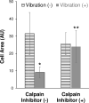
Fig 5. Inhibition of calpain activity attenuates vibration induced cell shape change.
HeLa cells were pre-treated overnight with DMSO (solvent control) [calpain inhibitor (-)] or with calpain inhibitor, PD150606 [calpain inhibitor (+)]. HeLa cells were washed and re-treated immediately prior to treated without vibration (-) [striped bar] or with vibration (+) [solid bar]. The perimeter of each cell (≥100) was delineated including any protrusions and the cell area was measured (AU). Data shown as average cell area (AU) for all cells measured (±sem for three independent experiments). * = p < 0.05 Vibration (+)/Calpain(-) [solid bar] vs. Vibration (-)/Calpain(-) [striped bar]. ** = p < 0.05 Vibration (+)/Calpain Inhibitor (+) vs. Vibration (+)/Calpain Inhibitor (-). No significant difference was noted between Vibration (+)/Calpain Inhibitor (+) and other sample groups [Vibration (-)/Calpain Inhibitor (+) or Vibration (-) / Calpain Inhibitor (-)].
Fig 6
Fig 6. Vibration alters calpain gene expression.
RNA isolated from HeLa cells without vibration or with vibration was subjected to reverse transcription. Resultant cDNA was used as a template for qPCR using primers for calpain and E-cadherin, an epithelial marker. Threshold cycles were normalized to GAPDH, a loading control. A fold difference of expression comparing vibration to no vibration of HeLa was calculated with the ΔΔCT method where no change is indicated by “1”. No reverse transcriptase and no template controls showed no product.

References

    1. Magos L. Preliminary Communication: A Physiological Study of the Effects of Vibration on the Fingers. Br J Ind Med. 1961. Apr;18(2):157–8. doi: 10.1136/oem.18.2.157 - DOI - PMC - PubMed
    1. Olsson AR, Skogh T, Axelson O, Wingren G. Occupations and exposures in the work environment as determinants for rheumatoid arthritis. Occup Environ Med. 2004. Mar;61(3):233–8. doi: 10.1136/oem.2003.007971 - DOI - PMC - PubMed
    1. Smith KC, Comite SL, Balasubramanian S, Carver A, Liu JF. Vibration anesthesia: a noninvasive method of reducing discomfort prior to dermatologic procedures. Dermatol Online J. 2004. Oct 15;10(2):1. - PubMed
    1. Juto J-E, Hallin RG. Kinetic oscillation stimulation as treatment of acute migraine: a randomized, controlled pilot study. Headache. 2015. Jan;55(1):117–27. doi: 10.1111/head.12485 - DOI - PMC - PubMed
    1. Ahmad M, Mohmand MH. Clinical significance of Vibration Anesthesia on reducing pain of Ring-Block (Subcutaneous Injections) in the patients undergoing Hair Restoration Surgery. Ann Dermatol Res. 2017. Oct 18;1(1):001–5.