. 2022 Aug 3;14(656):eabo0718.
doi: 10.1126/scitranslmed.abo0718. Epub 2022 Aug 3.

Mutations in the SARS-CoV-2 RNA-dependent RNA polymerase confer resistance to remdesivir by distinct mechanisms

Affiliations

Mutations in the SARS-CoV-2 RNA-dependent RNA polymerase confer resistance to remdesivir by distinct mechanisms

Laura J Stevens et al. Sci Transl Med. .

Abstract

The nucleoside analog remdesivir (RDV) is a Food and Drug Administration-approved antiviral for treatment of severe acute respiratory syndrome coronavirus 2 (SARS-CoV-2) infections. Thus, it is critical to understand factors that promote or prevent RDV resistance. We passaged SARS-CoV-2 in the presence of increasing concentrations of GS-441524, the parent nucleoside of RDV. After 13 passages, we isolated three viral lineages with phenotypic resistance as defined by increases in half-maximal effective concentration from 2.7- to 10.4-fold. Sequence analysis identified nonsynonymous mutations in nonstructural protein 12 RNA-dependent RNA polymerase (nsp12-RdRp): V166A, N198S, S759A, V792I, and C799F/R. Two lineages encoded the S759A substitution at the RdRp Ser759-Asp-Asp active motif. In one lineage, the V792I substitution emerged first and then combined with S759A. Introduction of S759A and V792I substitutions at homologous nsp12 positions in murine hepatitis virus demonstrated transferability across betacoronaviruses; introduction of these substitutions resulted in up to 38-fold RDV resistance and a replication defect. Biochemical analysis of SARS-CoV-2 RdRp encoding S759A demonstrated a roughly 10-fold decreased preference for RDV-triphosphate (RDV-TP) as a substrate, whereas nsp12-V792I diminished the uridine triphosphate concentration needed to overcome template-dependent inhibition associated with RDV. The in vitro-selected substitutions identified in this study were rare or not detected in the greater than 6 million publicly available nsp12-RdRp consensus sequences in the absence of RDV selection. The results define genetic and biochemical pathways to RDV resistance and emphasize the need for additional studies to define the potential for emergence of these or other RDV resistance mutations in clinical settings.


Figures
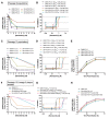
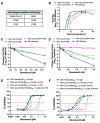
Fig. 1.
Fig. 1.. SARS-CoV-2 RDV resistance develops after serial passaging.
SARS-CoV-2 was serially passaged in the presence and absence of GS-441524 in Vero-E6 cells in triplicate lineages. (A) Sensitivity of P9 lineages (Lin) to RDV in A549-hACE2 cells was determined by change in genome copy number. (B) Percent inhibition was calculated from genome copy number (A) and fold-change in EC50 compared to vehicle (DMSO)-passaged lineage 1 at P9. (C) Sensitivity of P13 lineages to RDV in A549-hACE2 cells was determined by change in genome copy number. (D) Percent inhibition calculated from genome copy number (C) and fold-change in EC50 compared to vehicle-passaged lineage 1 at P13. (E) Replication kinetics of P13 drug-passaged viral lineages are shown compared to vehicle-passaged lineage 1. (F) Sensitivity to RDV is shown for plaque-pick (PP) isolates from GS-441524-passaged lineage 3 and vehicle-passaged lineage 1 population viruses and input virus in A549-hACE2 cells, as determined by change in genome copy number. Plaque-picks (PP) from lineage 3 were isolated and expanded in presence of 1μM GS-441524. (G) Percent inhibition was calculated from raw genome copy number (F) and fold-change in EC50 compared to vehicle DMSO-passaged lineage 1. (H) Replication kinetics of plaque isolates tested in (F) and (G). Error bars indicate standard deviation.
Fig. 2.
Fig. 2.. Evolution and intramolecular linkage of nsp12 mutations.
SARS-CoV-2 was passaged 13 times in increasing concentrations of GS-441524 in 3 lineages. RNA from infected cell monolayers was subjected to Illumina RNA sequencing and Oxford nanopore MinION sequencing. (A to C) RNA-seq was used to measure the percent of nsp12 mutations in lineages 1 (A), 2 (B), and 3 (C). (D to F) Nanopore amplicon sequencing was used to measure percent of nsp12 mutations (MUT) in lineages 1 (D), 2 (E), and 3 (F) relative to wild-type (WT) nsp12. (G to I) The frequency of single and combined sets of nsp12 mutations is shown for in single viral genomes in lineages 1 (G), 2 (H), and 3 (I). Variants were mapped according to their genomic position and frequency, expressed as a percentage of the total reads mapped to that position.
Fig. 3.
Fig. 3.
Structural modelling predictions of identified nsp12-RdRp mutations. (A) Observed nsp12 amino acid substitutions were mapped on a model of the SARS-CoV-2/RDV-TP pre-incorporation complex. nsp12 is shown in white, nsp7 in pink, nsp8 in cyan, the primer strand in yellow, the template strand in orange, RDV-TP in magenta, and mutations in green. S759A is in the active site, whereas V166A, V792A and C799F/R are adjacent to the active site, clustered around motif D (in blue). Details of the RDV-TP pre-incorporation model are shown, highlighting the polar residues that interact with the 2’OH and the 1’CN. (B) S759 is seen to be in close contact with the 1’CN, forming a favorable interaction. (C) Substitution of the serine with an alanine increases the distance between residue 759 and the 1’CN, resulting in a loss of favorable interaction. N198S does not appear to impact either the NiRAN or Pol sites. (D) A model of the lineage 1 mutations V166A and S759A (green) overlaid on the wild-type structure (white) is shown. V166A is in direct contact with V792 and may impact the dynamics of motif D. (E) A model of the similar lineage 3 mutations V792I and S759A (green) overlaid on the wild-type structure (white) is shown. (F) A model of the lineage 2 mutation C799R (green) overlaid on the wild-type structure (white) is shown. The mutation is predicted to alter the conformation of motif D, impacting how K798 interacts with the substrate γ-phosphate. (G) A model of the lineage 1 mutations V166A and C799F is shown, which are also observed to alter the conformation of motif D and the position of K798.
Fig. 4.
Fig. 4.
SARS-CoV-2 resistance mutations confer RDV resistance in MHV. (A) Candidate resistance mutations identified in SARS-CoV-2 were engineered at conserved homologous positions in the MHV infectious clone. Wild-type MHV and mutant viruses were tested against RDV in murine delayed brain tumor (DBT) cells. (B) MHV replication kinetics. (C) Change in infectious viral titers as measured by plaque assay. Changes are shown relative to DMSO control treatment. (D) Change in MHV genome copy number as measured using qRT-PCR. (E) Percent inhibition and EC50 values calculated using infectious virus titers from (B). (F) Percent inhibition and EC50 values calculated using genome copy numbers from (C).
Fig. 5.
Fig. 5.. RDV-TP is differentially incorporated by wild-type, S759A, and V792I mutant SARS-CoV-2 RdRp complexes.
(A) Graphical representation of ATP or RDV-TP single nucleotide incorporation during RNA synthesis as a function of their respective concentrations shown in fig. S2. Best fit lines illustrate fitting of the data points to Michaelis-Menten kinetics function using GraphPad Prism 7.0. Error bars illustrate the standard deviation of the data. All data represent at least three independent experiments. (B) The efficiencies of incorporation (ATP and RDV-TP) and selectivity (ATP over RDV-TP) of the mutant enzymes were quantified and corrected for differences in ATP incorporation. (C) Selectivity and discrimination values were calculated for RDV-TP across mutant enzymes.
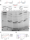
Fig. 6.
Fig. 6.
RNA synthesis differs between SARS-CoV-2 wild-type and mutant S759A, V792I, and S759A+V792I RdRp complexes. (A) The RNA primer/template sequences used are shown. (B) RDV-MP is embedded at position 11 in the template R strand while AMP is in the same position on the template A strand. RNA products were synthesized by the wild-type or mutant SARS-CoV-2 RdRps in a reaction mixture containing the primer/template pair, MgCl2, and indicated NTP concentrations. G (red) indicates the incorporation of [α-32P] GTP at position 5 and 4 indicates the migration pattern of 5′-32P-labeled 4-nt primer is used as a size marker. The 0 point in red indicates a reaction where [α-32P] GTP was the only NTP present to control for contaminating NTPs in the template preparations. (C and D) The fraction of RNA synthesis beyond position 11 with respect to total RNA products formed was quantified. (C) The comparison of reactions using template A and template R with wild-type RdRp and increasing concentrations of UTP is shown. (D) Comparisons of RNA synthesis by wild-type and mutant enzymes are shown. Data corresponding to UTP = 33 and 100 μM were excluded to focus on the differences in the lower concentration range.

References

    1. CDC, COVID Data Tracker, Cent. Dis. Control Prev . (2020), (available at https://covid.cdc.gov/covid-data-tracker).
    1. W. H. O. Coronavirus, (COVID-19) Dashboard, (available at https://covid19.who.int).
    1. Warren T. K., Jordan R., Lo M. K., Ray A. S., Mackman R. L., Soloveva V., Siegel D., Perron M., Bannister R., Hui H. C., Larson N., Strickley R., Wells J., Stuthman K. S., Van Tongeren S. A., Garza N. L., Donnelly G., Shurtleff A. C., Retterer C. J., Gharaibeh D., Zamani R., Kenny T., Eaton B. P., Grimes E., Welch L. S., Gomba L., Wilhelmsen C. L., Nichols D. K., Nuss J. E., Nagle E. R., Kugelman J. R., Palacios G., Doerffler E., Neville S., Carra E., Clarke M. O., Zhang L., Lew W., Ross B., Wang Q., Chun K., Wolfe L., Babusis D., Park Y., Stray K. M., Trancheva I., Feng J. Y., Barauskas O., Xu Y., Wong P., Braun M. R., Flint M., McMullan L. K., Chen S.-S., Fearns R., Swaminathan S., Mayers D. L., Spiropoulou C. F., Lee W. A., Nichol S. T., Cihlar T., Bavari S., Therapeutic efficacy of the small molecule GS-5734 against Ebola virus in rhesus monkeys. Nature 531, 381–385 (2016). 10.1038/nature17180 - DOI - PMC - PubMed
    1. Sheahan T. P., Sims A. C., Graham R. L., Menachery V. D., Gralinski L. E., Case J. B., Leist S. R., Pyrc K., Feng J. Y., Trantcheva I., Bannister R., Park Y., Babusis D., Clarke M. O., Mackman R. L., Spahn J. E., Palmiotti C. A., Siegel D., Ray A. S., Cihlar T., Jordan R., Denison M. R., Baric R. S., Broad-spectrum antiviral GS-5734 inhibits both epidemic and zoonotic coronaviruses. Sci. Transl. Med. 9, eaal3653 (2017). 10.1126/scitranslmed.aal3653 - DOI - PMC - PubMed
    1. Agostini M. L., Andres E. L., Sims A. C., Graham R. L., Sheahan T. P., Lu X., Smith E. C., Case J. B., Feng J. Y., Jordan R., Ray A. S., Cihlar T., Siegel D., Mackman R. L., Clarke M. O., Baric R. S., Denison M. R., Coronavirus Susceptibility to the Antiviral Remdesivir (GS-5734) Is Mediated by the Viral Polymerase and the Proofreading Exoribonuclease. mBio 9, e00221–e18 (2018). 10.1128/mBio.00221-18 - DOI - PMC - PubMed

Publication types