Review
. 2022 Apr 29;130(9):1404-1422.
doi: 10.1161/CIRCRESAHA.121.319970. Epub 2022 Apr 28.

Group 3 Pulmonary Hypertension: From Bench to Bedside

Affiliations
Review

Group 3 Pulmonary Hypertension: From Bench to Bedside

Navneet Singh et al. Circ Res. .

Abstract

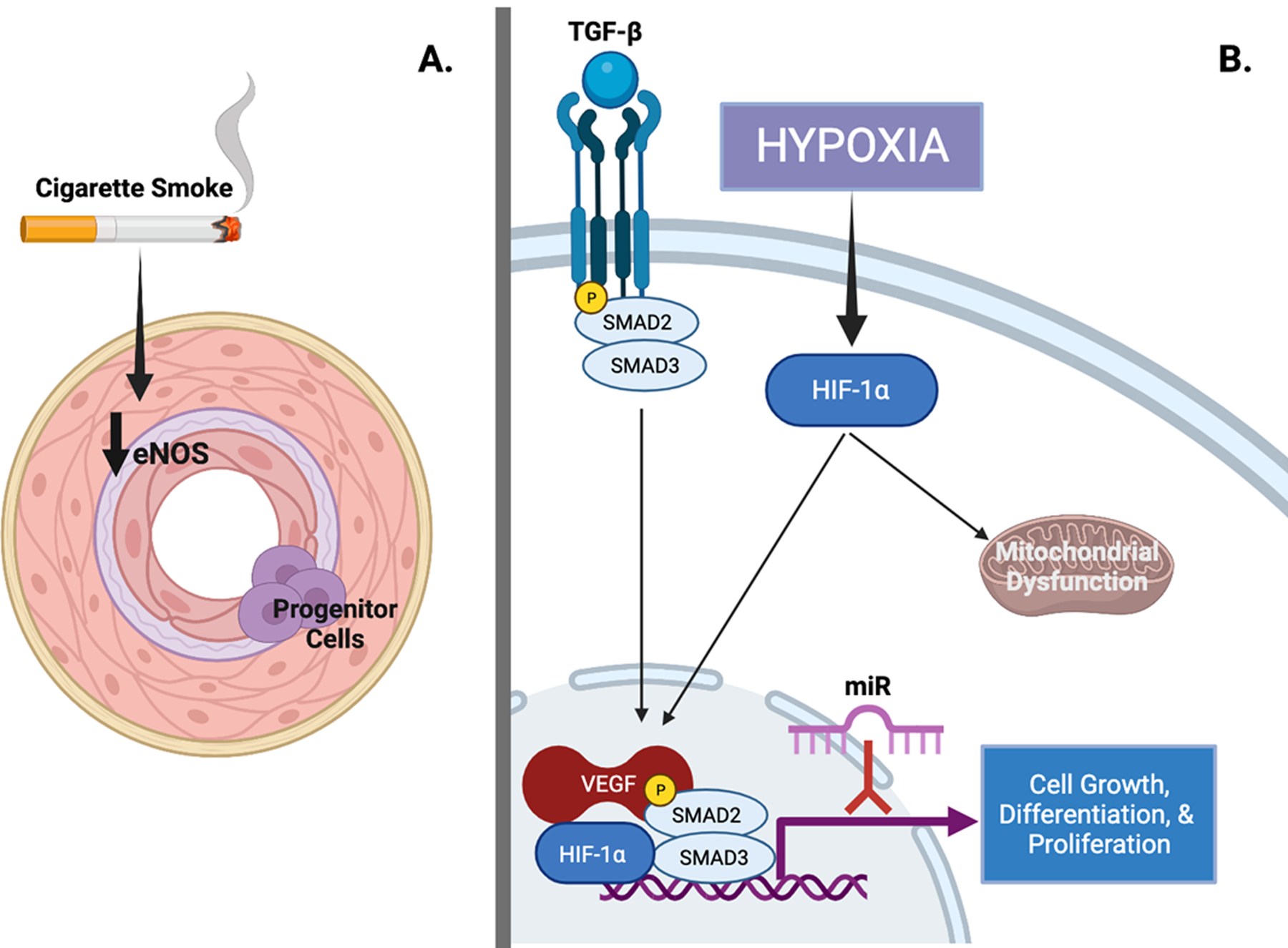
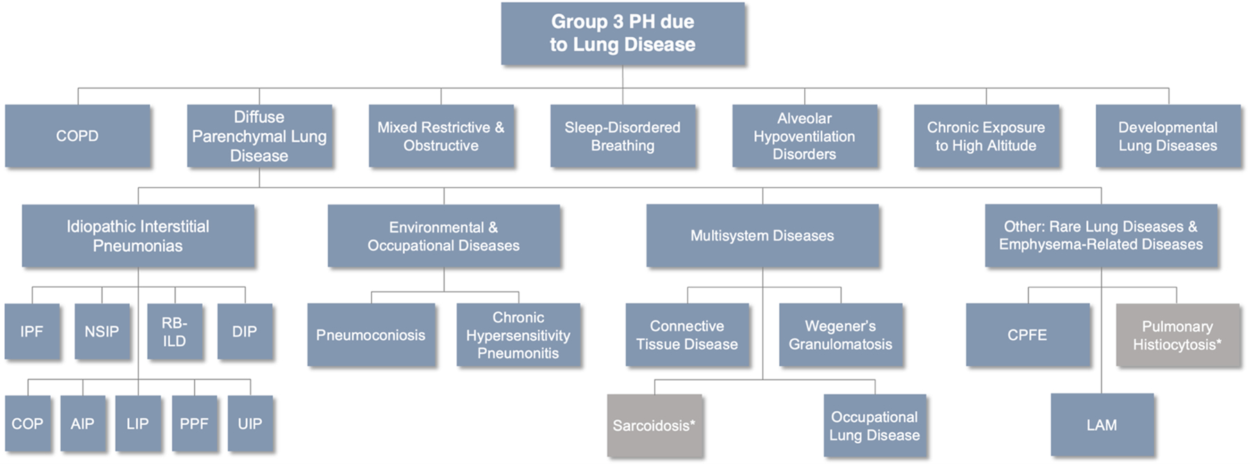
Pulmonary hypertension (PH) because of chronic lung disease is categorized as Group 3 PH in the most recent classification system. Prevalence of these diseases is increasing over time, creating a growing need for effective therapeutic options. Recent approval of the first pulmonary arterial hypertension therapy for the treatment of Group 3 PH related to interstitial lung disease represents an encouraging advancement. This review focuses on molecular mechanisms contributing to pulmonary vasculopathy in chronic hypoxia, the pathology and epidemiology of Group 3 PH, the right ventricular dysfunction observed in this population and clinical trial data that inform the use of pulmonary vasodilators in Group 3 PH.

Keywords: hypertension, pulmonary; hypoxia; lung diseases, interstitial; prevalence; ventricular dysfunction, right.


Figures

Figure 1.
Figure 1.
Molecular mechanisms of pulmonary vasculopathy in Group 3 pulmonary hypertension (PH). A. Cigarette smoke leads to decreased expression of endothelial nitric oxide (eNOS) along with intimal hyperplasia, increased expression of vascular endothelial growth factor (VEGF), inflammatory infiltrate, and metabolic dysfunction. Poorly differentiated progenitor cells have been observed in the intima of pulmonary arteries; their origin and role in the development of pulmonary vasculopathy remains unclear. B. Hypoxia inducible factor-1 alpha (HIF-1α) and transforming growth factor-beta (TGF-β) signaling are recognized to regulate growth, differentiation, and proliferation of almost all cell types in hypoxia-induced PH. VEGF expression is induced by hypoxia via HIF-1α signaling. HIF-1α is also thought to play a central role in the development of mitochondrial dysfunction that may explain a Warburg-like shift to anaerobic glycolysis. microRNAs (miRs) are small noncoding RNAs that regulate gene expression and in mouse models inhibition of miRs prevent development of pulmonary vasculopathy. Figure created with biorender.com. (Illustration Credit: Ben Smith).
Fig. 2.
Fig. 2.
Pulmonary vascular changes in patients with lung disease and Group 3 pulmonary hypertension (PH). A. Pulmonary artery (PA) and its adjacent airway in a patient with idiopathic pulmonary fibrosis (IPF) and PH; note the constrictive intimal fibrosis of the vessel (straight arrows) within the fibrosing area (upper part) and the slender walls of the PA branching out of this area into preserved lung areas (flexuous arrow) with near-normal architecture (lower part). B. Smooth muscle cell hyperplasia within the interstitium (arrows) and within the wall of a small PA, in a patient with IPF-PH; smooth muscle actin-staining. C and D: Capillary density of a patient with chronic obstructive pulmonary disease (COPD) displaying moderate PH (C) and a patient with COPD and severe PH (D): note the loss of ERG-stained endothelial cells within the alveolar septa, corresponding to a decrease of vascular density; photos reprinted with regard to the STM permission guidelines 2022 from Bunel et al. CHEST. 2019. E. PA in a patient with COPD-PH, with severe intimal fibrosis; note the perivascular lymphocytic infiltrate (small blue dots, center). F. Remodeled arterioles in a COPD-PH patient with dense CD5-positive peri-vascular lymphocytic infiltrate (red dots), a feature that can also be observed in pulmonary arterial hypertension.
Figure 3.
Figure 3.
Clinical classification of diseases causing Group 3 pulmonary hypertension (PH). Aside from chronic obstructive pulmonary disease (COPD), many diseases responsible for Group 3 PH belong to the diffuse parenchymal lung diseases (DPLDs) and are further classified based on their etiology. Idiopathic interstitial pneumonias (IIPs) are idiopathic diseases of varying prevalence but represent an important group of diseases that contribute to the development of PH. *Belongs to Group 5 PH. IPF = idiopathic pulmonary fibrosis. NSIP = nonspecific interstitial pneumonitis. RB-ILD = respiratory bronchiolitis-interstitial lung disease. DIP = desquamative interstitial pneumonia. COP = cryptogenic organizing pneumonia. AIP = acute interstitial pneumonia. LIP = lymphocytic interstitial pneumonia. PPF = pleuroparenchymal fibroelastosis. UIP = usual interstitial pneumonia. CPFE = combined pulmonary fibrosis and emphysema. LAM = lymphangioleiomyomatosis. Adapted from Nathan, SD. “Inhaled Treprostinil in Interstitial Lung Disease Associated Pulmonary Hypertension: The INCREASE Study.” Breaking News: Clinical Trial Results in Pulmonary Medicine. ATS Meeting. May 28, 2020.

References

    1. Simonneau G, Montani D, Celermajer DS, Denton CP, Gatzoulis MA, Krowka M, Williams PG and Souza R. Haemodynamic definitions and updated clinical classification of pulmonary hypertension. European Respiratory Journal. 2019;53. - PMC - PubMed
    1. Prins KW, Duval S, Markowitz J, Pritzker M and Thenappan T. Chronic use of PAH-specific therapy in World Health Organization Group III Pulmonary Hypertension: a systematic review and meta-analysis. Pulmonary circulation. 2017;7:145–155. - PMC - PubMed
    1. Prins KW, Rose L, Archer SL, Pritzker M, Weir EK, Kazmirczak F, Misialek JR and Thenappan T. Disproportionate Right Ventricular Dysfunction and Poor Survival in Group 3 Pulmonary Hypertension. Am J Respir Crit Care Med. 2018;197:1496–1499. - PMC - PubMed
    1. Chen L, Einbinder E, Zhang Q, Hasday J, Balke CW and Scharf SM. Oxidative stress and left ventricular function with chronic intermittent hypoxia in rats. American journal of respiratory and critical care medicine. 2005;172:915–920. - PubMed
    1. Silverman NA, Kohler J, Levitsky S, Pavel DG, Fang RB and Feinberg H. Chronic hypoxemia depresses global ventricular function and predisposes to the depletion of high-energy phosphates during cardioplegic arrest: implications for surgical repair of cyanotic congenital heart defects. The Annals of thoracic surgery. 1984;37:304–308. - PubMed

MeSH terms

Substances