. 2022 Apr 28;18(4):e1010496.
doi: 10.1371/journal.ppat.1010496. eCollection 2022 Apr.

The Giardia ventrolateral flange is a lamellar membrane protrusion that supports attachment

Affiliations

The Giardia ventrolateral flange is a lamellar membrane protrusion that supports attachment

William R Hardin et al. PLoS Pathog. .

Abstract

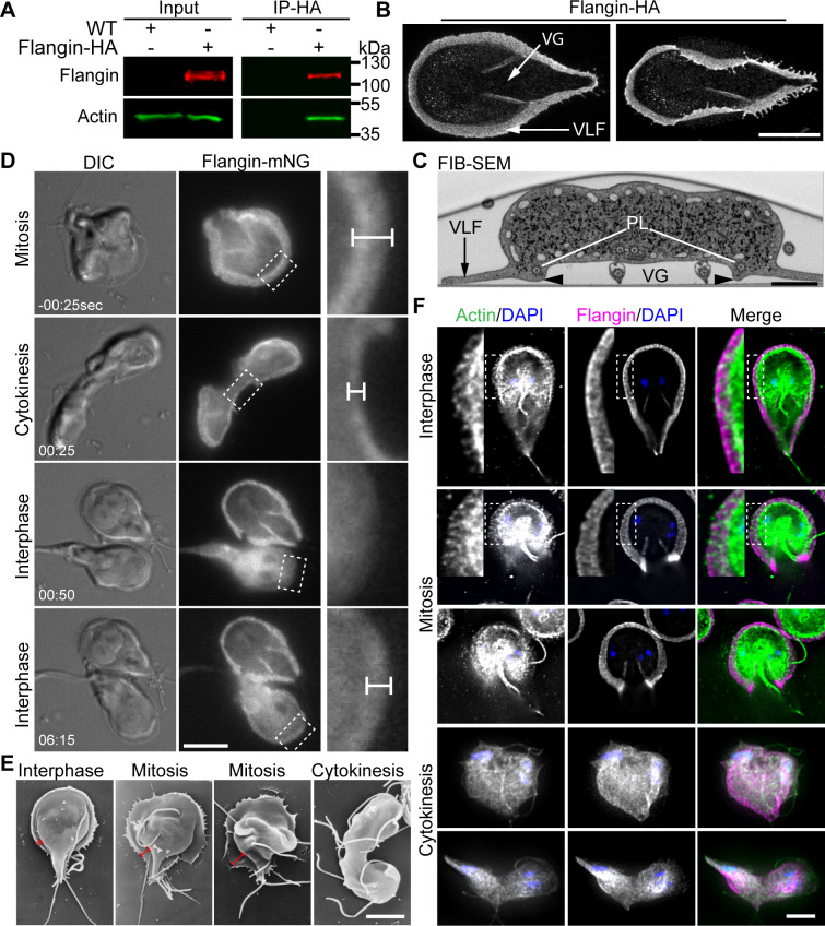
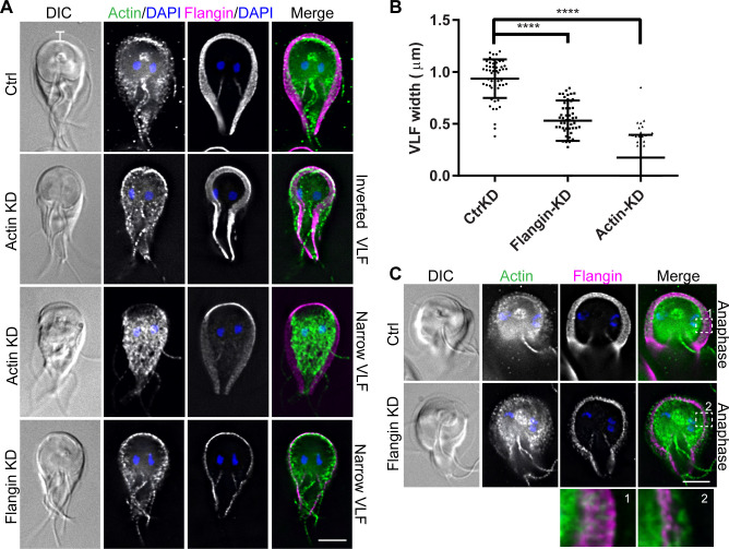
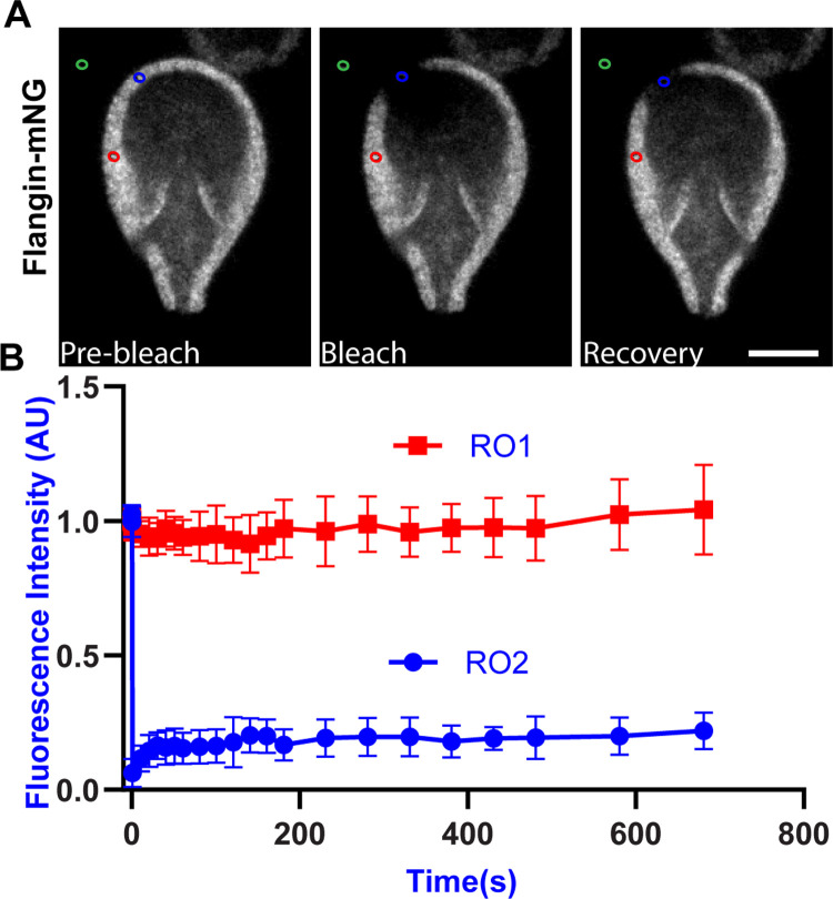
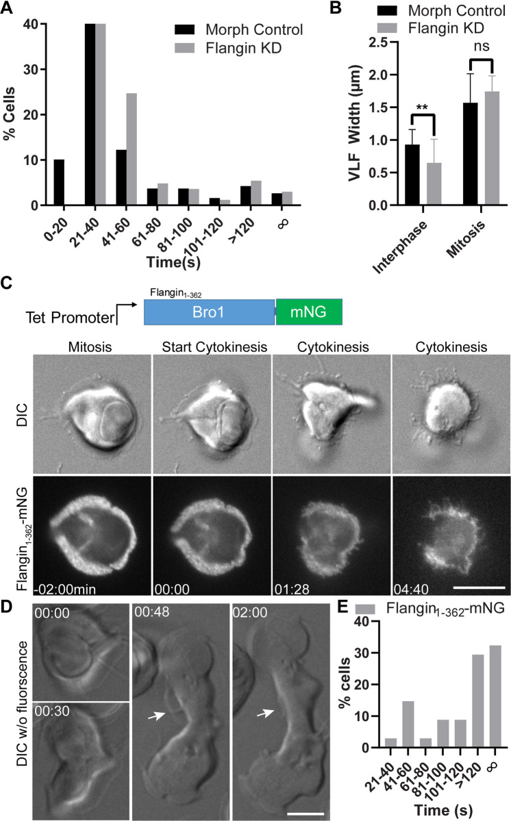
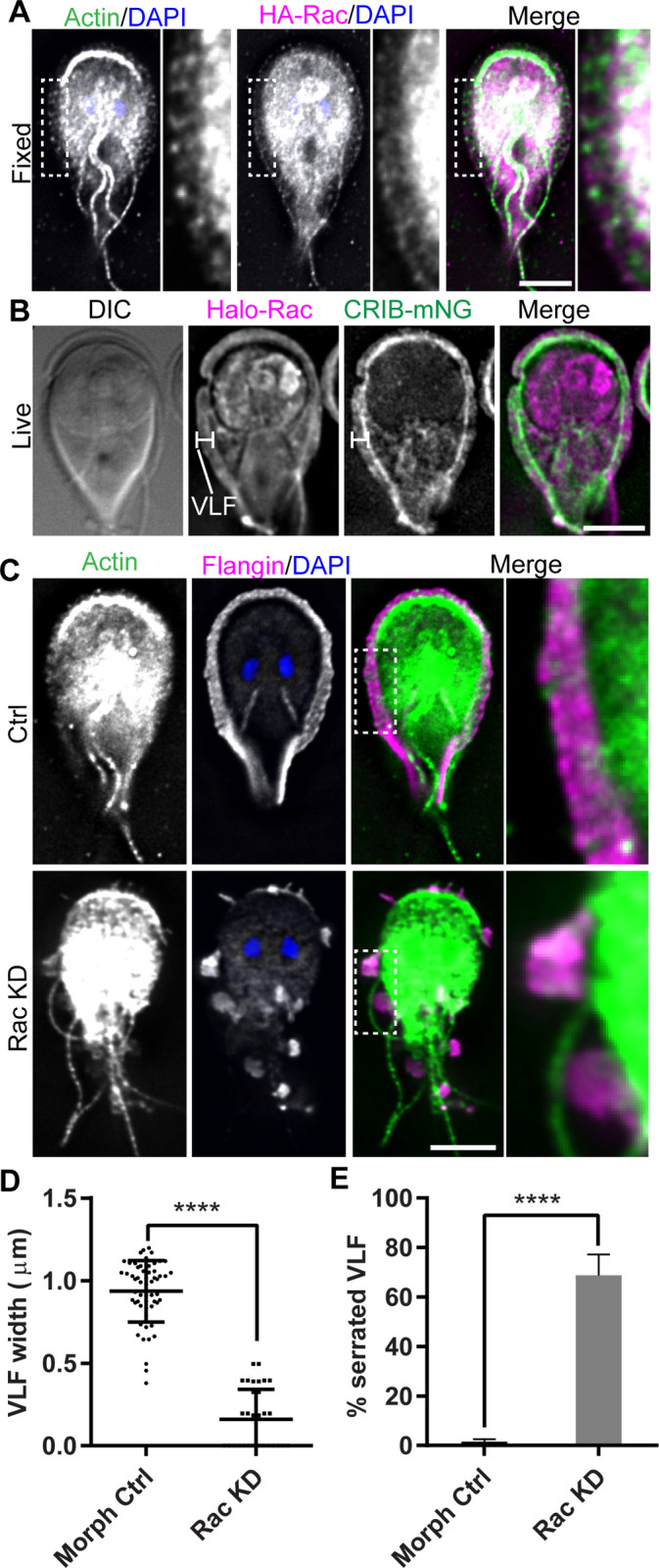
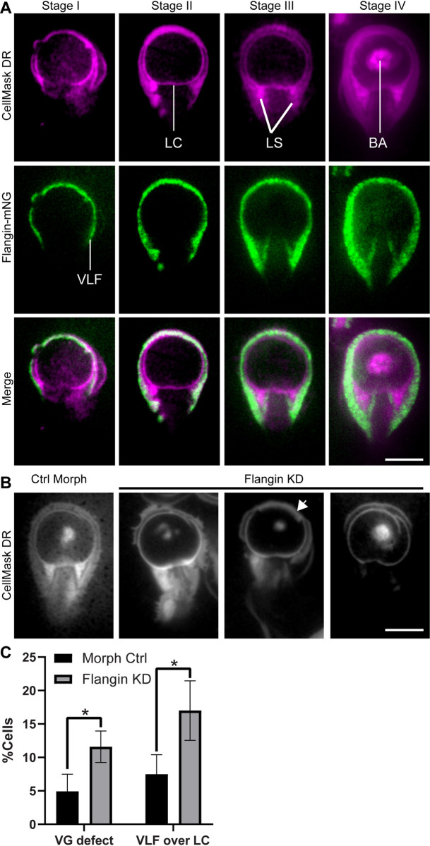
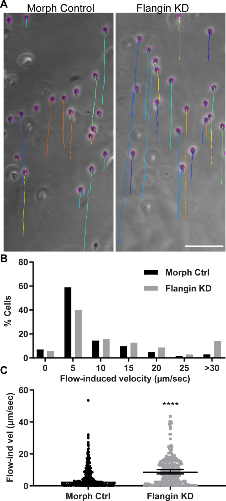
Attachment to the intestinal epithelium is critical to the lifestyle of the ubiquitous parasite Giardia lamblia. The ventrolateral flange is a sheet-like membrane protrusion at the interface between parasites and attached surfaces. This structure has been implicated in attachment, but its role has been poorly defined. Here, we identified a novel actin associated protein with putative WH2-like actin binding domains we named Flangin. Flangin complexes with Giardia actin (GlActin) and is enriched in the ventrolateral flange making it a valuable marker for studying the flanges' role in Giardia biology. Live imaging revealed that the flange grows to around 1 μm in width after cytokinesis, then remains uniform in size during interphase, grows in mitosis, and is resorbed during cytokinesis. A flangin truncation mutant stabilizes the flange and blocks cytokinesis, indicating that flange disassembly is necessary for rapid myosin-independent cytokinesis in Giardia. Rho family GTPases are important regulators of membrane protrusions and GlRac, the sole Rho family GTPase in Giardia, was localized to the flange. Knockdown of Flangin, GlActin, and GlRac result in flange formation defects. This indicates a conserved role for GlRac and GlActin in forming membrane protrusions, despite the absence of canonical actin binding proteins that link Rho GTPase signaling to lamellipodia formation. Flangin-depleted parasites had reduced surface contact and when challenged with fluid shear force in flow chambers they had a reduced ability to remain attached, confirming a role for the flange in attachment. This secondary attachment mechanism complements the microtubule based adhesive ventral disc, a feature that may be particularly important during mitosis when the parental ventral disc disassembles in preparation for cytokinesis. This work supports the emerging view that Giardia's unconventional actin cytoskeleton has an important role in supporting parasite attachment.


Conflict of interest statement

The authors have declared that no competing interests exist.

Figures

Fig 1
Fig 1. Giardia possesses a lamellipodium-like structure that contains GlActin and Flangin.
(A) Immunoprecipitation of Flangin-HA from cell extracts, followed by anti-GlActin Western blotting demonstrates Flangin associates with GlActin. (B) Maximum projections of expanded trophozoites imaged with confocal fluorescence microscopy. Flangin-HA marks the ventrolateral flange (VLF) and membrane protrusions associated with the cytoplasmic portions of the posterolateral flagella axonemes that bound the ventral grove (VG). The left image shows that the flange can completely encircle the base of the cell and the image on the right shows that the flange is thin and flexible. (C) Transverse section of wild-type Giardia (posterior to the ventral flagella exit point) as viewed with Focused Ion Beam-Scanning Electron Microscopy (FIB-SEM). Arrowhead marks the membrane protrusion associated with Flangin and the cytoplasmic axonemes of the posterolateral (PL) axonemes that bound the ventral groove. (D) Live cell imaging of Flangin-mNG during cytokinesis. Compared with interphase cells the flange is wider in mitosis and was resorbed during cytokinesis. Upon cytokinesis, the flange disassembled and Flangin-mNG translocated to the cytoplasm. Daughter cells lack a flange, but assembly was initiated almost immediately after division. Flangin-mNG was recruited back to the flange as it reformed (see S1 Movie). Magnified views of the boxed area show Flangin is at the leading edge of the flange. Dimension bars in C and D point out flange width. (E) Scanning electron microscopy shows an expanded flange in mitosis and the lack of a flange during cytokinesis. (F) Immunofluorescence localization of GlActin (green), Flangin-HA (magenta), and DNA (blue), throughout the cell cycle (see S3 Fig for tubulin staining). GlActin and Flangin accumulate in the flange during interphase and mitosis. The insets show a magnified view of GlActin and Flangin in the flange. During cytokinesis flange localized Flangin and GlActin translocated to cytoplasm as the flange is disassembled. Note that the interphase and mitotic cells are partial projections optimized to show GlActin localization, while the entire Z-stack was projected for the cells in cytokinesis to show that the flange has been resorbed. Scale bars = 5 μm except (C) = 1 μm.
Fig 2
Fig 2. Flangin and GlActin are necessary for flange assembly.
(A) Trophozoites were stained for GlActin (green), Flangin-HA (magenta), and DNA (blue). Note that images are scaled to display the remaining protein and are not intended to show differences in protein levels. GlActin knockdown (KD) resulted in inverted and collapsed flange morphology. Flangin-HA KD similarly resulted in a thin flange phenotype. (B) Quantification of flange width when measured at the cell anterior from three independent experiments; control (n = 57), Flangin-HA (n = 54), and actin (n = 51). Statistical significance was evaluated for Flangin-KD and Actin-KD respectively, t test. ****, P<0.0001. (C) Mitotic Flangin depleted cells were capable of extending their flange beyond the leading edge of Flangin. The insets show actin beyond the leading edge of Flangin-HA in Flangin-HA KD cells during mitotic flange extension. Scale bars = 5 μm.
Fig 3
Fig 3. Flangin is part of a stable structure.
(A) Fluorescence Recovery after Photobleaching (FRAP) was performed on the flange of Flangin-mNG cells, Blue circle bleached ROI 01, Red circle non-bleached ROI 02, and Green circle background ROI 03. (B) ROI 01 showed minimal post-bleaching recovery after 12min, n = 4 independent experiments. A lack of fluorescence recovery was also observed in 29 additional FRAP experiments of various lengths and regions of the cell including the small membrane protrusion associated with the posterolateral axonemes. Scale bar = 5 μm. See S3 Movie for confirmation of viability.
Fig 4
Fig 4. Flange breakdown is necessary for cleavage furrow progression.
(A) Histogram showing the timing of cytokinesis for morpholino control (black) and Flangin KD cells (gray). See S4 and S5 Movies. Cytokinesis median time: control 31 sec (n = 188) and Flangin KD 35 sec (n = 166). Due to sampling frequency (timing between image stacks) the difference in median values in this experiment is not meaningful, similar to our previous study, more than 90% of the cells in both groups complete cytokinesis by 120 seconds indicating that there is no cytokinesis defect. (B) Flangin KD cells grow their flange before cytokinesis similar to control cells (See S5 Movie). (C) A tet inducible N-terminal truncation of Flangin interferes with flange retraction during cytokinesis. This blocks furrow progression and the ability of the parasite to lift off the cover glass (Compare S1 and S6 Movies). (D) DIC imaging of the same cell line without fluorescence. This is an example of a moderate defect where the flange persists and cytokinesis took 8 minutes to complete (see S7 Movie). White arrow points to regions of the flange that should have been resorbed by this point in cytokinesis. (E) Cytokinesis timing for Flangin1-362-mNG filmed with DIC alone (n = 34), only 38.3% of cells completed cytokinesis within 120 seconds. Scale bar = 5 μm.
Fig 5
Fig 5. GlRac signaling is required for flange assembly.
(A) Immunofluorescence localization of GlActin (green), HA-Rac (magenta), and DNA (blue). GlActin and GlRac localize to the flange. (B) Live cell imaging of Halo-Rac (magenta) and CRIB-mNG (green) indicate that GlRac is actively signaling in the flange. (C) GlRac KD resulted in a serrated flange phenotype not observed in control cells. GlActin and Flangin localize to the remaining sporadic flange protrusions. (D) Quantification of flange width measured at cell anterior from three independent experiments: control (n = 57) and GlRac KD (n = 32). Statistical significance was evaluated using the t-test. ****, P<0.0001. (E) Quantification of serrated cells. Cells with three or more flange gaps were defined as serrated; control (n = 596) and GlRac KD (n = 618). Statistical significance was evaluated using the t-test. ****, P< 0.0001. Scale bars = 5 μm.
Fig 6
Fig 6. The flange makes intimate contact with attached surfaces and is required for normal ventral groove morphology.
(A) Two color total internal reflection (TIRF) microscopy. Membranes are stained with CellMask Deep Red (magenta) and the flange is labeled with Flangin-mNG. As attachment progresses the flange becomes intimately associated with the substrate surface. (B) Knockdown of Flangin with translation blocking morpholinos uncouples the normal progression of attachment. Note that the ventral disc and bare area continues to make contact with the surface, but lateral shields (LS) do not make their typical contact with the attachment surface. This results in disruption of ventral groove channel important for fluid flows under the cell. Arrowhead points to an example of the flange being improperly positioned under the lateral crest (LC) where it may interfere with normal attachment. (C). Quantification of cells with ventral groove (VG) defects and cells with the flange positioned under the LC. Data are from three independent replicates including at least 100 cells each.
Fig 7
Fig 7. The flange has a role in attachment.
(A) Flow chamber assay to test the role of the flange in attachment. A flow rate of 100μl/s induced sliding. The paths of cells were followed with TrackMate, the paths at the end of the 10s challenge are indicated by the colored lines. See S9 Movie. (B) Histogram of flow induced velocities for the standard morpholino control n = 268 and anti-Flangin morpholino treated cells n = 248 analyzed from six challenge assays. (C) Plot of measured values where error bars are median with 95% confidence interval; statistical significance determined with a Mann-Whitney U test. ****, P<0.0001. Scale bar = 100 μm.

Similar articles

Cited by

References

    1. Lane S, Lloyd D. Current trends in research into the waterborne parasite Giardia. Crit Rev Microbiol. 2002;28(2):123–47. Epub 2002/07/12. doi: 10.1080/1040-840291046713 . - DOI - PubMed
    1. Baldursson S, Karanis P. Waterborne transmission of protozoan parasites: review of worldwide outbreaks—an update 2004–2010. Water Res. 2011;45(20):6603–14. doi: 10.1016/j.watres.2011.10.013 . - DOI - PubMed
    1. Dawson SC, Paredez AR. Alternative cytoskeletal landscapes: cytoskeletal novelty and evolution in basal excavate protists. Curr Opin Cell Biol. 2013;25(1):134–41. doi: 10.1016/j.ceb.2012.11.005 . - DOI - PMC - PubMed
    1. Nosala C, Hagen KD, Dawson SC. ’Disc-o-Fever’: Getting Down with Giardia’s Groovy Microtubule Organelle. Trends Cell Biol. 2018;28(2):99–112. Epub 2017/11/21. doi: 10.1016/j.tcb.2017.10.007 . - DOI - PMC - PubMed
    1. Steele-Ogus MC, Johnson RS, MacCoss MJ, Paredez AR. Identification of Actin Filament-Associated Proteins in Giardia lamblia. Microbiol Spectr. 2021:e0055821. Epub 2021/07/22. doi: 10.1128/Spectrum.00558-21 . - DOI - PMC - PubMed

Publication types