. 2022 Jun 14;55(6):1096-1104.e4.
doi: 10.1016/j.immuni.2022.04.002. Epub 2022 Apr 7.

Analysis of mRNA vaccination-elicited RBD-specific memory B cells reveals strong but incomplete immune escape of the SARS-CoV-2 Omicron variant

Affiliations

Analysis of mRNA vaccination-elicited RBD-specific memory B cells reveals strong but incomplete immune escape of the SARS-CoV-2 Omicron variant

Aurélien Sokal et al. Immunity. .

Abstract

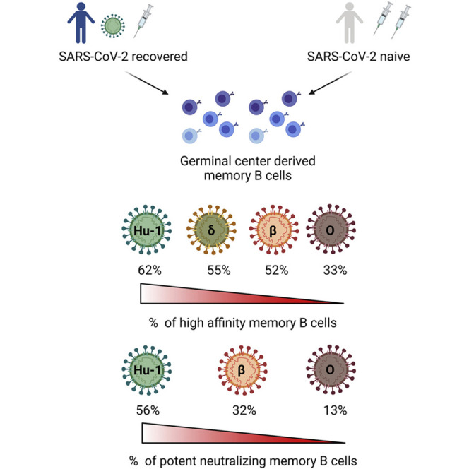
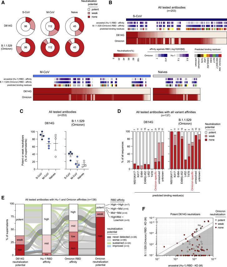
The SARS-CoV-2 Omicron variant can escape neutralization by vaccine-elicited and convalescent antibodies. Memory B cells (MBCs) represent another layer of protection against SARS-CoV-2, as they persist after infection and vaccination and improve their affinity. Whether MBCs elicited by mRNA vaccines can recognize the Omicron variant remains unclear. We assessed the affinity and neutralization potency against the Omicron variant of several hundred naturally expressed MBC-derived monoclonal IgG antibodies from vaccinated COVID-19-recovered and -naive individuals. Compared with other variants of concern, Omicron evaded recognition by a larger proportion of MBC-derived antibodies, with only 30% retaining high affinity against the Omicron RBD, and the reduction in neutralization potency was even more pronounced. Nonetheless, neutralizing MBC clones could be found in all the analyzed individuals. Therefore, despite the strong immune escape potential of the Omicron variant, these results suggest that the MBC repertoire generated by mRNA vaccines still provides some protection against the Omicron variant in vaccinated individuals.

Keywords: B cells; COVID-19; MBC; Omicron; SARS-CoV-2; VOC; affinity; germinal center; mRNA vaccine; memory B cells; variants.


Conflict of interest statement

Declaration of interests Outside of the submitted work, M. Mahévas received research funds from GSK and personal fees from LFB and Amgen. J.-C.W. received consulting fees from Institut Mérieux. P.B. received consulting fees from Regeneron Pharmaceuticals. J.-M.P. received personal fees from Abbvie, Gilead, Merck, and Siemens Healthcare. F.A.R. is a member of the board of MELETIOS Therapeutics and of the Scientific Advisory Board of eureKARE.

Figures

None
Graphical abstract
Figure 1
Figure 1
The memory B cell pool of vaccinated individuals contains a reduced frequency of high-affinity clones against the Omicron RBD (A) RBD (extracted from the PDB:6XR8 spike protein trimer structure) shown in three orthogonal views with the ACE2-receptor-binding motif highlighted in yellow and the residues found mutated in at least one of the Alpha, Beta, Gamma, Kappa, or Delta variants (L452, K417, T478, E484, and N501) highlighted in black. Residues specifically mutated in Omicron (G339, S371, S373, N440K, G446, S477, Q493, G496, Q498, and Y505H) are highlighted in red. Single or groups of mutations predicted as key binding residues for particular antibodies are further highlighted by colored ovals according to the color scheme used in (B) and Figure 2B. (B) The distribution of known mutations in the RBD domain between B.1.1.7 (Alpha), B.1.351 (Beta), P.1 (Gamma), B.1.617.1 (Kappa), B.1.617.2 (Delta), and B.1.1.529 (Omicron) SARS-CoV-2 variants. (C) Histograms showing the binding affinity distribution of monoclonal antibodies from single-cell culture supernatants of RBD-specific MBCs isolated from vaccinated SARS-CoV-2 recovered (n = 225) and vaccinated naive donors (n = 88) against the ancestral (Hu-1) RBD and the B.1.1.529 (Omicron) RBD variant, defined as follows: high (KD < 10−9 M), mid (10−9 ≤ KD < 10−8 M), and low (10−8 ≤ KD < 10−7). (D) Histograms showing the binding affinity distribution of monoclonal antibodies from the same single-cell culture supernatants against the ancestral (Hu-1) RBD and the B.1.351 (Beta), P.1 (Gamma), B.1.617.1 (Kappa), B.1.617.2 (Delta), and B.1.1.529 (Omicron) RBD variants, defined as follows: high (KD < 10–9 M), mid (10−9 ≤ KD < 10–8 M), and low (10−8 ≤ KD < 10–7). The clones with KD ≥ 10−7 reflecting an undetectable binding using the BLI were defined as “non-binders.” All the data from (D) come from previously published affinity measurements (Sokal et al., 2021b), whereas all the data in (C), including the Hu-1 RBD measurement, represent new measurements on these supernatants. (C and D) Bars indicate mean ± SEM. See also Table S1.
Figure 2
Figure 2
Omicron RBD evades a large share of Hu-1 selected hypermutated and high-affinity memory B cell-derived antibodies (A) The measured KD (M) against the ancestral (Hu-1) (top panel) or B.1.1.529 (Omicron) RBD (bottom panel) versus the number of VH mutations for all the tested monoclonal antibodies from the single-cell culture supernatants of the RBD-specific MBCs isolated from the vaccinated SARS-CoV-2 recovered (n = 225) and vaccinated naive donors (n = 75) with available VH sequence from the SARS-CoV-2 recovered (dark blue) and naive (white) donors (the Spearman correlations for all sequences are as follow: VH mutation/Hu-1 KD: r = 0.3791, p < 0.0001; VH mutation/B.1.1.529 KD: r = 0.1597, p = 0.0058). (B and C) (B) Pie charts showing the binding affinity distribution of all tested monoclonal antibodies against the ancestral (Hu-1) RBD and B.1.1.529 (Omicron) RBD variants, and (C) pie charts showing the binding affinity distribution of the same supernatants against the ancestral (Hu-1), B.1.1.7 (Alpha), B.1.351 (Beta), P.1 (Gamma), B.1.617.1 (Gamma), and B.1.617.2 (Delta) RBD variants, when available. In both cases, the monoclonal antibodies are grouped according to their overall number of mutations as follows: low (<10 mutations, upper panel), intermediate (<20 and ≥10, mid panel), or high VH mutation numbers (≥20, lower panel). All the data from (C) come from previous affinity measurements (Sokal et al., 2021b), whereas all the data in (B), including the Hu-1 RBD measurement, represent new measurements on these supernatants. (B and C) The affinity groups are defined in Figure 1C and the numbers at the center of each pie chart indicate the total number of tested monoclonal antibodies in each group. See also Table S1.
Figure 3
Figure 3
Omicron-specific RBD mutations expand their overall escape of memory B cell-derived antibodies (A) Dot plot representing the KDs for B.1.1.529 (Omicron) RBD versus ancestral (Hu-1) RBD for all tested monoclonal antibodies from the SARS-CoV-2 recovered (dark blue dots) and naive donors (white dots). The red shaded area indicates monoclonal antibodies with at least two-fold increased KD for B.1.1.529 than for Hu-1 (termed “B.1.1529-affected antibodies” herein) (left panel). Dot plots representing KDs against the B.1.1.7 (Alpha), B.1.351 (Beta), P.1 (Gamma), B.1.617.1 (Kappa), and B.1.617.2 (Delta) RBD versus the ancestral (Hu-1) RBD for all tested antibodies (right panels). B.1.1.529-affected antibodies are highlighted as larger size red dots (corresponding to clones present in the red sector in the left panel). The percentages indicate the proportion of B.1.1.529-affected monoclonal antibodies also affected by the indicated RBD variant (gray zone). (B) The frequencies of antibodies targeting one of the predicted essential binding residue groups in the Omicron-affected antibodies (left panel) and in all the tested antibodies (right panel), as defined by RBD variants recognition profile in BLI, among all tested antibodies for each of the 11 individuals from whom memory B cells were assayed. The numbers of monoclonal antibodies for each donor are indicated on top of each histogram in black. The number of antibodies affected by Omicron-specific mutations is detailed in red. S-CoV, patients who recovered from severe COVID-19; M-CoV, patients who recovered from mild COVID-19; Na, patients naive from COVID-19. (C) The proportion of IGHV1-2, IGHV1-69, IGHV3-30, IGHV3-53, and IGHV3-66 usage among all the tested monoclonal antibodies with available VH sequence and grouped based on their predicted essential binding residues, as defined in (B). The numbers of tested monoclonal antibodies from all donors are indicated on top of each histogram. (D and E) The ratio of Hu-1 over B.1.1.529 (Omicron) RBD KD (D) or B.1.351 (Beta) over B.1.1.529 (Omicron) KD for all the monoclonal antibodies tested, grouped based on their neutralization potency against D614G (D) or B.1.351 SARS-CoV-2 (E) (refer to Sokal et al., 2021b). The numbers on top indicate the numbers of monoclonal antibodies with a [KD for Hu-1 RBD / KD for B.1.1.529 RBD] ratio <0.5 (D) or a [KD for B.1.351 / KD for B.1.1.529 RBD] ratio <0.5 (E). Values above 10 or below 0.001 were plotted on the axis. See also Table S1.
Figure 4
Figure 4
The Omicron VOC evades neutralization from a large proportion of memory B cell-derived monoclonal antibodies (A) Pie charts showing the proportion of single-cell culture supernatants of RBD-specific MBCs isolated from SARS-CoV-2 recovered (S-CoV, n = 96; M-CoV, n = 112) and naive donors (n = 45) displaying potent, weak, or no neutralization potency (none) against D614G SARS-CoV-2 and B.1.1.529 (Omicron) SARS-CoV-2 variant. Potent neutralizers are defined as >80% neutralization at 16 nM and weak neutralizers as neutralization between 25% and 80% at 16 nM. None neutralizers are defined as neutralization <25% at 16 nM. (B) A heatmap showing the in vitro neutralization of D614G SARS-CoV-2 and Omicron variant at 16 nM for all cultured supernatants tested. KD (M) against the ancestral (Hu-1) and B.1.1.529 (Omicron) RBD for tested monoclonal antibodies are represented on top along with predicted binding residues. (C) The percentage of potent neutralizers against SARS-CoV-2 D614G or variant B.1.1.529 (Omicron) viruses among monoclonal antibodies analyzed for each donor in each group. (D) The proportion of potent, weak, or non-D614G or B.1.1.529 (Omicron) SARS-CoV-2 neutralizers among all tested monoclonal antibodies, grouped based on their predicted binding residues, as defined in Figure 3B. (E) A river plot connecting affinity for the ancestral (Hu-1) and B.1.1.529 (Omicron) RBD with neutralization potency for D614G and B.1.1.529 (Omicron) SARS-CoV-2. The binding and neutralizing affinity groups are defined in Figures 1C and 4A. Clones are connected with colored lines. The line colors indicate the evolution of the neutralization potency toward B.1.1.529 (Omicron) SARS-CoV-2 as follows: green indicates potent or weak clones remaining respectively potent or weak; light gray indicates clones with diminished neutralization potency (potent to weak, none, or weak to none); dark gray indicates clones that do not neutralize any of the 2 viruses; and blue indicates clones with improved neutralization toward B.1.1.529 (only none to weak). (F) KD (M) for the B.1.1.529 (Omicron) RBD versus the ancestral (Hu-1) RBD for all D614G SARS-CoV-2 potent neutralizers monoclonal antibodies. The dot color indicates the neutralization potency against B.1.1.529 SARS-CoV-2 variant. The gray-shaded area highlights binding-impaired clones against the B.1.1.529 RBD variant as defined in Figure 3A. See also Table S1.

Comment in

References

    1. Barnes C.O., West A.P., Huey-Tubman K.E., Hoffmann M.A.G., Sharaf N.G., Hoffman P.R., Koranda N., Gristick H.B., Gaebler C., Muecksch F., et al. Structures of human antibodies bound to SARS-CoV-2 spike reveal common epitopes and recurrent features of antibodies. Cell. 2020;182:828–842.e16. - PMC - PubMed
    1. Cai Y., Zhang J., Xiao T., Peng H., Sterling S.M., Walsh R.M., Rawson S., Rits-Volloch S., Chen B. Distinct conformational states of SARS-CoV-2 spike protein. Science. 2020;369:1586–1592. - PMC - PubMed
    1. Cameroni E., Bowen J.E., Rosen L.E., Saliba C., Zepeda S.K., Culap K., Pinto D., VanBlargan L.A., De Marco A., di Iulio J., et al. Broadly neutralizing antibodies overcome SARS-CoV-2 Omicron antigenic shift. Nature. 2022;602:664–670. - PMC - PubMed
    1. Carreño J.M., Alshammary H., Tcheou J., Singh G., Raskin A.J., Kawabata H., Sominsky L.A., Clark J.J., Adelsberg D.C., Bielak D.A., et al. Activity of convalescent and vaccine serum against SARS-CoV-2 Omicron. Nature. 2022;602:682–688. - PubMed
    1. Cerutti G., Guo Y., Liu L., Liu L., Zhang Z., Luo Y., Huang Y., Wang H.H., Ho D.D., Sheng Z., Shapiro L. Cryo-EM structure of the SARS-CoV-2 Omicron spike. Cell Rep. 2022;38:110428. - PMC - PubMed

Publication types

Supplementary concepts