. 2022 May 10;55(5):895-911.e10.
doi: 10.1016/j.immuni.2022.04.001. Epub 2022 Apr 27.

Tissue remodeling by an opportunistic pathogen triggers allergic inflammation

Affiliations

Tissue remodeling by an opportunistic pathogen triggers allergic inflammation

Karen Agaronyan et al. Immunity. .

Abstract

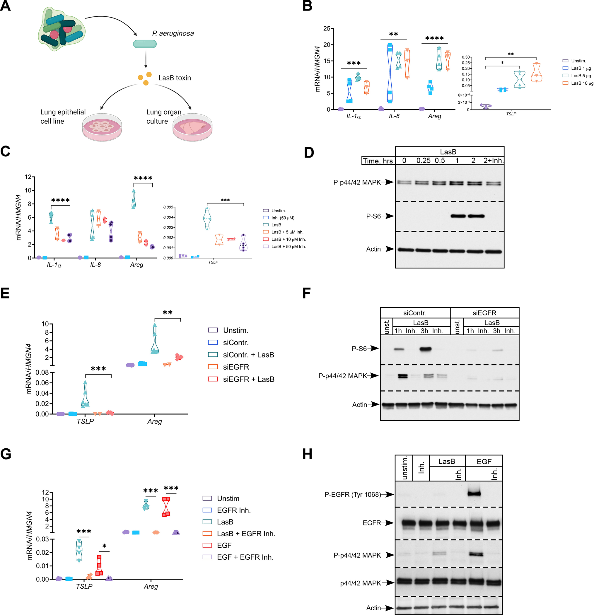
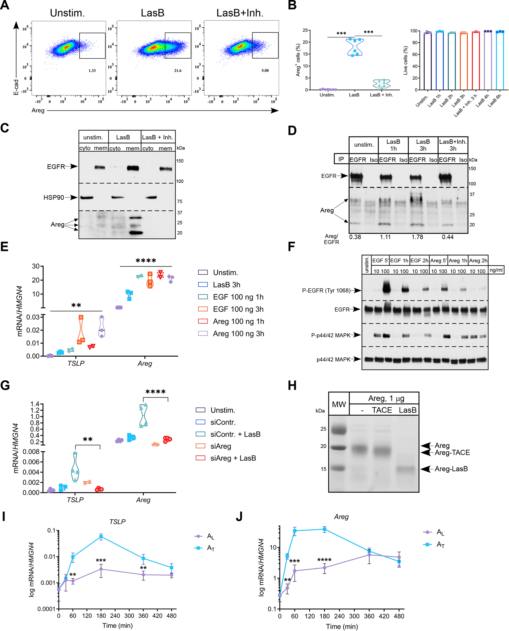
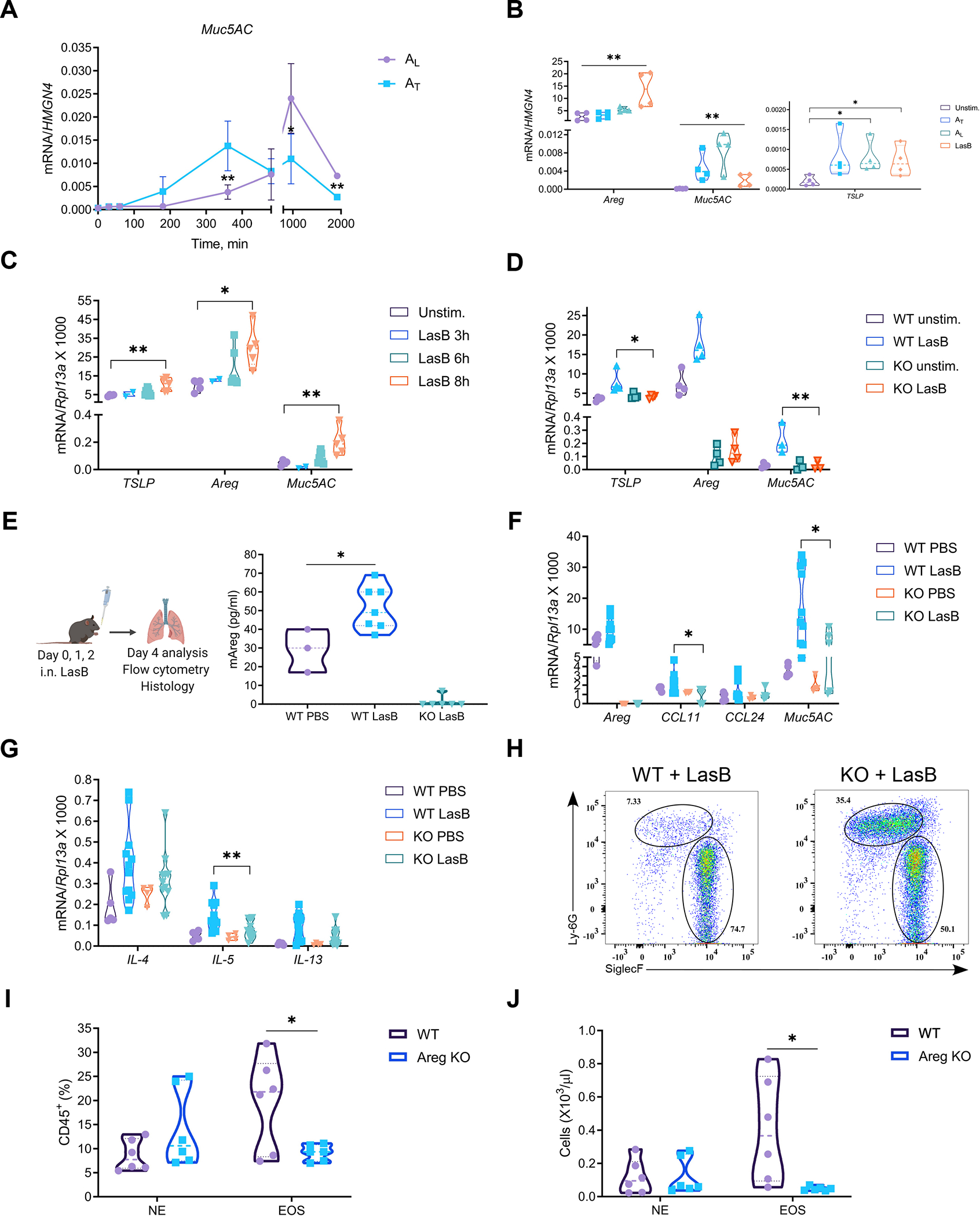
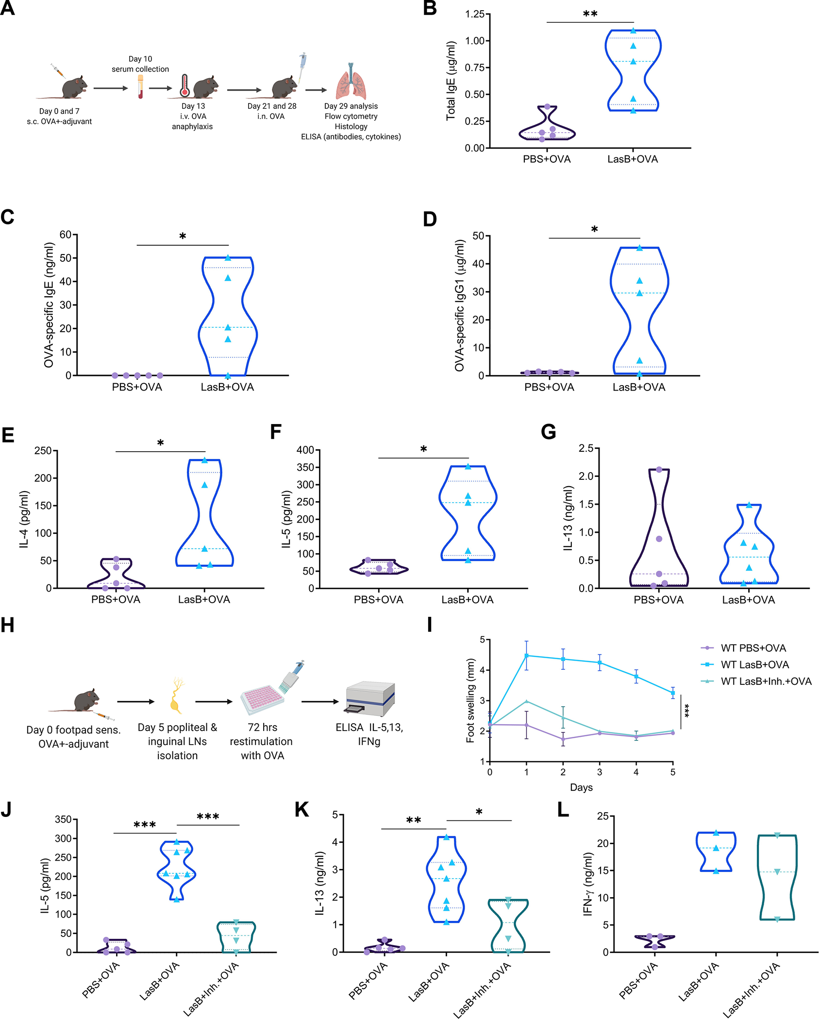
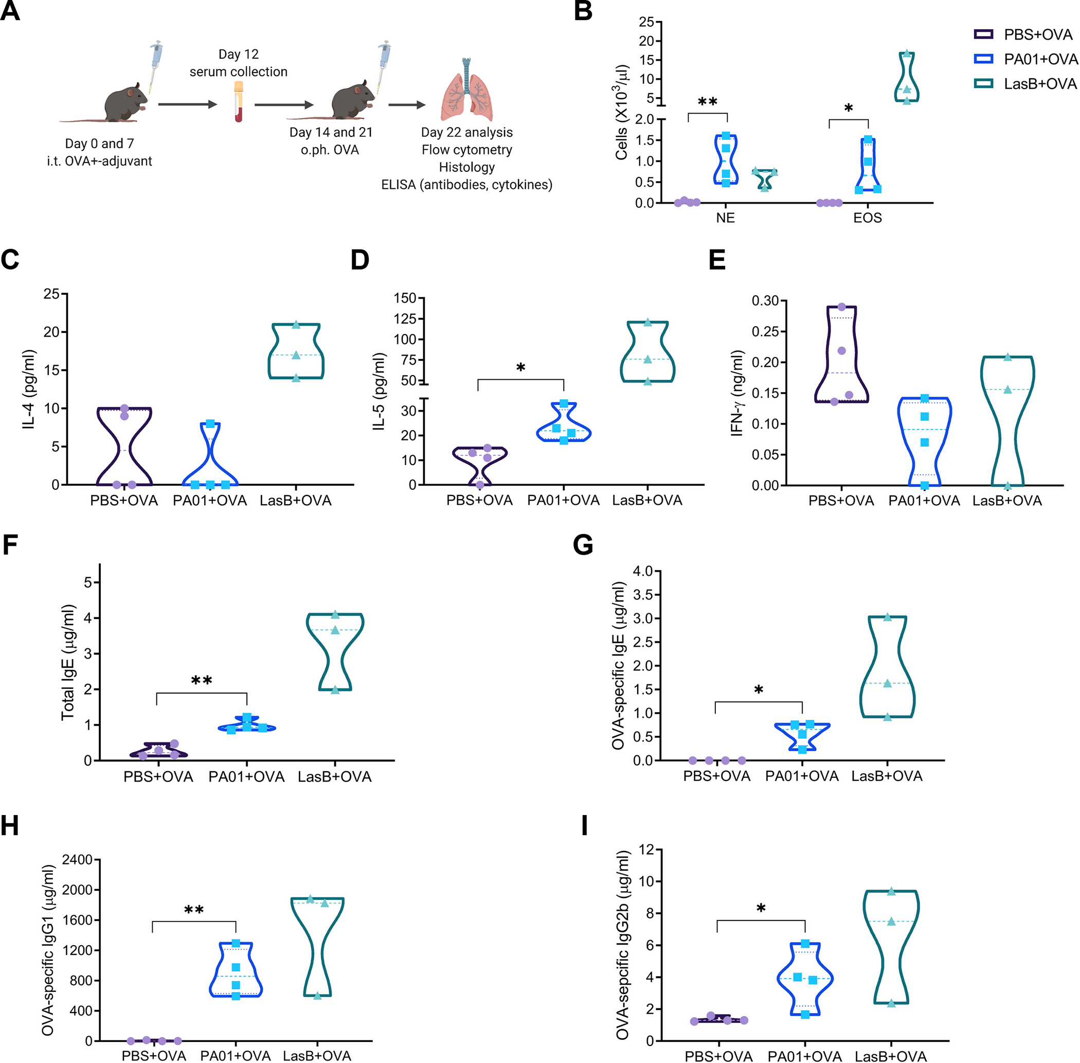
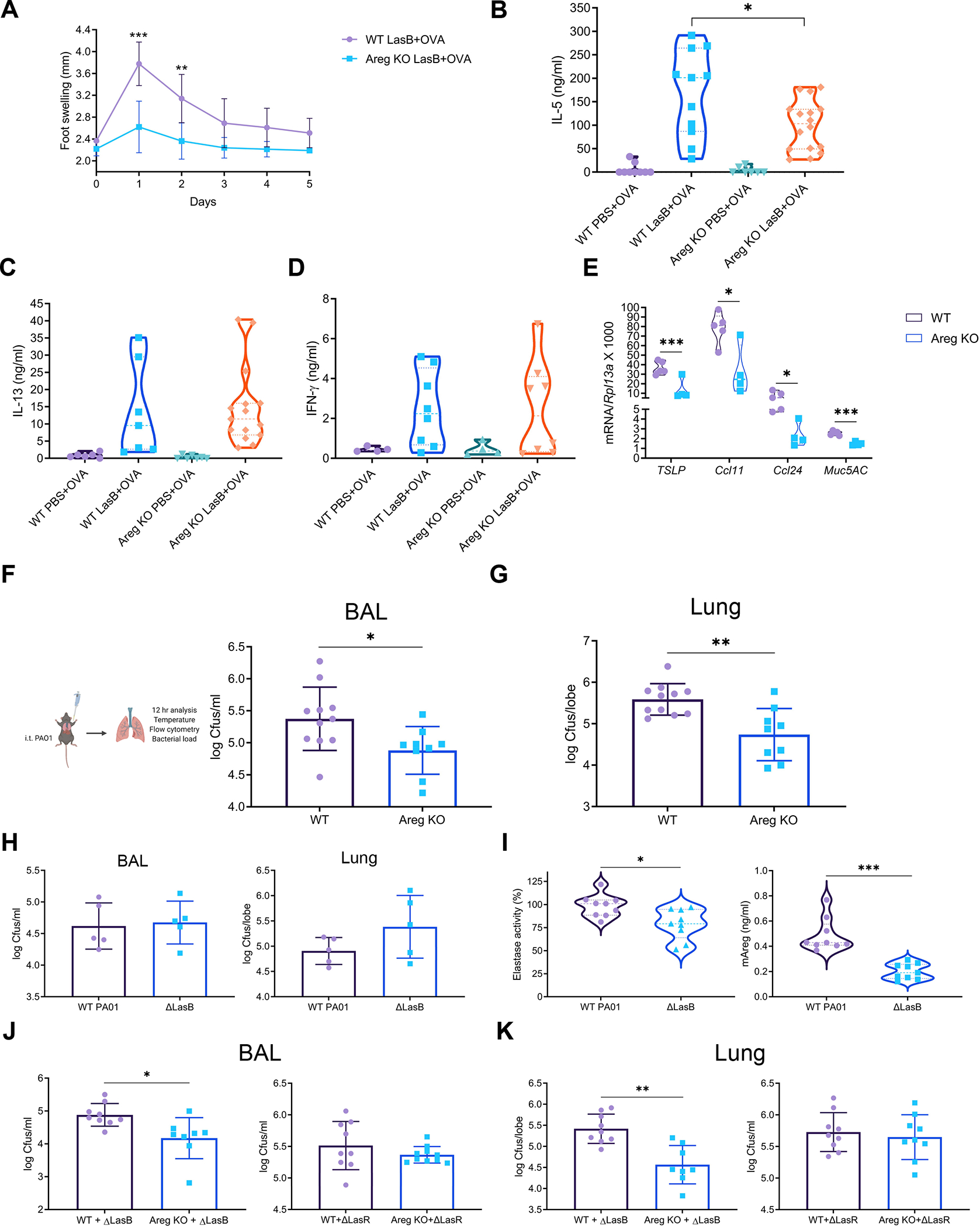
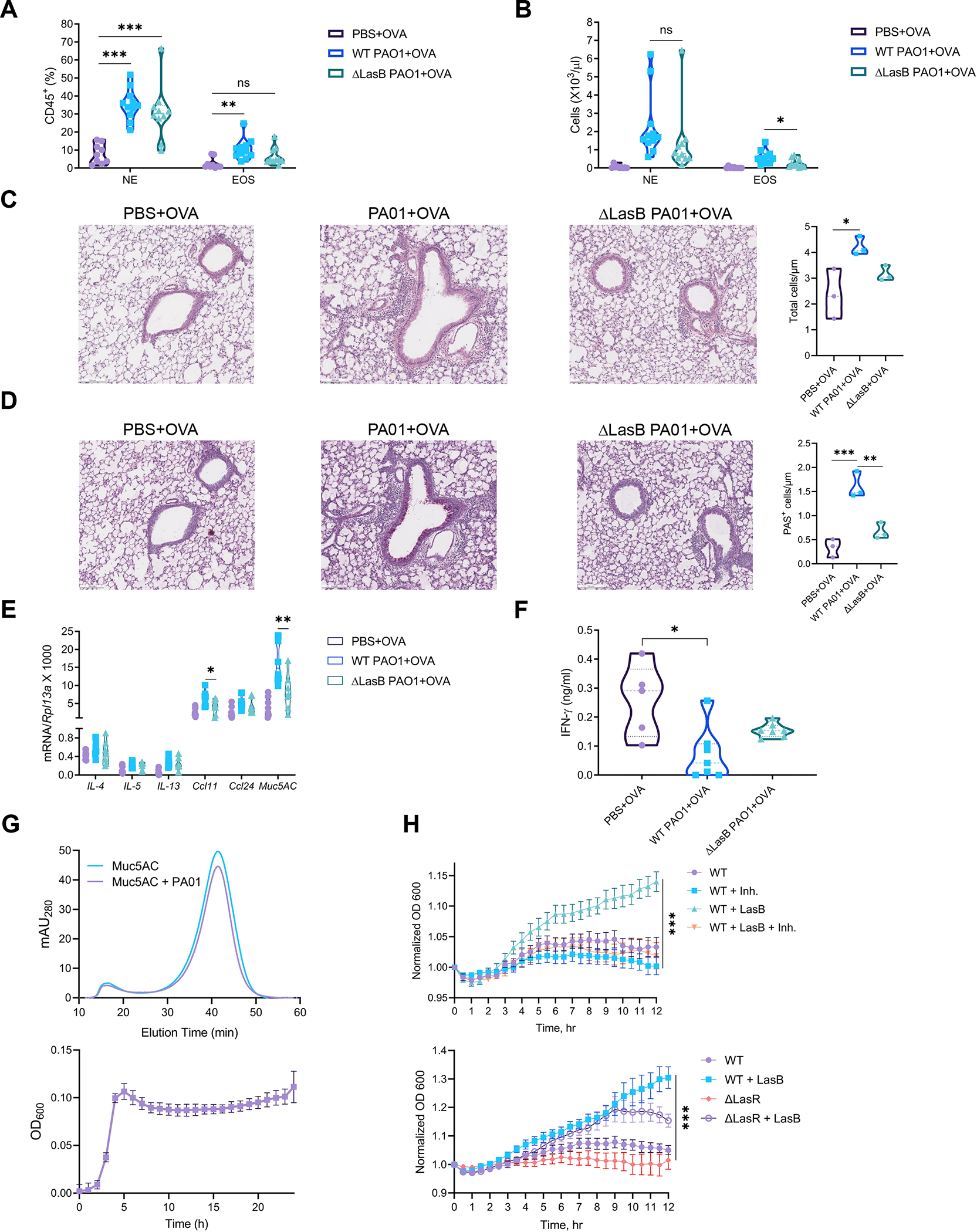
Different effector arms of the immune system are optimized to protect from different classes of pathogens. In some cases, pathogens manipulate the host immune system to promote the wrong type of effector response-a phenomenon known as immune deviation. Typically, immune deviation helps pathogens to avoid destructive immune responses. Here, we report on a type of immune deviation whereby an opportunistic pathogen, Pseudomonas aeruginosa (P. aeruginosa), induces the type 2 immune response resulting in mucin production that is used as an energy source by the pathogen. Specifically, P. aeruginosa-secreted toxin, LasB, processed and activated epithelial amphiregulin to induce type 2 inflammation and mucin production. This "niche remodeling" by P. aeruginosa promoted colonization and, as a by-product, allergic sensitization. Our study thus reveals a type of bacterial immune deviation by increasing nutrient supply. It also uncovers a mechanism of allergic sensitization by a bacterial virulence factor.

Keywords: LasB; P. aeruginosa; allergic inflammation; amphiregulin; immune deviation; lung; mucin; opportunistic pathogen; type 2 immunity.


Conflict of interest statement

Declaration of interests All authors declare no competing interests. B.V. is currently an employee of EMD Serono.

Figures

Figure 1.
Figure 1.. P. aeruginosa toxin induces tissue repair gene program in epithelial cells that is dependent on EGFR pathway
(A) Schematics of in vitro experimental system. (B and C) Relative mRNA expression of pro-inflammatory (IL-1a, IL-8) and tissue repair (TSLP, Areg) genes in H292 cells treated with indicated amounts of LasB (B) or LasB (2 μg/ml) +/− indicated concentrations of inhibitor (C) for 3 hr (n=3–4). (D) Immunoblot analysis of P-p44/42 MAPK and P-S6 in H292 cells treated with LasB (2 μg/ml) +/− inhibitor (50 μM) at different time points (n=4). (E and F) Relative mRNA expression of TSLP and Areg (E) and immunoblot analysis of P-p44/42 MAPK and P-S6 (F) in H292 cells treated with control or EGFR siRNA and stimulated with LasB (2 μg/ml) +/−inhibitor (50 μM) for 1 or 3 hr (n=6). (G and H) Relative mRNA expression of TSLP and Areg (G) and immunoblot analysis of P-EGFR Tyr1068 and P-p44/42 MAPK (H) in H292 cells pre-treated with EGFR kinase inhibitor (20 μM) for 20 min and stimulated with LasB (5 μg/ml) or EGF (10 ng/ml) for 3hr (n=4). mRNA expression was measured relative to high mobility group nucleosome-binding domain-containing protein 4 (HMGN4). ß-actin, total EGFR and p44/42 MAPK were used as loading controls. n = independent experiments. Data were analyzed by one-way ANOVA with Tukey’s multiple comparisons test. *p < 0.05; **p < 0.01; ***p < 0.001; ****p < 0.0001. See also Figure S1.
Figure 2.
Figure 2.. P. aeruginosa toxin utilizes amphiregulin to activate epithelial cells
(A and B) Representative FACS plots (n=6) of frequency of Areg+ H292 cells (A) stimulated with LasB (2 μg/ml) +/− inhibitor (50 μM) for 3hr and quantification (B) of Areg+ (left) and total live cells (right) (n=3–6). (C and D) Immunoblot analysis of Areg in cytosolic and membrane fractions (C) and after immunoprecipitation with anti-EGFR antibodies (D) in H292 cells stimulated with LasB (3 μg/ml) +/− inhibitor (50 μM) for 3hr (n=3). (E) Relative mRNA expression of TSLP and Areg in H292 cells stimulated with LasB (2 μg/ml) for 3hr, EGF and Areg (100 ng/ml) for 1 or 3 hr (n=3). (F) Immunoblot analysis of P-EGFR Tyr1068 and P-p44/42 MAPK in H292 cells stimulated with EGF and Areg (10 or 100 ng/ml) at indicated time points (n=3). (G) Relative mRNA expression of TSLP and Areg in H292 cells treated with control or Areg siRNA and stimulated with LasB (5 μg/ml) for 3hr (n=4). (H) In vitro cleavage of recombinant human Areg (1 μg) by LasB and ADAM17 or TACE (0.5 μg) for 30 min at RT (n=4). (I and J) Kinetics of relative mRNA expression of TSLP (I) and Areg (J) in H292 cells treated with AL or AT (50 ng/ml) (n=3). mRNA expression was measured relative to HMGN4. ß-actin, total EGFR and p44/42 MAPK and HSP90 were used as loading controls. n = independent experiments. Data are mean ± SD and were analyzed by one-way ANOVA with Tukey’s multiple comparisons test (B, E, G) or two-tailed unpaired Student’s t test (I, J). *p < 0.05; **p < 0.01; ***p < 0.001; ****p < 0.0001. See also Figures S1 and S2.
Figure 3.
Figure 3.. Amphiregulin is required for mucin expression and recruitment of eosinophils
(A) Kinetics of relative mRNA expression of Muc5AC in H292 cells treated with 50 ng/ml of AL or AT (n=3). (B) Relative mRNA expression of TSLP, Areg and Muc5AC in H292 cells stimulated with LasB (0.5 μg/ml), AL or AT (50 ng/ml) for 12hr (n=4). (C) Relative mRNA expression of TSLP, Areg and Muc5AC in ex vivo mouse lung organ cultures stimulated with LasB (5 μg/ml) for indicated times (n=5). (D) Relative mRNA expression of TSLP, Areg and Muc5AC in WT and Areg−/− (KO) mouse lung organ cultures stimulated with LasB (5 μg/ml) for 8 hr (n=3–4). (E) (Left) Experimental protocol for the development of innate airway inflammation and (Right) ELISA of Areg concentrations in the BAL of WT and Areg−/− (KO) mice (combined of 3 experiments yielding 3–7 mice per group). (F and G) Relative mRNA expression of Areg, Ccl11, Ccl24 and Muc5AC (F) and type 2 cytokines (IL-4, 5, 13) (G) in the lungs of LasB-treated WT and Areg−/− (KO) mice (combined of 3 experiments yielding 4–12 mice per group). (H-J) Representative FACS plots (n=5) (H) and graphs showing cell frequency (I) and numbers (J) of eosinophils (CD45+MHCII–?CD11b+Siglec-F+) and neutrophils (CD45+MHCII–CD11b+Ly6G+) out of CD45+ cells in the BAL of LasB-treated WT and Areg−/− (KO) mice (combined of 2 experiments yielding 6 mice per group). mRNA expression was measured relative to HMGN4 in H292 or Rpl13a in mouse lung organ cultures/lungs. n = independent experiments. Data are mean ± SD and were analyzed by two-tailed unpaired Student’s t test (A, I, J) or one-way ANOVA with Tukey’s multiple comparisons test (B-G). *p < 0.05; **p < 0.01; ***p < 0.001. See also Figure S3 and S4.
Figure 4.
Figure 4.. LasB is an adjuvant for allergic response
(A) Experimental protocol of LasB allergic airway inflammation. (B-D) ELISA of total IgE (B), OVA-specific IgE (C) and OVA-specific IgG1 (D) amounts in the serum of mice immunized with OVA +/− LasB. (E-G) ELISA of IL-4 (E), IL-5 (F) and IL-13 (G) amounts in the BAL of mice immunized with OVA +/− LasB. (H) Experimental protocol of skin allergic inflammation. (I) Kinetics of swelling in the left footpad of mice immunized with LasB/OVA +/− LasB inhibitor (combined of 2 experiments yielding 3–6 mice per group). (J-L) IL-5 (J), IL-13 (K) and IFN-γ (L) production after ex vivo stimulation of total lymph node cells from mice immunized as in I (J and K - combined of 2 experiments with 4–7 mice per group, L - representative of 3 experiments, 3 mice per group). For (B-G) data combined of 2 experiments yielding 5 mice per group. Data are mean ± SD and were analyzed by two-tailed unpaired Student t-test (B-G) or one-way ANOVA with Tukey’s multiple comparisons test (I-L). *p < 0.05; **p < 0.01; ***p < 0.001. See also Figures S3 and S5.
Figure 5.
Figure 5.. P. aeruginosa can serve as an adjuvant for an allergic response
(A) Experimental protocol of PA01 allergic airway inflammation. (B) Cell frequency of eosinophils (CD45+MHCII–CD11b+CD64-Siglec-F+) and neutrophils (CD45+MHCII–CD11b+CD64-Ly6G+) out of CD45+ cells in the BAL of mice immunized with OVA +/− PA01 or LasB. (C-E) ELISA of IL-4 (C), IL-5 (D) and IFN-γ amounts in the BAL of mice immunized as in (A). (F-I) ELISA of total IgE (F), OVA-specific IgE (G), OVA-specific IgG1 (H) and IgG2b (I) amounts in the serum of mice immunized as in (A). For (B-I) representative of 3 independent experiments, 3–4 mice per group. Data were analyzed by two-tailed unpaired Student’s t test. *p < 0.05; **p < 0.01; ***p < 0.001. See also Figure S4.
Figure 6.
Figure 6.. Amphiregulin is required for successful P. aeruginosa infection
(A) Kinetics of left footpad swelling of WT and Areg−/− (KO) mice immunized with LasB/OVA (combined of 2 experiments yielding 4–7 mice per group). (B-D) IL-5 (B), IL-13 (C) and IFN-γ (D) production after ex vivo stimulation of total lymph node cells from WT and Areg−/− (KO) mice immunized as in A (B and C - combined of 3 experiments yielding 6–16 mice per group, D - combined of 2 experiments yielding 4–8 mice per group). (E) Relative mRNA expression of TSLP, Ccl11, Ccl24 and Muc5AC in WT and Areg/ (KO) lungs infected with WT PA01 for 12 hr (representative of 5 experiments, 4–5 mice per group). (F and G) Experimental protocol of WT PA01 infection (F, left), BAL (F, right) and lung (G) bacterial loads of WT PA01 in WT and Areg−/− (KO) mice 12 hr after infection (combined of 2 experiments yielding 9–11 mice per group). (H and I) BAL (H, left) and lung (H, right) bacterial loads and BAL elastase activity (I, left) and Areg amounts (I, right) of WT and ΔLasB PA01 in WT mice 12 hr after infection (H - representative of 3 experiments, 5 mice per group; I - combined of 2 experiments yielding 9 mice per group). (J and K) BAL (G) and lung (H) bacterial loads of ΔLasB and ΔLasR PA01 in WT and Areg−/− (KO) mice 12 hr after infection (combined of 2 experiments yielding 8–10 mice per group). mRNA expression was measured relative to Rpl13a. Data are mean ± SD and were analyzed by two-tailed unpaired Student’s t test (A, E-K) or one-way ANOVA with Tukey’s multiple comparisons test (B-D). *p < 0.05; **p < 0.01; ***p < 0.001. See also Figure S5.
Figure 7.
Figure 7.. LasB-deficient P. aeruginosa have reduced adjuvant activity to stimulate eosinophil recruitment and mucin production
(A and B) Cell frequency (A) and numbers (B) of eosinophils (CD45+MHCII–CD11b+CD64-Siglec-F+) and neutrophils (CD45+MHCII–CD11b+CD64-Ly6G+) out of CD45+ cells in the BAL of mice immunized with OVA +/− WT or ΔLasB PA01 (combined of 3 experiments yielding 9–11 mice per group). (C and D) H&E (C) and PAS (D) staining in the lungs of mice immunized as in (A and B) (representative of 3 experiments, 4 mice per group, scale bar = 500 μm). (E) Relative mRNA expression of type 2 cytokines (IL-4, 5, 13), eotaxins (Ccl11, 24) and Muc5AC in the lungs of mice immunized as in (A and B) (combined of 2 experiments yielding 7–9 mice per group). (F) ELISA of IFN-γ amounts in the BAL of mice immunized with OVA +/− WT or ΔLasB PA01 (combined of 2 experiments yielding 5–7 mice per group). (G) (Top) Representative fast protein liquid chromatography (FPLC) profile (n=3) of Muc5AC before and after incubation with PA01 and (bottom) growth curves of PA01 on Muc5AC (growth curve data are mean ± SEM, n=3; FPLC data are the mean of n=3 traces). (H) (Top) PA01 growth +/− LasB (3 μg/ml) and/or phosphoramidon (50 mM) and (bottom) WT and ΔLasR PA01 growth +/− LasB (3 μg/ml) on human sputum samples for 12 hr at 37 °C (data are mean ± SEM, n=4). mRNA expression was measured relative to Rpl13a. Data were analyzed by one-way ANOVA with Tukey’s multiple comparisons test. *p < 0.05; **p < 0.01; ***p < 0.001. See also Figures S6 and S7.

Comment in

References

    1. Abebe F (2019). Synergy between Th1 and Th2 responses during Mycobacterium tuberculosis infection: A review of current understanding. Int Rev Immunol 38, 172–179. - PubMed
    1. Afgan E, Baker D, Batut B, van den Beek M, Bouvier D, Cech M, Chilton J, Clements D, Coraor N, Gruning BA, et al. (2018). The Galaxy platform for accessible, reproducible and collaborative biomedical analyses: 2018 update. Nucleic acids research 46, W537–W544. - PMC - PubMed
    1. Allen JE, and Wynn TA (2011). Evolution of Th2 Immunity: A Rapid Repair Response to Tissue Destructive Pathogens. PLoS pathogens 7. - PMC - PubMed
    1. Annunziato F, Romagnani C, and Romagnani S (2015). The 3 major types of innate and adaptive cell-mediated effector immunity. The Journal of allergy and clinical immunology 135, 626–635. - PubMed
    1. Arabyan N, Park D, Foutouhi S, Weis AM, Huang BHC, Williams CC, Desai P, Shah J, Jeannotte R, Kong N, et al. (2016). Salmonella Degrades the Host Glycocalyx Leading to Altered Infection and Glycan Remodeling. Scientific reports 6. - PMC - PubMed

Publication types