Review
. 2022 May 5;29(5):692-721.
doi: 10.1016/j.stem.2022.04.002. Epub 2022 Apr 27.

Biomaterials to enhance stem cell transplantation

Affiliations
Review

Biomaterials to enhance stem cell transplantation

Bhushan N Kharbikar et al. Cell Stem Cell. .

Abstract

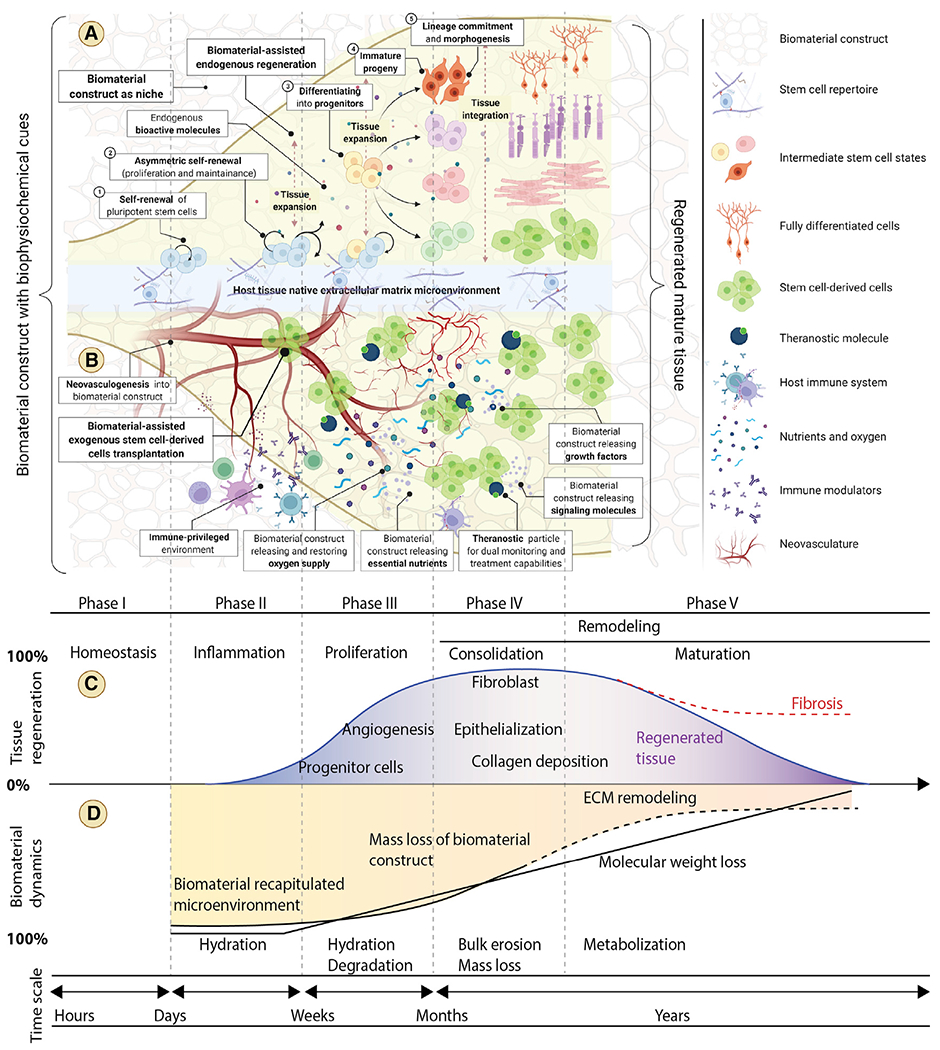
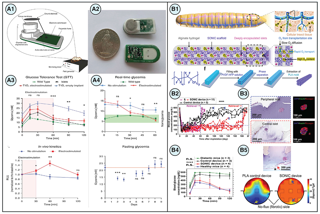
The successful transplantation of stem cells has the potential to transform regenerative medicine approaches and open promising avenues to repair, replace, and regenerate diseased, damaged, or aged tissues. However, pre-/post-transplantation issues of poor cell survival, retention, cell fate regulation, and insufficient integration with host tissues constitute significant challenges. The success of stem cell transplantation depends upon the coordinated sequence of stem cell renewal, specific lineage differentiation, assembly, and maintenance of long-term function. Advances in biomaterials can improve pre-/post-transplantation outcomes by integrating biophysiochemical cues and emulating tissue microenvironments. This review highlights leading biomaterials-based approaches for enhancing stem cell transplantation.


Conflict of interest statement

Declaration of interests The authors declare the following competing financial interest(s): T.A.D. is a scientific founder of Encellin, a cell therapy device company, and she is listed as an inventor of a macro-encapsulation technology (US Patent #10,865,378) described in this paper.

Figures

Figure 1.
Figure 1.. Biomaterial-facilitated stem cell transplantation with engineered biophysiochemical traits for tissue regeneration.
(A) Biomaterial recapitulated microenvironments present essential and complex biophysiochemical cues to retain stemness, direct differentiation, promote reprogramming, manipulate genomic and epigenomic traits, and select for functional phenotypes while dictating stem cell fate during regeneration and repair. (B) Optimal biomaterial-based methods of stem cell administration by injection or transplantation may improve cell retention and integration with host tissue by allowing for the migration of transplanted and host cells. The intrinsic biomaterial properties (bioinert, bioactive, and biotolerant) and the engineered extrinsic bioactive properties, including biophysical (porosity, pressure, elasticity, force, topography, etc.), biochemical (hormones, cytokines, peptides, growth factors, and immune modulators), and physiochemical (hydrophilicity, temperature, pH, oxygen, nutrients, charge, light, and magnetic field), of the material can protect stem cells after transplantation from stress, hypoxia, starvation, and immune attack, thus facilitating long-term viability and maintenance of the graft. (C and D) Optimally designed biomaterial constructs should possess dynamic properties that closely align with the different phases of tissue regeneration after implantation. Matching the appropriate timescale of material characteristics including hydration, degradation, bulk erosion, mass loss, and metabolization to regenerative and reparative processes can be beneficial to facilitate tissue regeneration and enable new tissue to overtake functions initially provided by the scaffold while replacing damaged host tissue.
Figure 2.
Figure 2.. Biomaterial-facilitated stem cell-based regenerative therapies for cardiovascular applications
(A) (A1) An engineered design of a 4D biomaterial patch with enhanced biomechanical properties using stretchable architecture to accommodate changes in cardiac tissue curvature during diastole and systole. (A2–A4) In vivo implantation of the 4D patches in rodent models of ischemia reperfusion MI demonstrated high engraftment of cardiomyocytes on the patch at week 3. Scale bars, 100 μm. (A5) Immunostaining of α-actinin (green) and human-specific CD31 (red) showed cellularization of the patch after 4 months. Scale bars, 50 μm. (A6) Quantification of von Willebrand factor staining depicted increased vascularization of the patch from 10 weeks to 4 months. Data are presented as means ± SD, *p < 0.05 and **p < 0.01 (Cui et al., 2020). (B) (B1) Microneedle (MN) patches integrated with cardiac stromal cells (CSCs) is a promising strategy for cardiac regeneration after MI. (B2) DiO-labeling of CSCS (green) demonstrated successful incorporation of the cells onto the MN patch (red). Scale bars, 500 μm. (B3 and B4) Treatment with MN patches in porcine models of acute MI improved ejection fraction and fractional shortening after 48 h. Data are presented as means ± SD, *p < 0.05 and **p < 0.01. (B5 and B6) immunostaining demonstrated an increased presence of proliferating cardiomyocytes and vasculature in post-MI rat hearts treated with MN-CSCs. Data are presented as means ± SD, *p < 0.05. Scale bars, 200 μm (Tang et al., 2018). Figures reproduced with permission.
Figure 3.
Figure 3.. Biomaterial-facilitated stem cell-based regenerative therapies for central nervous system applications.
(A) (A1 and A2) SMART spheroids were developed to improve cell-cell and cell-matrix interactions and achieve controlled drug release to enhance in vivo neuronal differentiation of transplanted stem cells, thereby leading to functional recovery in models of SCI. (A3) Injection of SMART neurospheres (spheroids assembled from NSCs) achieved long-term stem cell survival and neuronal differentiation along with reduced glial scar and functional recovery 1 month postinjection. Data are presented as means ± SEM, *p < 0.05 and **p < 0.01. (A4) Treatment with SMART neurospheres resulted in faster recovery rates at 1 month based on the Basso mouse scale (BMS) scoring. Data are presented as means ± SEM, *p < 0.05 (Rathnam et al., 2021). (B) (B1 and B2) SHIELD, an injectable shear-thinning hydrogel, was designed to improve cell survival and engraftment after transplantation by incorporating celladhesive ligands and employing self-healing and thixotropic characteristics. (B3) immunostaining quantification of the lesion and perilesion regions in spinal cord sections revealed a significant reduction of the pan-macrophage marker ED1 in animals treated with Schwann cells (SCs) in SHIELD compared with injury only controls, whereas no significant differences were observed between the groups for Iba1, microglia marker, or Tomato lectin, vasculature marker. Data are presented as means ± SEM, *p < 0.05. (B4) Forelimb coordination significantly increased in SHIELD-delivered SCs-treated animals after 4 weeks as measured by a decrease in the percentage of missed steps with the horizontal ladder walk test. Data are presented as means ± SEM, *p < 0.05 and $p = 0.970 comparison between before injury and 4-week SCs in SHIELD (Marquardt et al., 2020). Figures reproduced with permission.
Figure 4.
Figure 4.. Biomaterial-facilitated stem cell-based regenerative therapies for ocular tissues
(A) (A1) A grafting strategy devised to introduce tissue-engineered human embryonic stem cell-derived retinal pigment-epithelial (hESC-RPE) cell sheets to the subretinal space of the eye via injection while maintaining polarity of the hESC-RPE cell sheet. (A2) Immunostaining of the tissue construct demonstrated the organization of hESC-RPE cells in a monolayer (TYRP1 = red, DAPI/nuclei = blue). Scale bars, 50 μm. (A3) An optokinetic test determined that treatment with transplanted hESC-RPE cell sheets significantly improved visual acuity compared with sham at various time points post-transplantation (4, 6, and 13 weeks). Data are presented as means ± SEM, *p < 0.05, **p < 0.01, and ***p < 0.001. (A4) Outer nuclear layer (ONL) thickness was increased in rats treated with hESC-RPE cell sheets. Data are presented as means ± SEM, *p < 0.05 and **p < 0.01. (A5) Histological analysis confirmed that more photoreceptor cell nuclei were preserved after transplantation in rat eyes with hESC-RPE cell sheets compared with hESC-RPE cell suspensions. Scale bars, 50 μm (M’Barek et al., 2017). (B) (B1) Human RPE stem cell-derived RPE monolayers grown on PET membranes were being evaluated for their potential as a cell-replacement therapy for age-related macular degeneration. (B2) After 1 week, retinal tissue loss was observed over the implant center but remained stable for the duration of later time points, pointing to a future challenge that remains for hRPE xenografts. Scale bars, 200 μm (rows 1–3) and 250 μm (row 4). (B3) Immunostaining for human-specific marker SC121 (red) confirmed survival of the human RPE monolayer subretinally for 1 month, although costaining of SC121 with apical membrane markers MCT1 and ezrin (top left and right, respectively) (green) confirmed that the RPE was still polarized. The absence of Ki67, phosphohistone H3, and caspase-3 (bottom, from left to right, respectively) indicated that neither proliferation nor apoptosis was occurring. (B4) TEM imaging revealed polarized fetal and adult hRPE cells on the PET carriers. Scale bars, 2 μm. Inset scale bars, 0.2 μm (Stanzel et al., 2014). Figures reproduced with permission.
Figure 5.
Figure 5.. Biomaterial-facilitated stem cell-based regenerative therapies for pancreatic tissues
(A) (A1 and A2) A strategy employing wireless electrical stimulation of engineered electrosensitive beta-cells (Electroβ cells) housed inside of a bioelectronic device enabled electrogenic control of insulin release from cells that could be used for type 1 diabetes therapy. The bioelectronic implant was placed subcutaneously in a mouse, whereas a field generator provided the necessary wireless energy transmission. (A3) Electroβ cells re-established postprandial glucose metabolism and achieved fast vesicular secretion after electrostimulation in insulin-deficient type 1 diabetic mice. Data are presented as means ± SEM, *p < 0.05, **p < 0.01, and ***p < 0.001. (A4) Moreover, it was found that blood glucose levels could be quickly restored to normoglycemia after electrostimulation and that glycemia could be controlled over long periods of time without experiencing hypoglycemia. Data are presented as means ± SEM, *p < 0.05, **p < 0.01, and ***p < 0.001 (Krawczyk et al., 2020). (B) (B1) A novel biomimetic scaffold design called SONIC utilized continuous air channels to improve oxygen diffusivity within cell encapsulation systems and was inspired by the tracheal systems in mealworms. (B2) Diabetic C57BL/6 mice implanted with SONIC devices with rat islets demonstrated long-term controlled blood glucose readings spanning 6 months until device retrieval, where blood glucose levels returned to hyperglycemia. Individual device data are presented, ****p < 0.0001. (B3) Histological evaluation and immunostaining of insulin (green) and glucagon (red) confirmed islet viability and function from cells in retrieved devices on day 60. Scale bars, 200 μm (left) and 100 μm (right). (B4) Intraperitoneal glucose tolerance tests were conducted on day 180 postimplantation, and the results showed animals treated with the SONIC device had glycemic profiles similar to that of healthy mice, with blood glucose levels returning to normoglycemia within 2 h. Data are presented as means ± SD, ****p < 0.0001 (diabetic mice versus SONIC device-treated mice, diabetic mice versus healthy mice, control device-treated mice versus SONIC device-treated mice, and control device-treated mice versus healthy mice). (B5) Histology confirmed that islets near regions of fibrosis remained healthy and corroborated findings from computational simulations of fibrosis where control devices were found to be hypoxic with high levels of islet necrosis, whereas SONIC devices were sufficiently oxygenated throughout the implant. Scale bars, 200 μm (Wang et al., 2021b). Figures reproduced with permission.

References

    1. Abdeen AA, and Saha K (2017). Manufacturing cell therapies using engineered biomaterials. Trends Biotechnol. 35, 971–982. 10.1016/J.TIBTECH.2017.06.008. - DOI - PMC - PubMed
    1. Adu-Berchie K, and Mooney DJ (2020). Biomaterials as local niches for immunomodulation. Acc. Chem. Res 53, 1749–1760. 10.1021/ACS.ACCOUNTS.0C00341. - DOI - PubMed
    1. Aguado BA, Mulyasasmita W, Su J, Lampe KJ, and Heilshorn SC (2012). Improving viability of stem cells during syringe needle flow through the design of hydrogel cell carriers. Tissue Eng. Part A 18, 806–815. 10.1089/ten.TEA.2011.0391. - DOI - PMC - PubMed
    1. Ahn CB, Lee J-H, Kim JH, Kim TH, Jun H-S, Son KH, and Lee JW (2022). Development of a 3D subcutaneous construct containing insulin-producing beta-cells using bioprinting. Bio-Des. Manuf 5, 265–276. 10.1007/S42242-021-00178-9/FIGURES/8. - DOI
    1. Ali M, and Payne SL (2021). Biomaterial-based cell delivery strategies to promote liver regeneration. Biomater. Res 25, 5. 10.1186/S40824-021-00206-W. - DOI - PMC - PubMed

Publication types

Substances