. 2022 May 12;185(10):1709-1727.e18.
doi: 10.1016/j.cell.2022.03.043. Epub 2022 Apr 27.

Maladaptive innate immune training of myelopoiesis links inflammatory comorbidities

Affiliations

Maladaptive innate immune training of myelopoiesis links inflammatory comorbidities

Xiaofei Li et al. Cell. .

Abstract

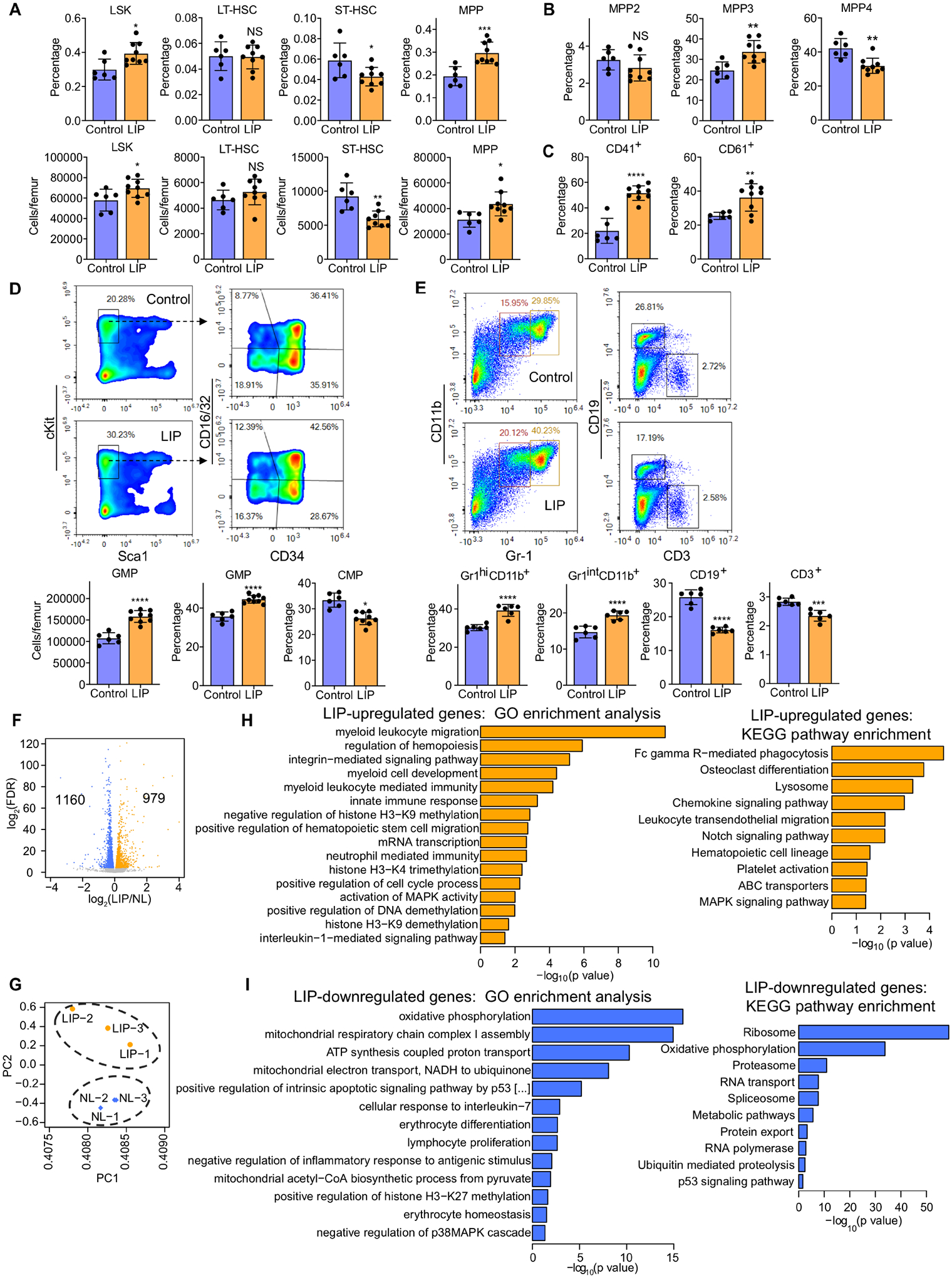
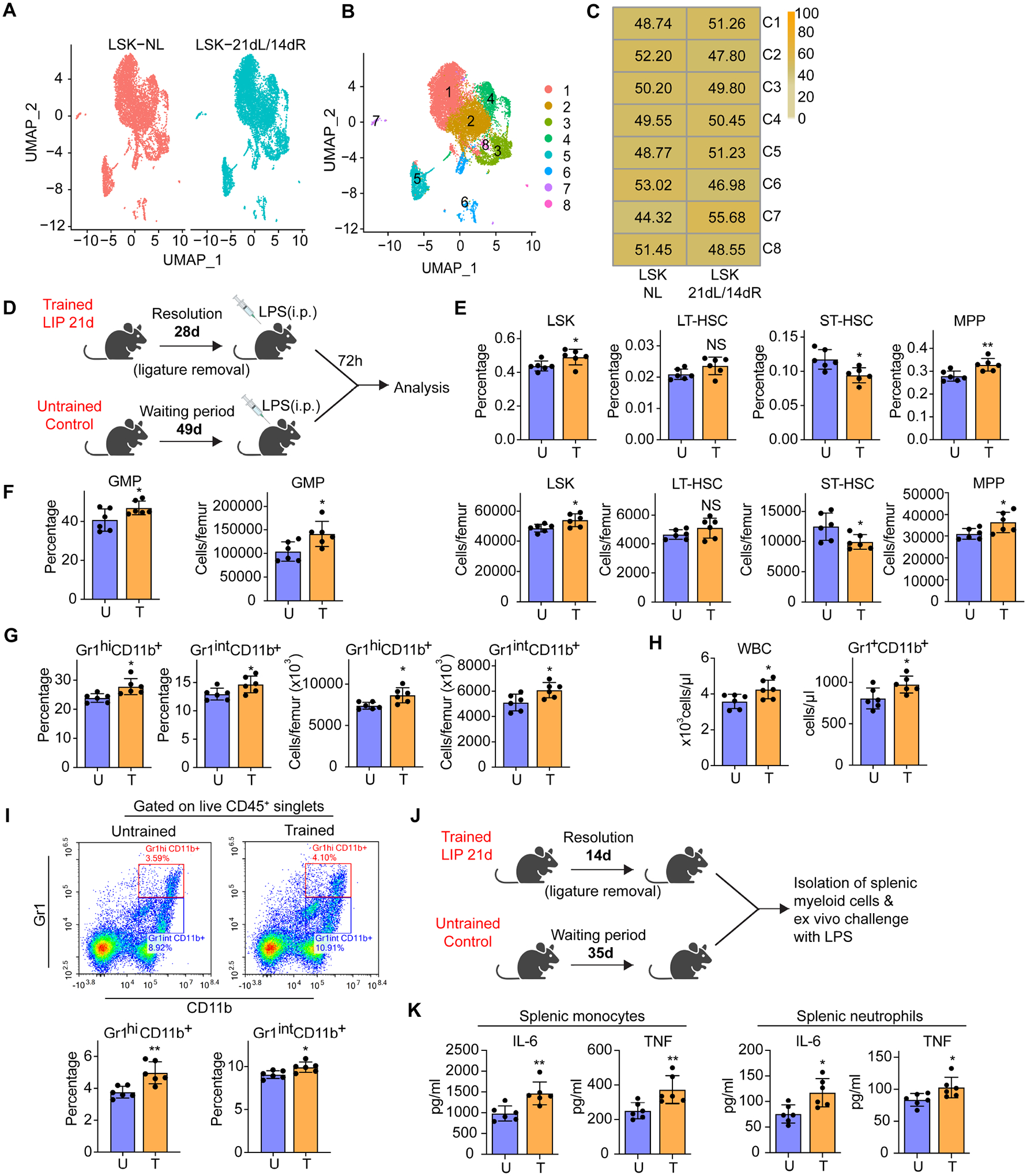
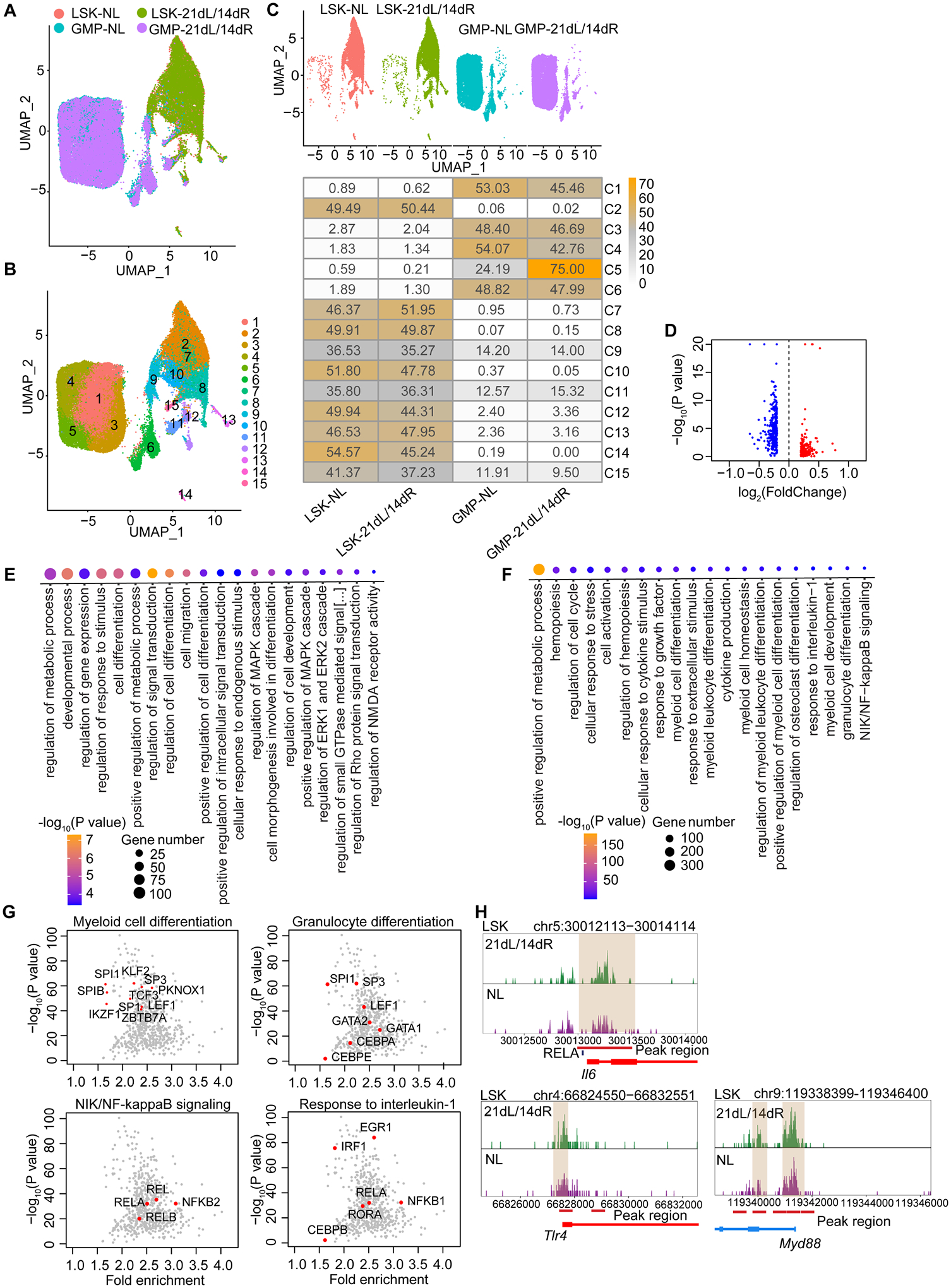
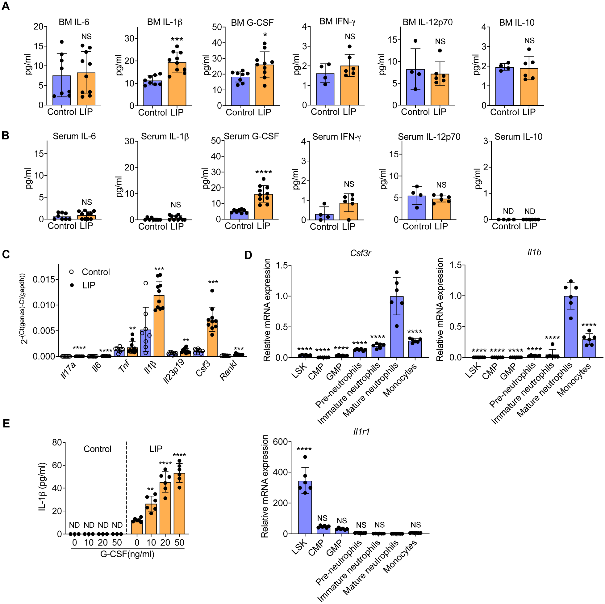
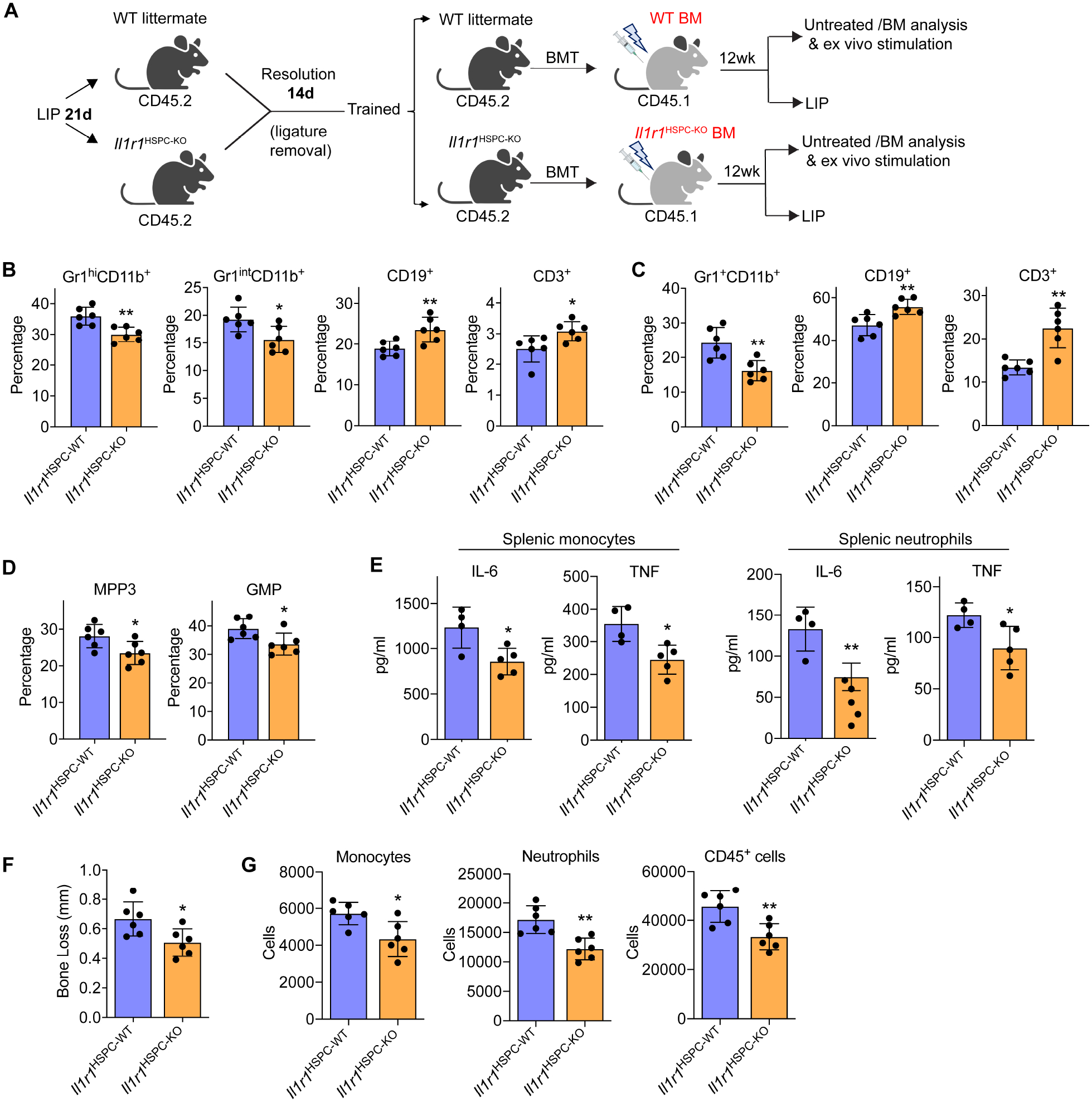
Bone marrow (BM)-mediated trained innate immunity (TII) is a state of heightened immune responsiveness of hematopoietic stem and progenitor cells (HSPC) and their myeloid progeny. We show here that maladaptive BM-mediated TII underlies inflammatory comorbidities, as exemplified by the periodontitis-arthritis axis. Experimental-periodontitis-related systemic inflammation in mice induced epigenetic rewiring of HSPC and led to sustained enhancement of production of myeloid cells with increased inflammatory preparedness. The periodontitis-induced trained phenotype was transmissible by BM transplantation to naive recipients, which exhibited increased inflammatory responsiveness and disease severity when subjected to inflammatory arthritis. IL-1 signaling in HSPC was essential for their maladaptive training by periodontitis. Therefore, maladaptive innate immune training of myelopoiesis underlies inflammatory comorbidities and may be pharmacologically targeted to treat them via a holistic approach.

Keywords: arthritis; bone marrow transplantation; comorbidities; epigenetic rewiring; hematopoietic stem and progenitor cells; inflammation; myelopoiesis; periodontitis; trained immunity.


Conflict of interest statement

Declaration of interests M.G.N. is the scientific founder and member of the Scientific Advisory Board of Trained Therapeutics and Discovery (TTxD). M.G.N. has two patents: US18/61935 (Targeted nanoimmunotherapy to increase trained immunity) and US18/61939 (Targeted nanoimmunotherapy for inhibition of trained immunity). The other authors declare no competing interests.

Figures

Figure 1.
Figure 1.. LIP causes a sustained increase in myelopoiesis.
Mice were subjected, or not (control), to LIP followed by BM analysis after 21 days. (A) Frequencies (top) of LSK, LT-HSC, ST-HSC and MPP in total BM cells and absolute cell numbers (bottom) of the same populations. (B) Frequencies of MPP subsets in LSK in the BM of mice. (C) Frequency of CD41+ and CD61+ LT-HSC in total LT-HSC cells in the BM. (D, top) Representative FACS plots for GMP and CMP in the BM; (D, bottom) absolute cell numbers of GMP (left) and frequency within the MyP pool of GMP (middle) and CMP (right) in the BM. (E, top) Representative FACS plots to identify Gr1hiCD11b+ granulocytes, Gr1intCD11b+ myeloid cells, CD19+ B cells and CD3+ T cells and (E, bottom) frequencies of these populations in CD45+ cells in the BM. (F-I) Mice were subjected, or not (control), to LIP and BM cells were harvested on day 7. FACS-sorted LSK were subjected to RNA-sequencing analysis. (F) Differential gene expression in LSK from LIP-subjected mice vs. non-ligated controls. Volcano plot showing the distribution of the adjusted P values (−log2(FDR)) and the fold changes (log2 fold change). Significant changes are shown in blue (down-regulated) or orange (up-regulated) (FDR<0.05), n=3. (G) PCA plot of LSK samples from ligated and non-ligated (NL) groups. Top overrepresented GO terms and KEGG pathways including upregulated (orange) (H) or downregulated (blue) genes (I) in LSK from LIP-subjected mice vs. NL controls. (A-D) Control, n=6 mice/group; LIP, n=9 mice/group and (E) n=6 mice/group. Data are means±SD. *P<0.05, **P<0.01, ***P<0.001, ****P<0.0001, NS, not significant vs. control mice; two-tailed Student’s t-test. See Figure S1.
Figure 2.
Figure 2.. LIP-experienced mice display increased myelopoiesis after LPS challenge.
(A-C) BM LSK were sorted from mice subjected to 21dL/14dR or not (NL) and scRNA-seq was performed. (A,B) Two-dimensional UMAP representation of 15257 cells, according to (A) sample origin and (B) results of clustering. (C) Heat map visualization of the distribution of cells within each of the identified clusters, normalized for the number of cells per sample in the dataset. (D) Experimental design. (E) Frequencies (top) of LSK, LT-HSC, ST-HSC and MPP in total BM cells and absolute cell numbers (bottom) of the same populations. (F) Frequency (left) within the MyP pool of GMP and absolute numbers (right) of GMP in the BM. (G) Frequency of Gr1hiCD11b+ granulocytes and Gr1intCD11b+ myeloid cells in CD45+ cells (left) and cell numbers (right) of the same populations in the BM. (H) Total white blood cell (WBC) count (left) and Gr1+CD11b+ cell counts (right) in the peripheral blood. (I) Representative FACS plots (left) to identify Gr1hiCD11b+ granulocytes and Gr1intCD11b+ myeloid cells and frequency (right) of the same populations in CD45+ cells in the lungs of mice. (J) Experimental design. (K) Isolated splenic monocytes and neutrophils were re-stimulated ex vivo with LPS (10ng/ml) for 24h. The supernatant was collected for measuring IL-6 and TNF. Data are means±SD (n=6 mice/group) (E-I and K). *P<0.05, **P<0.01, NS, not significant vs. untrained mice; two-tailed Student’s t-test. U, untrained; T, trained. See Figure S2.
Figure 3.
Figure 3.. Epigenetic rewiring of trained LSK and GMP.
BM LSK and GMP were sorted from mice subjected to 21dL/14dR or not (NL) and scATAC-seq was performed. (A, B) Two-dimensional UMAP representation of 37,903 cells, according to (A) sample origin and (B) results of clustering. (C) UMAP (up) of the distribution of cells from the four different samples (LSK and GMP subjected to 21dL/14dR or not) and heat map visualization (bottom) within each of the identified clusters, normalized for the number of cells per sample in the dataset are shown. (D) Volcano plots displaying differential accessibility analysis results (blue, lower vs. red, greater differential chromatin accessibility) for LSK subjected to 21dL/14dR vs. their counterparts from control mice (abs(Log2FC) ≥ 0.2, P<0.05). (E) GO enrichment results of top 20 significantly enriched GO terms sorted by PANTHER based on genes annotated to regions more accessible due to 21dL/14dR treatment (Bonferroni-corrected, P<0.05). (F) GO enrichment analysis of enriched TF binding motifs in LSK subjected to 21dL/14dR vs. control (NL) group and the top 20 significantly enriched GO terms (Bonferroni-corrected, P<0.05). (G) Visualization of top enriched TF binding motifs in the indicated GO terms for the LIP specifically accessible regions in LSK using the homer TF motif database. (H) Genome browser track showing DAR in the Il6 gene locus and the RELA binding motifs within this region, as well as DARs close to the promoter regions of Tlr4 and Myd88 gene locus. See Figure S3.
Figure 4.
Figure 4.. LIP-induced trained myelopoiesis contributes to the periodontitis-arthritis comorbidity.
(A) CD45.2+ mice were either trained (T) by LIP for 21 days followed by 14-day resolution or left untrained (U). BM cells were isolated from trained and untrained mice and transferred to lethally irradiated congenic B6.SJL CD45.1+ mice. At 12 weeks post-BMT, groups of recipient CD45.1+ mice were subjected to LIP for 5 days (B-D) or CAIA for 14 days (E-G). (B) Bone loss, (C) relative gingival mRNA expression of indicated cytokines and (D) FACS analysis of gingival monocytes (live CD45+CD11cCD11b+Ly6GLy6C+), neutrophils (live CD45+CD11cCD11b+Ly6G+Ly6C) and total CD45+ cells in recipient CD45.1+ mice subjected to 5-day LIP. (E) Clinical arthritis score (left) and hind ankle joint thickness (right), (F) representative images of H&E (left) and Safranin-O staining (right) of tissue sections from knee joints harvested on day 7 (scale bars 500μm) and (G) quantification of monocytes (live CD45+CD11cCD11b+Ly6GLy6C+), neutrophils (live CD45+CD11cCD11b+Ly6G+Ly6C) and total CD45+ cells in the synovium of knee joints harvested on day 7. (H) 100 LT-HSC sorted from CD45.2+ mice subjected, or not (control), to LIP for 21 days followed by 14-day resolution– were co-transplanted with 5×10⁵ CD45.1+ BM competitors. (I) WBC count, donor-derived percentage, and lineage output (% of indicated cell types in donor-derived cells) in peripheral blood of mice receiving CD45.2+ LT-HSC. Data are means±SD (B and C, n=5 mice/group; D, G and I, n=6 mice/group; E n=7 mice/group). *P<0.05, **P<0.01, ***P<0.001, ****P<0.0001, NS, not significant vs. untrained mice. Two-tailed Student’s t-test (B,C,D,G) except for Rankl in panel C (two-tailed Mann-Whitney U test); two-way repeated measures ANOVA and Sidak’s multiple comparisons test (E,I). W, weeks. See Figure S4.
Figure 5.
Figure 5.. CAIA– or LIP–induced trained myelopoiesis contributes to the bidirectional arthritis-periodontitis comorbidity.
(A) CD45.2+ mice were either trained (T) by subjecting them to CAIA as indicated, or were left untrained (U; naïve controls). BM cells were isolated from trained and untrained mice and transferred to lethally irradiated congenic B6.SJL CD45.1+ mice. At 12 weeks post-BMT, groups of recipient CD45.1+ mice were subjected to LIP for 5 days (B-D) or CAIA for 14 days (E-G). (B) Bone loss, (C) relative gingival mRNA expression of indicated cytokines and (D) FACS analysis of gingival monocytes, neutrophils and total CD45+ cells in recipient CD45.1+ mice subjected to 5-day LIP. (E) Clinical arthritis score (left) and hind ankle joint thickness (right), (F) quantification of monocytes, neutrophils and total CD45+ cells in the synovium of knee joints harvested on day 7, and (G) representative images of H&E (left) and Safranin-O staining (right) of tissue sections from knee joints harvested on day 7 (scale bars 500μm). (H) CD45.1+ mice were transplanted with 200 CD45.2+ LT-HSC from LIP-trained or untrained control mice together with 8×10⁵ CD45.1+ BM competitor cells (left). Frequency of donor-derived cells in the BM of recipient mice (chimerism) 12 weeks post-BMT (right). (I-L) 12 weeks post-BMT, additional recipient CD45.1+ mice were subjected to CAIA. (I) Clinical arthritis score (left) and hind ankle joint thickness (right). (J) Quantification of total monocytes, neutrophils, and CD45+ leukocytes and (K) percentages of CD45.2+ cells in monocytes, neutrophils and CD45+ cells (top panel) and absolute numbers of CD45.2+ monocytes, neutrophils and total CD45.2+ cells (bottom panel) in the synovium of knee joints harvested on day 7. (L) Isolated splenic monocytes and neutrophils were stimulated with LPS (10 ng/ml) for 24h and IL-6 and TNF concentration in the supernatant was measured. Data are means±SD (B-D, F, I-K n=6 mice/group; E, n=7 mice/group; H right, L, n=4 mice/group). *P<0.05, **P<0.01, ***P<0.001, ****P<0.0001, NS, not significant vs. untrained mice. Two-tailed Student’s t-test (B-D,F,H,J-L); two-way repeated measures ANOVA and Sidak’s multiple comparisons test (E,I). See Figures S5 and S6.
Figure 6.
Figure 6.. Analysis of proinflammatory mediators in LIP-trained BM.
(A-E) Mice were subjected, or not (control) to LIP. After 14 days, indicated cytokines were analysed in BM extracellular fluid (A) and serum (B) (n=4–10 mice/group). (C) After 21 days, relative mRNA expression (normalized to Gapdh) of indicated cytokines was analysed in the gingiva (n=8–10 mice/group). (D,E) After 14 days, BM cells were harvested and the indicated cell types were FACS sorted (Gating strategies of LSK, CMP and GMP in Figures 1 and S1; gating strategies for pre-neutrophils, CD11b+CD115Gr-1+cKit+CXCR4+; immature neutrophils, CD11b+CD115Gr-1+cKitCXCR4Ly6G+CXCR2; mature neutrophils, CD11b+CD115Gr-1+cKitCXCR4Ly6G+CXCR2+; monocytes, CD11b+Ly6GLy6C+) and examined for Csf3r, Il1b and Il1r1 expression (D). Mature neutrophils isolated from the BM of LIP-trained or untrained controls were stimulated with recombinant mouse G-CSF for 24h and IL-1β was measured in culture supernatants (n=6 mice/group) (E). Data are means±SD. *P<0.05, **P<0.01, ***P<0.001, ****P<0.0001, NS, not significant vs. control (A-C, E) or mature neutrophils (D); two-tailed Student’s t-test (A-C), except for IL-12p70 in panel A (two-tailed Mann-Whitney U test); one-way ANOVA with Dunnett’s multiple-comparisons test (D,E).
Figure 7.
Figure 7.. IL-1-signaling in HSPC mediates LIP-induced maladaptive training of myelopoiesis.
(A) CD45.2+ Il1r1HSPC-KO and littermate controls with intact HSPC IL-1R expression were trained (21-day LIP and 14-day resolution) and used as donors for BMT to lethally irradiated congenic B6.SJL (CD45.1+) mice. (B-D) 12 weeks post-BMT, the frequencies of indicated myeloid cells and lymphocytes in CD45+ cells in BM (B) and peripheral blood (C) of recipients, and the frequencies of MPP3 (in LSK) and GMP (in MyP) (D) were determined by FACS. (E) Splenic monocytes (left) and neutrophils (right) isolated from recipient mice were stimulated with LPS (10ng/ml) for 24h and IL-6 and TNF concentrations in the supernatant were measured. (F,G) The recipient mice were subjected to 5-day LIP, as shown in (A) and assayed for bone loss (F) and abundance of gingival monocytes, neutrophils and total CD45+ cells by FACS (G). Data are means±SD (n=4–6 mice/group). *P<0.05, **P<0.01, NS, not significant vs. WT littermate controls; two-tailed Student’s t-test. T, Trained; U, Untrained. See Figure S7.

Comment in

References

    1. Abe T, and Hajishengallis G (2013). Optimization of the ligature-induced periodontitis model in mice. J Immunol Meth 394, 49–54. - PMC - PubMed
    1. Bajaj JS, Matin P, White MB, Fagan A, Deeb JG, Acharya C, Dalmet SS, Sikaroodi M, Gillevet PM, and Sahingur SE (2018). Periodontal therapy favorably modulates the oral-gut-hepatic axis in cirrhosis. Am J Physiol Gastrointest Liver Physiol 315, G824–G837. - PMC - PubMed
    1. Berendsen MLT, Schaltz-Buchholzer F, Bles P, Biering-Sørensen S, Jensen KJ, Monteiro I, Silva I, Aaby P, and Benn CS (2021). Parental Bacillus Calmette-Guérin vaccine scars decrease infant mortality in the first six weeks of life: A retrospective cohort study. EClinicalMedicine 39, 101049. - PMC - PubMed
    1. Bernelot Moens SJ, van der Valk FM, Strang AC, Kroon J, Smits LP, Kneepkens EL, Verberne HJ, van Buul JD, Nurmohamed MT, and Stroes ES (2016). Unexpected arterial wall and cellular inflammation in patients with rheumatoid arthritis in remission using biological therapy: a cross-sectional study. Arthritis Res Ther 18, 115. - PMC - PubMed
    1. Bomans K, Schenz J, Tamulyte S, Schaack D, Weigand MA, and Uhle F (2018). Paternal sepsis induces alterations of the sperm methylome and dampens offspring immune responses—an animal study. Clin Epig 10, 89. - PMC - PubMed

Publication types