. 2022 May 12;185(10):1745-1763.e22.
doi: 10.1016/j.cell.2022.03.041. Epub 2022 Apr 27.

Enhanced safety and efficacy of protease-regulated CAR-T cell receptors

Affiliations

Enhanced safety and efficacy of protease-regulated CAR-T cell receptors

Louai Labanieh et al. Cell. .

Abstract

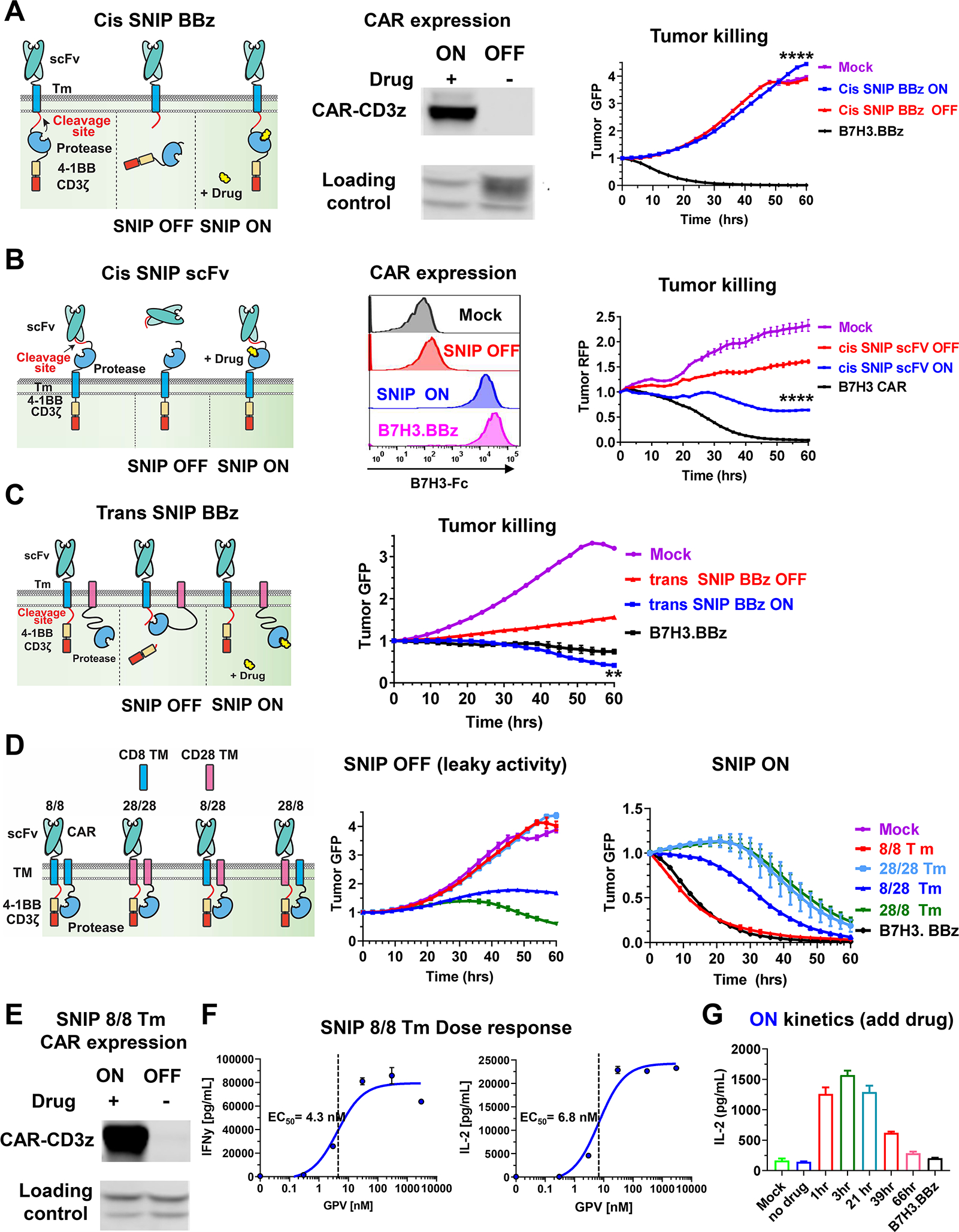
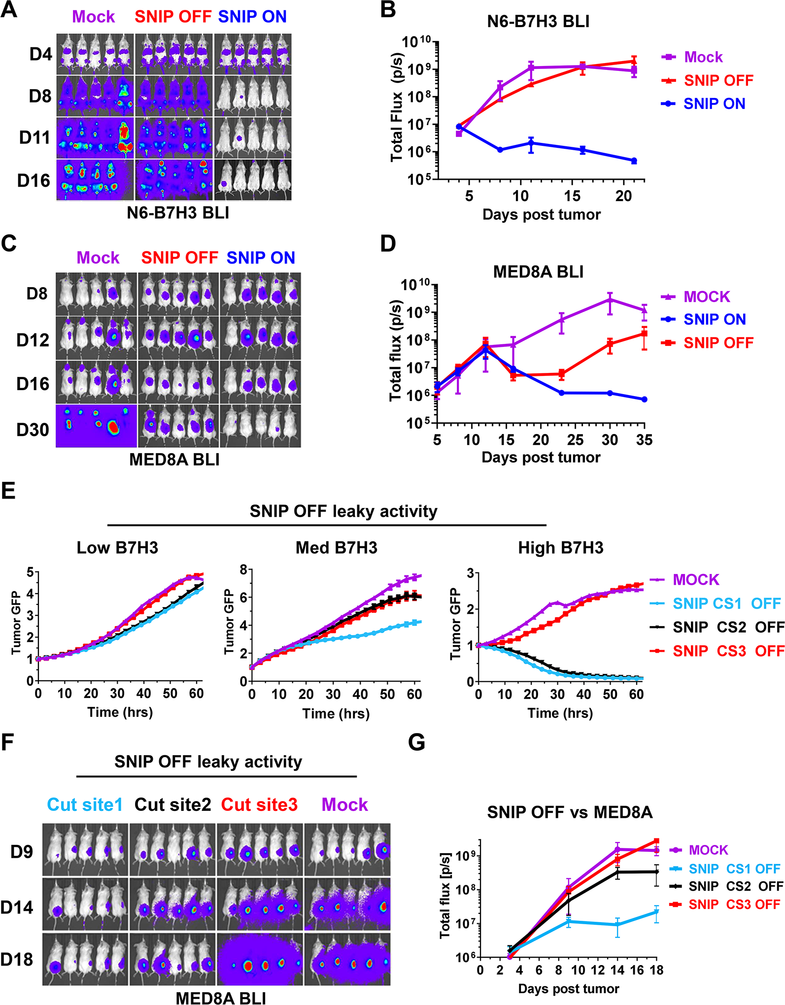
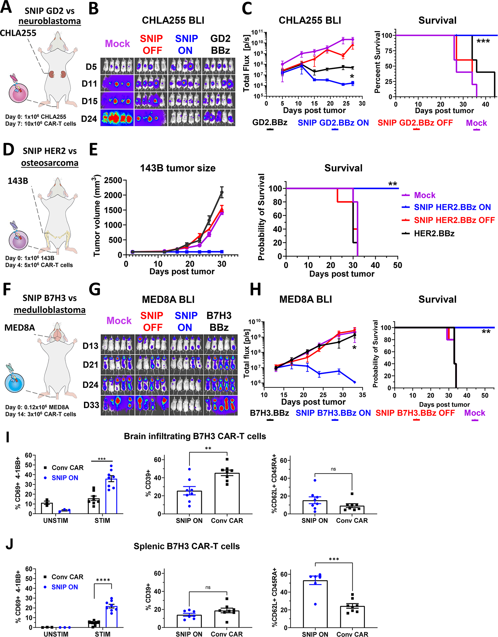
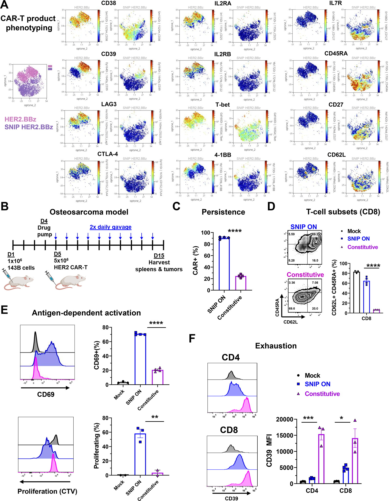
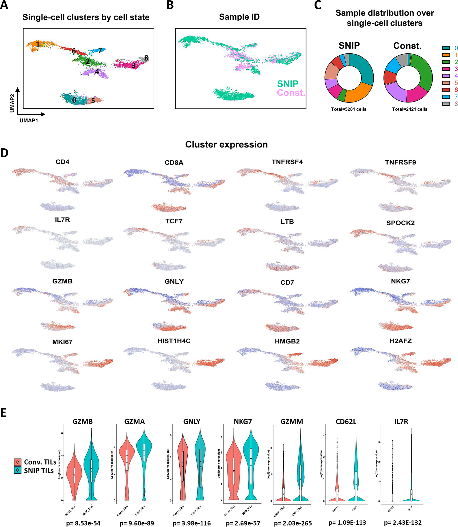
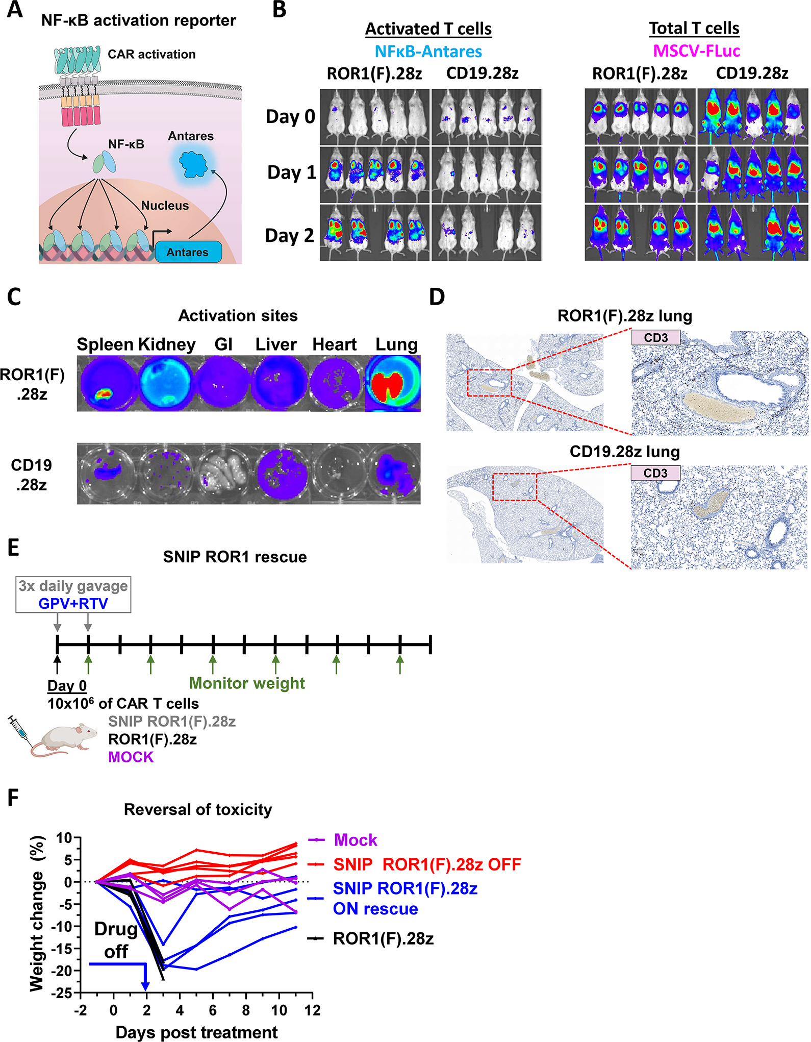
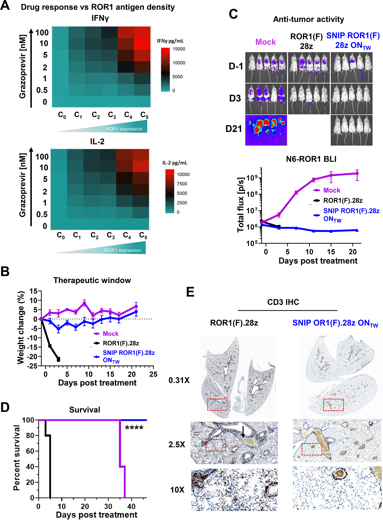
Regulatable CAR platforms could circumvent toxicities associated with CAR-T therapy, but existing systems have shortcomings including leakiness and attenuated activity. Here, we present SNIP CARs, a protease-based platform for regulating CAR activity using an FDA-approved small molecule. Design iterations yielded CAR-T cells that manifest full functional capacity with drug and no leaky activity in the absence of drug. In numerous models, SNIP CAR-T cells were more potent than constitutive CAR-T cells and showed diminished T cell exhaustion and greater stemness. In a ROR1-based CAR lethality model, drug cessation following toxicity onset reversed toxicity, thereby credentialing the platform as a safety switch. In the same model, reduced drug dosing opened a therapeutic window that resulted in tumor eradication in the absence of toxicity. SNIP CARs enable remote tuning of CAR activity, which provides solutions to safety and efficacy barriers that are currently limiting progress in using CAR-T cells to treat solid tumors.

Keywords: CAR-T therapy; T cell differentiation; T cell exhaustion; brain tumors; cellular immunotherapy; chimeric antigen receptors; immune cell engineering; solid cancers; synthetic biology.


Conflict of interest statement

Declaration of interests L.L., R.G.M., M.Z.L., and C.L.M. are coinventors on a patent related to this work. C.L.M. is a cofounder of Lyell Immunopharma, Syncopation Life Sciences, and Link Cell Therapies, which are developing CAR-based therapies, and consults for Lyell, NeoImmune Tech, Apricity, Nektar, Immatics, Ensoma, Mammoth, Glaxo Smith Kline, and Bristol Myers Squibb. L.L., R.G.M., E.S., and E.W.W. are consultants for and hold equity in Lyell Immunopharma. L.L. is a cofounder of, consults for, and holds equity in Syncopation Life Sciences. R.G.M. is a cofounder of, consults for, and holds equity in Syncopation Life Sciences and Link Cell Therapies. R.G.M. is a consultant for Illumina Radiopharmaceuticals, NKarta, ImmunAI, Arovella Therapeutics, Zai Lab, and Aptorum Group. R.G.M. serves on the Data and Safety Monitoring Board for Fate Therapeutics. J.T. is a consultant for Dorian Therapeutics. E.W.W. consults for and holds equity in VISTAN Health. A.T.S. is a founder of Immunai and Cartography Biosciences and receives research funding from Arsenal Biosciences, Allogene Therapeutics, and 10x Genomics. K.R.P. is a cofounder and employee of Cartography Biosciences. H.Y.C. is a cofounder of Accent Therapeutics, Boundless Bio, and Cartography Biosciences and is an advisor to 10x Genomics, Arsenal Biosciences, and Spring Discovery. J.R.C. is a cofounder and equity holder of Trapeze Therapeutics, Combangio, and Virsti Therapeutics; he has financial interests in Aravive, Xyence Therapeutics, and Syncopation Life Sciences; and he is a member of the Board of Directors of Ligand Pharmaceuticals and Revel Pharmaceuticals. S.A.Y.-H. is a consultant for Trapeze Therapeutics and Xyence Therapeutics.

Figures

Figure 1.
Figure 1.. The optimized SNIP design, trans SNIP BBz, confers robust control over CAR-T cytotoxicity.
A) Schematic illustration of the Cis SNIP BBz system (left). CAR-CD3ζ immunoblots of protein lysates from anti-B7H3 Cis SNIP BBz CAR-T cells ± 3μM GPV (middle). Cytotoxicity of day 10 anti-B7H3 Cis SNIP BBz CAR-T cells (right). (B) Schematic illustration of the Cis SNIP scFv system (left), flow cytometry of surface B7H3 CAR (middle), and tumor killing (right) (C) Schematic illustration of the Trans SNIP BBz system (left) and tumor killing (right). (D) Schematic of various combinations of CD8α and CD28 Tm for the CAR and protease modules (left). Tumor killing in the absence (SNIP OFF, middle) or presence (SNIP ON, right) of GPV. (E) CD3ζ immunoblots of the 8/8 Tm configuration. (F) Cytokine production of SNIP 8/8 CAR-T cells after coculture with 143B cells treated with various concentrations of GPV. (G) Cytokine secretion of SNIP CAR-T cells against N6-B7H3 cells incubated in 3μM GPV for the indicated amount of time prior to the coculture. Mock, no drug, and B7H3.BBz are shared controls and duplicated between the ON and OFF kinetics in Fig. S1F. In B, B7H3.BBz and Mock are shared controls for CAR expression and are duplicated in Fig. S1A. Data are mean ± s.e.m. of triplicate wells. In A-C Statistical significance was computed at the final time point by unpaired two-tailed t-tests. In A-G, reproducible in three independent experiments with different donors. See also Figure S1.
Figure 2.
Figure 2.. Leaky cytotoxic activity is driven by high antigen density on target lines and ameliorated by substituting cleavage sites with high catalytic activity
(A-B) Tumor BLI of mice inoculated with Nalm6-B7H3 leukemia cells then treated with B7H3.BBz CAR or Mock T cells four days later. SNIP-treated mice were given 50mg/kg GPV and 25mg/kg ritonavir (RTV) (SNIP ON) or vehicle control (SNIP OFF) three times per day by oral gavage. RTV is a pharmacokinetic enhancer of GPV. (C-D) Tumor BLI of mice inoculated with MED8A cells in the right flank and cerebellum then treated with B7H3.BBz CAR or Mock T cells five days later. (E) Cytotoxic activity of SNIP B7H3.BBz bearing different cleavage sites (CS) against GFP-labeled tumor cells in the absence of GPV (SNIP OFF). Data are mean ± s.e.m. of triplicate wells. (F-G) Tumor BLI of mice inoculated with MED8A cells in the right flank then treated with B7H3.BBz or Mock T cells on day 5. GPV was not administered to mice (SNIP OFF). In A-D and F-G, experiments were reproducible in two independent experiments with separate donors. See also Figure S2.
Figure 3.
Figure 3.. SNIP CAR-T cells display enhanced anti-tumor efficacy in orthotopic solid tumor models
(A) Schematic of the CHLA255 neuroblastoma kidney capsule tumor model using GD2-specific CARs. (B) Tumor progression of CHLA255 by BLI, (C) Quantification of BLI (left) and survival curves of mice in the CHLA255 neuroblastoma model (right). (D) Schematic of the 143B osteosarcoma tumor model using HER2-specific CARs. (E) Tumor progression in the 143B osteosarcoma was monitored by tumor volume measurements (left) and survival curves in the 143B model (right). (F) Schematic of the MED8A medulloblastoma brain tumor model using B7H3-specific CARs. (G) Tumor progression of MED8A BLI, (H) quantification of MED8A medulloblastoma BLI (left), and survival curves of mice in the MED8A medulloblastoma model (right). (I/J) Expression of surface markers on SNIP or conventional B7H3.BBz CAR-T cells harvested from (I) spleens and (J) brains ten days after treatment in the medulloblastoma model shown in Fig. S3D–S3I (N=8 mice per group). Data are mean ± s.e.m. of N=8 mice. P values determined by unpaired two-tailed t-tests. In C, E, and H, tumor quantification data are mean ± s.e.m. of n=5 mice in each group. P values were determined by unpaired two-tailed t-tests. Survival curves were compared by the log-rank Mantel-Cox test. Reproducible in two independent experiments with different donors. See also Figure S3.
Figure 4.
Figure 4.. SNIP CAR-T cells in culture and isolated from treated mice are less exhausted, develop larger fractions of TSCM subsets, and are more responsive to ex vivo stimulation than constitutive CAR-T cells
(A) Mass cytometry analysis of levels of expression of exhaustion, activation and TSCM-associated markers on CD8+ constitutive HER2.BBz and SNIP HER2.BBz CAR-T cells after the 10-day manufacturing period. (B) Schematic of the 143B osteosarcoma model. (C) Persistence of SNIP CAR-T cells as measured by frequency of CAR+ cells gated on live CD45+ cells from spleens of treated mice. (D) Representative flow plots showing surface expression of CD45RA and CD62L on splenic CD8+ HER2 CAR-T cells (left) and quantification of TSCM subsets on splenic CAR-T cells (right). (E) Splenic CAR-T cells were stimulated with plate bound HER2 then analyzed for CD69 expression (top) and proliferation (bottom). (F) Surface expression of the exhaustion marker CD39 on CAR+ TILs. P values were determined by unpaired two-tailed t-tests. In D-F, Bar graph data are mean of individual mice ± s.e.m. of each group. See also Figures S4 and S5.
Figure 5.
Figure 5.. scRNAseq analysis identifies unique subsets in SNIP CAR TILs that include CD4+ memory cells and CD8+ cells with high expression of cytotoxic genes.
HER2.BBz CAR TILs were harvested from mice in the 143B osteosarcoma model ten days after T cell administration. (A-C) UMAP dimensionality reduction and clustering of CAR TILs by transcriptomic states. (D) Select gene expression features in UMAP analysis. (E) Violin plots showing the distribution of expression of cytotoxic and memory associated markers. P-values are indicated below each plot and were determined by unpaired two-tailed Mann–Whitney U test. See also Figure S5.
Figure 6.
Figure 6.. ROR1(F).28Z CAR-T cells are rapidly activated in vivo in non-tumor bearing mice. SNIP ROR1(F).28z CAR-T toxicity is reversed after drug withdrawal.
(A) Schematic of the NF-KB Antares reporter for imaging activation of T cells in vivo. (B) Non-tumor bearing NSG mice were treated with 10×106 ROR1(F).28z CAR, or CD19.28z CAR T cells by tail vein injection. Activation of T cells was monitored daily by BLI using the NF-KB-Antares reporter (left). BLI of total T cells using the constitutive MSCV-Fluc reporter (right). (C) Two days after T cell administration, mouse organs were explanted and probed for the presence of activated T cells using the NF-kB-Antares reporter system. (D) CD3+ infiltrates in mouse lungs identified by IHC analysis. (E) Schematic of the SNIP toxicity rescue model. Non-tumor bearing NSG mice were engrafted with 10×106 ROR1(F).28z CAR, SNIP ROR1(F).28z CAR, or Mock T cells. Mice in the SNIP group were administered 50 mg/kg GPV and 25 mg/kg RTV by oral gavage three time per day for the first two days (SNIP ON) or vehicle control (SNIP OFF). Treatment-related toxicity was monitored by weight change. (F) SNIP ROR1(F).28z toxicity, as measured by weight change, is reversed after cessation of drug treatment (ON rescue). See also Figure S6.
Figure 7.
Figure 7.. Modified drug dosing opens a therapeutic window for SNIP ROR1(F).28z CAR-T cells resulting in potent antitumor efficacy without toxicity.
(A) IFNγ and IL-2 secretion from SNIP ROR1(F).28z CAR-T cells cocultured with various ROR1-expressing lines (see Fig. S7B) in the presence of various concentrations of GPV. (B) 1×10⁶ Nalm6-ROR1 leukemia cells, corresponding to the C5 cell line in Fig. S7B, were engrafted in NSG mice by tail vein injection. 10×106 SNIP ROR1(F).28z CAR, constitutive ROR1(F).28z CAR, or Mock T cells were administered by tail vein injection at day 1 post tumor engraftment, and an additional dose at day 7 for the SNIP and Mock groups. Mice in the SNIP ROR1(F).28z ONTW group were given 25 mg/kg doses of GPV and 2.5mg/kg RTV by oral gavage once per day. Treatment-related toxicities were monitored by weight change. (C) Tumor progression was monitored by BLI of Nalm6-ROR1 leukemia cells. (D) Survival curves of mice shown in (C) (E) CD3 Immunohistochemical staining of lungs from mice treated with ROR1(F).28z CAR-T cells (left) or SNIP ROR1(F).28z ONTW CAR-T cells (right). Dashed rectangles indicate inset field of view. Black arrow in 2.5x micrograph indicates edematous region surrounding a blood vessel. See also Figure S7.

Comment in

Similar articles

Cited by

References

    1. Adachi K, Kano Y, Nagai T, Okuyama N, Sakoda Y, and Tamada K (2018). IL-7 and CCL19 expression in CAR-T cells improves immune cell infiltration and CAR-T cell survival in the tumor. Nat. Biotechnol. - PubMed
    1. Ali N, Flutter B, Sanchez Rodriguez R, Sharif-Paghaleh E, Barber LD, Lombardi G, and Nestle FO (2012). Xenogeneic Graft-versus-Host-Disease in NOD-scid IL-2Rγnull Mice Display a T-Effector Memory Phenotype. PLoS One. - PMC - PubMed
    1. Ali SA, Shi V, Maric I, Wang M, Stroncek DF, Rose JJ, Brudno JN, Stetler-Stevenson M, Feldman SA, Hansen BG, et al. (2016). T cells expressing an anti-B-cell maturation antigen chimeric antigen receptor cause remissions of multiple myeloma. Blood 128, 1688–1700. - PMC - PubMed
    1. Belkina AC, Ciccolella CO, Anno R, Halpert R, Spidlen J, and Snyder-Cappione JE (2019). Automated optimized parameters for T-distributed stochastic neighbor embedding improve visualization and analysis of large datasets. Nat. Commun. - PMC - PubMed
    1. Benjamin R, Graham C, Yallop D, Jozwik A, Mirci-Danicar OC, Lucchini G, Pinner D, Jain N, Kantarjian H, Boissel N, et al. (2020). Genome-edited, donor-derived allogeneic anti-CD19 chimeric antigen receptor T cells in paediatric and adult B-cell acute lymphoblastic leukaemia: results of two phase 1 studies. Lancet 396, 1885–1894. - PMC - PubMed

Publication types

Substances