. 2022 Jul;102(1):121-135.
doi: 10.1016/j.kint.2022.02.037. Epub 2022 Apr 25.

Blocking ribosomal protein S6 phosphorylation inhibits podocyte hypertrophy and focal segmental glomerulosclerosis

Affiliations

Blocking ribosomal protein S6 phosphorylation inhibits podocyte hypertrophy and focal segmental glomerulosclerosis

Fang Li et al. Kidney Int. 2022 Jul.

Abstract

Ribosomal protein S6 (rpS6) phosphorylation mediates the hypertrophic growth of kidney proximal tubule cells. However, the role of rpS6 phosphorylation in podocyte hypertrophy and podocyte loss during the pathogenesis of focal segmental glomerulosclerosis (FSGS) remains undefined. Here, we examined rpS6 phosphorylation levels in kidney biopsy specimens from patients with FSGS and in podocytes from mouse kidneys with Adriamycin-induced FSGS. Using genetic and pharmacologic approaches in the mouse model of FSGS, we investigated the role of rpS6 phosphorylation in podocyte hypertrophy and loss during development and progression of FSGS. Phosphorylated rpS6 was found to be markedly increased in the podocytes of patients with FSGS and Adriamycin-induced FSGS mice. Genetic deletion of the Tuberous sclerosis 1 gene in kidney glomerular podocytes activated mammalian target of rapamycin complex 1 signaling to rpS6 phosphorylation, resulting in podocyte hypertrophy and pathologic features similar to those of patients with FSGS including podocyte loss, leading to segmental glomerulosclerosis. Since protein phosphatase 1 is known to negatively regulate rpS6 phosphorylation, treatment with an inhibitor increased phospho-rpS6 levels, promoted podocyte hypertrophy and exacerbated formation of FSGS lesions. Importantly, blocking rpS6 phosphorylation (either by generating congenic rpS6 knock-in mice expressing non-phosphorylatable rpS6 or by inhibiting ribosomal protein S6 kinase 1-mediated rpS6 phosphorylation with an inhibitor) significantly blunted podocyte hypertrophy, inhibited podocyte loss, and attenuated formation of FSGS lesions. Thus, our study provides genetic and pharmacologic evidence indicating that specifically targeting rpS6 phosphorylation can attenuate the development of FSGS lesions by inhibiting podocyte hypertrophy and associated podocyte depletion.

Keywords: focal segmental glomerulosclerosis (FSGS); podocyte hypertrophy; ribosomal protein S6 (rpS6).


Conflict of interest statement

DISCLOSURE

All the authors declared no competing interests. The abstract of this paper has been selected for presentation at the World Congress of Nephrology 2022 (WCN’22) scheduled for February 24–27, 2022. This abstract would be published electronically in Kidney International Reports, and WCN’22’s searchable database.

Figures

Figure 1 |
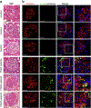
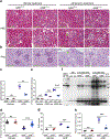
Figure 1 |. Phosphorylated ribosomal protein S6 (p-rpS6) was markedly increased in the renal glomeruli of patients with focal segmental glomerulosclerosis (FSGS).
(a) Glomerulus showing FSGS-like glomerular lesions, which manifested as regional sclerosis of the glomerular space in patients with primary FSGS, compared to para-carcinoma normal kidney tissues (control). (b) Double immunofluorescence staining of human kidney biopsy samples demonstrated significantly elevated p-rpS6 expression (green) in podocytes (double staining with p-rpS6 and podocin, shown in yellow, white arrow) of patients with FSGS (FSGS ≥ 10; control n = 5; bar = 25 μm). H&E, hematoxylin and eosin.
Figure 2 |
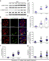
Figure 2 |. Phosphorylated ribosomal protein S6 (p-rpS6) was markedly increased in podocytes of Adriamycin (ADR)-induced FSGS mice.
(a) Western blot analysis of isolated podocyte revealed a gradually increased p-rpS6 level as well as phosphorylated substrate S6 kinase 1 (p-S6K1) expression from the fifth week after ADR treatment. (b,c) Immunofluorescence staining with the indicated antibodies confirmed the markedly enhanced phosphorylation of rpS6 (green) in some podocin-positive cells (red) of mice treated with ADR for 5 weeks and 10 weeks, whereas only slight p-rpS6 expression was detected in control mice (white arrows highlight merged areas; 20 glomeruli per mouse; n ≥ 5 male mice per group; bar 50 = μm; ***P < 0.001). (d,e) ADR injection significantly induced hypertrophy of surviving podocytes compared with podocytes in vehicle-injected control mice, for which the hypertrophy of podocytes was demonstrated by both the upregulated protein/DNA ratio and the marked increases of glomerular volume per podocyte (GV/P). (n ≥ 5 male mice per group; **P < 0.01, ***P < 0.001). (f,g) The averege podocyte size was estimated by measuring the snaptopodin-positive area per Wilms’ tumor-1 protein (WT-1)–positive nucleus, as well as estimating the podocin-positive area per WT-1-positive nucleus; an increase of podocyte size was revealed starting at 5 weeks after ADR treatment. (n ≥ 5 male mice per group; **P < 0.01, ***P < 0.001).
Figure 3 |
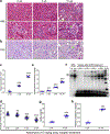
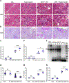
Figure 3 |. Characterization of Adriamycin (ADR)-induced focal segmental glomerulosclerosis.
(a–c) Hematoxylin and eosin (H&E) staining and periodic acid–Schiff (PAS) staining showed focal segmental glomerulosclerosis–like glomerular lesions in ADR-induced nephropathy (green arrows). (d) The podocyte number decreased in ADR nephropathy normalized by control, shown as the percentage of podocytes lost in glomeruli over the total number of podocytes in vehicle-treated mice. Values are mean from at least 40 glomeruli per mouse. (n ≥ 5 male mice per group). (e,f) Proteinuria increased over time as a marker for progressive glomerular damage after ADR injection (n ≥ 5 male mice per group; ***P < 0.001). (g,h) Both blood urea nitrogen (BUN) and plasma creatinine levels increased over time after ADR injection (n ≥ 5 male mice per group; bar 200 = μm in the upper row of (a); bar 100 μm in the lower row of (a) and in (b); *P < 0.05, **P < 0.01, ***P < 0.001. alb, albumin; BSA, bovine serum albumin.
Figure 4 |
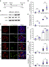
Figure 4 |. Tautomycetin (TC) markedly increased phosphorylated ribosomal protein S6 (p-rpS6) and significantly promoted podocyte hypertrophy.
(a,b) NPHS2-Cre; mT/mG mice with Adriamycin (ADR) injection were treated with either TC or vehicle (Veh) alone, every other day for 7 weeks, starting at 3 weeks after ADR injection. TC treatment significantly enhanced rpS6 phosphorylation in podocytes of mice injected with ADR, compared with Veh-treated mice injected with ADR (n ≥ 5 male mice per group). (c,d) Compared to Veh-treated mice, TC-treated mice had significantly increased p-rpS6–positive podocytes, with or without ADR injection (white arrows; 20 glomeruli per mouse; n ≥ 5 male mice per group; bar = 50 μm, ***P < 0.001). (e,f) TC-treated mice had significantly increased glomerular protein/DNA ratio as well as upregulated glomerular volume per podocyte (GV/P), compared with the Veh-treated group (n ≥ 5 male mice per group; *P < 0.05). (g,h) TC-treated mice had significant increases in both snaptopodin (snapt)-positive area per Wilms’ tumor-1 protein (WT-1)–positive nucleus and podocin-positive area per WT-1–positive nucleus, compared with Veh-treated mice, with or without ADR treatment. (n ≥ 5 male mice per group; *P < 0.05, **P < 0.01). t-rpS6, total-rpS6.
Figure 5 |
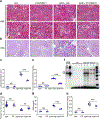
Figure 5 |. Tautomycetin (TC) leads to exacerbated focal segmental glomerulosclerosis (FSGS).
(a–c) In TC-treated mice injected with Adriamycin (ADR), the exacerbated FSGS lesions were indicated by (a) the increased glomerular lesions (green arrows) shown in the hematoxylin and eosin (H&E) staining, as well as (b,c) the increased glomerular sclerosis indicated by the periodic acid–Schiff (PAS) staining, compared with the vehicle (Veh)-treated group. After the treatment with TC, the mice injected with ADR showed (d) an increasing podocyte loss, (e,f) elevation of proteinuria, as well as (g) an increase of blood urea nitrogen (BUN) and (h) plasma creatinine (n ≥ 5 male mice per group; bar = 200 μm in the upper row of (a); bar = 100 μm in the lower row of (a) and in (b); **P < 0.01, ***P < 0.001. Alb, albumin; BSA, bovine serum albumin; NS, not significant: STD, standard deviation.
Figure 6 |
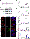
Figure 6 |. PF4708671 inhibited ribosomal protein S6 (rpS6) phosphorylation and blunted podocyte hypertrophy.
(a,b) NPHS2-Cre;mT/mG mice with Adriamycin (ADR) injection were treated with (a) either PF-4708671 or vehicle (Veh) alone, every other day for 7 weeks, starting at 3 weeks after ADR injection. (b) Western blot analysis of isolated podocyte showed decreased ribosomal protein S6 (rpS6) phosphorylation in PF4708671-treated mice injected with ADR, compared with the Veh group injected with ADR. (c,d) Immunofluorescence staining with the indicated antibodies confirmed that the phosphorylated (p)-rpS6-positive podocytes were significantly decreased in PF4708671-treated mice injected with ADR, compared with those in Veh-treated mice injected with ADR (white arrows; 20 glomeruli per mouse; n ≥ 5 male mice per group; bar = 50 μm; ***P < 0.001). (e–h) ADR-induced podocyte hypertrophy was significantly blunted by PF4708671. Values are means ± SEM (n ≥ 5 male mice per group, *P < 0.05, **P < 0.01, ***P < 0.001). GV/P, glomerular volume per podocyte; NS, not significant; PF, PF4708671; snapt, snaptopodin; t-rpS6, total-rpS6; WT-1, Wilms’ tumor-1 protein.
Figure 7 |
Figure 7 |. PF4708671 treatment attenuated focal segmental glomerulosclerosis (FSGS).
In PF4708671-treated mice injected with Adriamycin (ADR), the attenuated FSGS lesions were indicated by (a) the decreased glomerular lesions (green arrow) shown by the hematoxylin and eosin (H&E) staining, as well as (b,c) the decreased glomerulosclerosis indicated by the periodic acid–Schiff (PAS) staining, compared with the vehicle (Veh)-treated group. After the treatment with PF4708671, the mice injected with ADR showed (d) a decreased podocyte loss, (e,f) decreased proteinuria, as well as (g) a reduction of blood urea nitrogen (BUN) and (h) plasma creatinine level. (n ≥ 5 male mice per group; bar = 200 μm in the upper row of (a); bar = 100 μm in the lower row of (a) and in (b); **P < 0.01, ***P < 0.001). Alb, albumin; BSA, bovine serum albumin; PF, PF4708671; NS, not significant; STD, standard deviation.
Figure 8 |
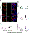
Figure 8 |. Congenic rpS6P−/− mice expressing nonphophorylatable ribosomal protein S6 (rpS6) showed significantly blunted podocyte hypertrophy.
(a,b) rpS6P+/+ and rpS6P−/− mice were simultaneously treated with Adriamycin (ADR; 17 mg/kg body weight) for 8 weeks. Immunofluorescence staining with the indicated antibodies confirmed complete deletion of rpS6 phosphorylation in the kidney (20 glomeruli per mouse; n ≥ 5 male mice per group; bar = 50 μm; ***P < 0.001). (c–e) ADR-induced podocyte hypertrophy was significantly blunted in rpS6P−/− mice normalized by their rpS6P+/+ littermates. Values are means ± SEM (n ≥ 5 male mice per group; ***P < 0.001). NS, not significant; p-rpS6, phosphorylated rpS6; snapt, snaptopodin; WT1, Wilms’ tumor-1 protein.
Figure 9 |
Figure 9 |. Genetic deletion of ribosomal protein S6 (rpS6) phosphorylation ameliorates focal segmental glomerulosclerosis (FSGS)–like disease.
rpS6P+/+ and rpS6P−/− mice were simultaneously treated with Adriamycin (ADR) for 8 weeks. FSGS lesions were ameliorated in rpS6P−/− mice group, as demonstrated by (a) the decreased glomerular lesions (green arrow) shown in the hematoxylin and eosin (H&E) staining, as well as (b,c) the decreased glomerulosclerosis indicated by the periodic acid–Schiff (PAS) staining, compared with that in the rpS6P+/+ mice. (d) A decrease in podocyte loss, (e,f) reduction of proteinuria, as well as the (g) decrease of BUN and (h) plasma creatinine level were demonstrated in the rpS6P−/− mice group treated with ADR, compared to their rpS6P+/+ littermates (n ≥ 5 male mice per group; bar = 200 μm in the upper row of (a); bar = 100 μm in the lower row of (a) and in (b); ***P < 0.001). Alb, albumin; BSA, bovine serum albumin; STD, standard deviation; Veh, vehicle.

References

    1. Jefferson JA, Shankland SJ. The pathogenesis of focal segmental glomerulosclerosis. Adv Chronic Kidney Dis. 2014;21:408–416. - PMC - PubMed
    1. Wharram BL, Goyal M, Wiggins JE, et al. Podocyte depletion causes glomerulosclerosis: diphtheria toxin-induced podocyte depletion in rats expressing human diphtheria toxin receptor transgene. J Am Soc Nephrol. 2005;16:2941–2952. - PubMed
    1. Kim YH, Goyal M, Kurnit D, et al. Podocyte depletion and glomerulosclerosis have a direct relationship in the PAN-treated rat. Kidney Int. 2001;60:957–968. - PubMed
    1. Matsusaka T, Xin J, Niwa S, et al. Genetic engineering of glomerular sclerosis in the mouse via control of onset and severity of podocyte-specific injury. J Am Soc Nephrol. 2005;16:1013–1023. - PubMed
    1. Wiggins RC. The spectrum of podocytopathies: a unifying view of glomerular diseases. Kidney Int. 2007;71:1205–1214. - PubMed

Publication types

LinkOut - more resources