Case Reports
. 2022 Apr 28;8(3):a006151.
doi: 10.1101/mcs.a006151. Print 2022 Apr.

Tumor-immune microenvironment revealed by Imaging Mass Cytometry in a metastatic sarcomatoid urothelial carcinoma with a prolonged response to pembrolizumab

Affiliations
Case Reports

Tumor-immune microenvironment revealed by Imaging Mass Cytometry in a metastatic sarcomatoid urothelial carcinoma with a prolonged response to pembrolizumab

Hussein Alnajar et al. Cold Spring Harb Mol Case Stud. .

Abstract

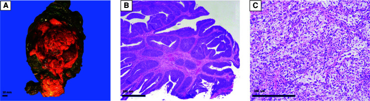
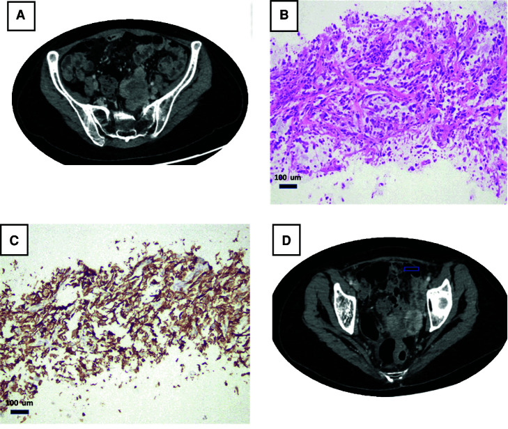
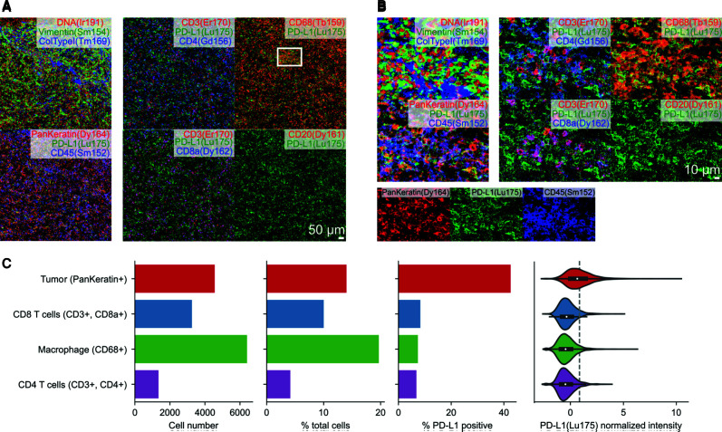
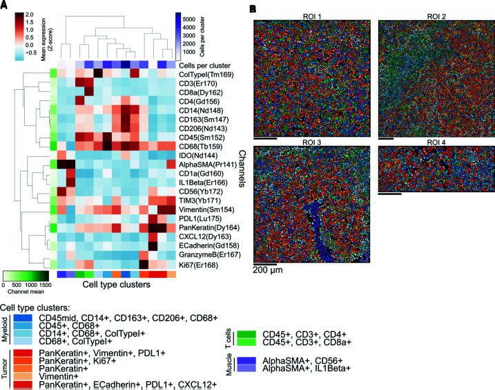
Sarcomatoid urothelial carcinoma (SUC) is a rare subtype of urothelial carcinoma (UC) that typically presents at an advanced stage compared to more common variants of UC. Locally advanced and metastatic UC have a poor long-term survival following progression on first-line platinum-based chemotherapy. Antibodies directed against the programmed cell death 1 protein (PD-1) or its ligand (PD-L1) are now approved to be used in these scenarios. The need for reliable biomarkers for treatment stratification is still under research. Here, we present a novel case report of the first Imaging Mass Cytometry (IMC) analysis done in SUC to investigate the immune cell repertoire and PD-L1 expression in a patient who presented with metastatic SUC and experienced a prolonged response to the anti-PD1 immune checkpoint inhibitor pembrolizumab after progression on first-line chemotherapy. This case report provides an important platform for translating these findings to a larger cohort of UC and UC variants.

Keywords: neoplasm of the genitourinary tract.


Figures

Figure 1.
Figure 1.
Histopathology of primary sarcomatoid urothelial carcinoma (UC) case. A radical cystectomy, hysterectomy and bilateral salpingo-oophorectomy, anterior vaginectomy showed a 6.5-cm bladder mass (A) with papillary high-grade urothelial carcinoma component (B) and an invasive malignant spindle cell component (C) diagnostic of sarcomatoid differentiation.
Figure 2.
Figure 2.
Clinicopathological presentation of metastatic sarcomatoid urothelial carcinoma (UC). Computed tomography (CT) with contrast showing an enhancing left pelvic mass (arrow) near the pelvic sidewall and near the left internal iliac vessels (A,B). Biopsy from that mass showed sarcomatoid carcinoma related to patient's known bladder primary (C). PDL-1 (SP263 antibody) shows a diffuse membranous staining, and the combined positive score (CPS) was 100% (D).
Figure 3.
Figure 3.
Tumor-immune microenvironment of metastatic sarcomatoid urothelial carcinoma (UC) revealed by Imaging Mass Cytometry. (A,B) Expression of PD-L1 in the context of coexpression of (A) tumor-specific or (B) immune-specific markers. (C) Quantification of cellular abundance in tumor and immune cells, and their expression of PD-L1.
Figure 4.
Figure 4.
Molecular phenotype of tumor and immune cell repertoire of sarcomatoid urothelial carcinoma (UC). (A) Phenotype of discovered cellular clusters based on the profiles of marker expression as assessed by Imaging Mass Cytometry. Clusters are grouped by broad cell type ontogeny. (B) Representation of cells in the sarcomatoid carcinoma case in which each single cell is labeled with the color of the phenotypic cluster from A.

References

    1. Al Zoughbi W, Fox J, Beg SB, Papp E, Hissong E, Ohara K, Keefer L, Sigouros M, Kane T, Bockelman D, et al. 2021. Validation of a ctDNA-based next-generation sequencing assay in a cohort of solid tumor patients: a proposed solution for decentralized plasma testing. Oncologist 26: e1971–e1981. 10.1002/onco.13905 - DOI - PMC - PubMed
    1. Beg S, Bareja R, Ohara K, Eng KW, Wilkes DC, Pisapia DJ, Al Zoughbi W, Kudman S, Zhang W, Rao R, et al. 2021. Integration of whole-exome and anchored PCR-based next generation sequencing significantly increases detection of actionable alterations in precision oncology. Transl Oncol 14: 1. 10.1016/j.tranon.2020.100944 - DOI - PMC - PubMed
    1. Berg S, Kutra D, Kroeger T, Straehle CN, Kausler BX, Haubold C, Schiegg M, Ales J, Beier T, Rudy M, et al. 2019. ilastik: interactive machine learning for (bio)image analysis. Nat Methods 16: 1226–1232. 10.1038/s41592-019-0582-9 - DOI - PubMed
    1. Carpenter AE, Jones TR, Lamprecht MR, Clarke C, Kang IH, Friman O, Guertin DA, Chang JH, Lindquist RA, Moffat J, et al. 2006. CellProfiler: image analysis software for identifying and quantifying cell phenotypes. Genome Biol 7: R100. 10.1186/gb-2006-7-10-r100 - DOI - PMC - PubMed
    1. Cheng L, Zhang S, Alexander R, MacLennan GT, Hodges KB, Harrison BT, Lopez-Beltran A, Montironi R. 2011. Sarcomatoid carcinoma of the urinary bladder: the final common pathway of urothelial carcinoma dedifferentiation. Am J Surg Pathol 35: e34–e36. 10.1097/PAS.0b013e3182159dec - DOI - PubMed

Publication types

MeSH terms

Substances