. 2022 Apr 28;8(3):a006182.
doi: 10.1101/mcs.a006182. Print 2022 Apr.

Tuberous sclerosis complex: a complex case

Affiliations

Tuberous sclerosis complex: a complex case

Ryan M Powell et al. Cold Spring Harb Mol Case Stud. .

Abstract

Tuberous sclerosis complex (TSC) is an inheritable disorder characterized by the formation of benign yet disorganized tumors in multiple organ systems. Germline mutations in the TSC1 (hamartin) or more frequently TSC2 (tuberin) genes are causative for TSC. The malignant manifestations of TSC, pulmonary lymphangioleiomyomatosis (LAM) and renal angiomyolipoma (AML), may also occur as independent sporadic perivascular epithelial cell tumor (PEComa) characterized by somatic TSC2 mutations. Thus, discerning TSC from the copresentation of sporadic LAM and sporadic AML may be obscured in TSC patients lacking additional features. In this report, we present a case study on a single patient initially reported to have sporadic LAM and a mucinous duodenal adenocarcinoma deficient in DNA mismatch repair proteins. Moreover, the patient had a history of Wilms' tumor, which was reclassified as AML following the LAM diagnosis. Therefore, we investigated the origins and relatedness of these tumors. Using germline whole-genome sequencing, we identified a premature truncation in one of the patient's TSC2 alleles. Using immunohistochemistry, loss of tuberin expression was observed in AML and LAM tissue. However, no evidence of a somatic loss of heterozygosity or DNA methylation epimutations was observed at the TSC2 locus, suggesting alternate mechanisms may contribute to loss of the tumor suppressor protein. In the mucinous duodenal adenocarcinoma, no causative mutations were found in the DNA mismatch repair genes MLH1, MSH2, MSH6, or PMS2 Rather, clonal deconvolution analyses were used to identify mutations contributing to pathogenesis. This report highlights both the utility of using multiple sequencing techniques and the complexity of interpreting the data in a clinical context.

Keywords: duodenal carcinoma; pulmonary lymphangiomyomatosis; renal angiomyolipoma.


Figures

Figure 1.
Figure 1.
Overview time line of the patient's clinical history.
Figure 2.
Figure 2.
Hematoxylin and eosin stain of formalin-fixed paraffin-embedded tissue samples. (A) Adjacent normal duodenal mucosa tissue (DAC N). (B) Mucinous duodenal adenocarcinoma with infiltration into the duodenal wall/muscularis layers (DAC 1). (C) Adenomatous polyp exhibiting high-grade dysplasia and mucinous foci (DAC 2). (D) Sessile polyp exhibiting high-grade dysplasia (DAC 3). (E) Sessile polyp with low-grade dysplasia associated with second tumor (DAC 4). (F) Polyp with low-grade dysplasia (DAC 5). (G) Duodenal lymph nodes with smooth-muscle infiltrate (LN). (H) Adjacent normal kidney tissue with tubules and glomeruli visible (AML N). (I) Angiomyolipoma with epithelioid differentiation (AML E). (J) Angiomyolipoma with smooth-muscle differentiation (AML S). (K) Lung tissue with proliferative smooth-muscle nodule and association thickening of the alveolar septa consistent with lymphangioleiomyomatosis (LAM 1). (L) Progressive lymphangioleiomyomatosis lung pathology with normal alveoli, thickened alveolar septa, LAM cell nodules, and cystic spaces (LAM 2). (Figure continues on following pages.)
Figure 2.
Figure 2.
Hematoxylin and eosin stain of formalin-fixed paraffin-embedded tissue samples. (A) Adjacent normal duodenal mucosa tissue (DAC N). (B) Mucinous duodenal adenocarcinoma with infiltration into the duodenal wall/muscularis layers (DAC 1). (C) Adenomatous polyp exhibiting high-grade dysplasia and mucinous foci (DAC 2). (D) Sessile polyp exhibiting high-grade dysplasia (DAC 3). (E) Sessile polyp with low-grade dysplasia associated with second tumor (DAC 4). (F) Polyp with low-grade dysplasia (DAC 5). (G) Duodenal lymph nodes with smooth-muscle infiltrate (LN). (H) Adjacent normal kidney tissue with tubules and glomeruli visible (AML N). (I) Angiomyolipoma with epithelioid differentiation (AML E). (J) Angiomyolipoma with smooth-muscle differentiation (AML S). (K) Lung tissue with proliferative smooth-muscle nodule and association thickening of the alveolar septa consistent with lymphangioleiomyomatosis (LAM 1). (L) Progressive lymphangioleiomyomatosis lung pathology with normal alveoli, thickened alveolar septa, LAM cell nodules, and cystic spaces (LAM 2). (Figure continues on following pages.)
Figure 3.
Figure 3.
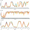
DNA methylation β values generated TSC-associated genes (A) TSC1, (B) TSC2, and (C) TBC1D7. The x-axis entails Illumina Infinium probe IDs. The y-axis shows the methylation β value.
Figure 4.
Figure 4.
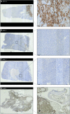
Immunohistochemistry demonstrating tuberin expression. (A) AML N—Adjacent normal kidney tissue with tuberin staining throughout tubules. (B) AML E—Epithelioid differentiated angiomyolipoma, no visible tuberin expression. (C) AML S—Smooth-muscle-differentiated angiomyolipoma, no visible tuberin expression. (D) LAM 1—Congested LAM lung tissue with visible tuberin staining in normal alveoli, but absent in proliferative smooth-muscle nodule (LAM). LAM 2—not shown, because of limited tissue samples.
Figure 5.
Figure 5.
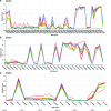
DNA methylation β values generated across the DNA mismatch repair genes (A) MLH1, (B) MSH2, and (C) MSH6 genes. Data are not shown for PMS2 because of the low number of methylation probes. The x-axis entails Illumina Infinium probe ID. The y-axis demonstrates methylation β value.
Figure 6.
Figure 6.
Subclone deconvolution analyses performed on duodenal samples using PhyloWGS. (A) Predicted clonal evolution within duodenal samples. (B) Predicted cellular prevalence of clones across duodenal samples.

References

    1. Binder ZA, Thorne AH, Bakas S, Wileyto EP, Bilello M, Akbari H, Rathore S, Ha SM, Zhang L, Ferguson CJ, et al. 2018. Epidermal growth factor receptor extracellular domain mutations in glioblastoma present opportunities for clinical imaging and therapeutic development. Cancer Cell 34: 163–177.e7. 10.1016/j.ccell.2018.06.006 - DOI - PMC - PubMed
    1. Crino PB, Nathanson KL, Henske EP. 2006. The tuberous sclerosis complex. N Engl J Med 355: 1345–1354. 10.1111/j.1749-6632.2009.05117.x - DOI - PubMed
    1. Dai L, Su X, Lu L, Lv D. 2018. Nonsmall cell lung cancer with rare exon 7 p.A289V mutation in the EGFR gene responds to Icotinib treatment: a case report. Med (United States) 97: 1–4. - PMC - PubMed
    1. Deshwar AG, Vembu S, Yung CK, Jang GH, Stein L, Morris Q. 2015. PhyloWGS: reconstructing subclonal composition and evolution from whole-genome sequencing of tumors. Genome Biol 16: 1–20. 10.1186/s13059-015-0602-8 - DOI - PMC - PubMed
    1. Fang S, Zhang Y, Xu M, Xue C, He L, Cai L, Xing X. 2016. Identification of damaging nsSNVs in human ERCC2 gene. Chem Biol Drug Des 88: 441–450. 10.1111/cbdd.12772 - DOI - PubMed

Publication types

Substances