Review
. 2022 Oct;40(10):1229-1247.
doi: 10.1016/j.tibtech.2022.03.009. Epub 2022 Apr 25.

In situ bioprinting: intraoperative implementation of regenerative medicine

Affiliations
Review

In situ bioprinting: intraoperative implementation of regenerative medicine

Mohamadmahdi Samandari et al. Trends Biotechnol. 2022 Oct.

Abstract

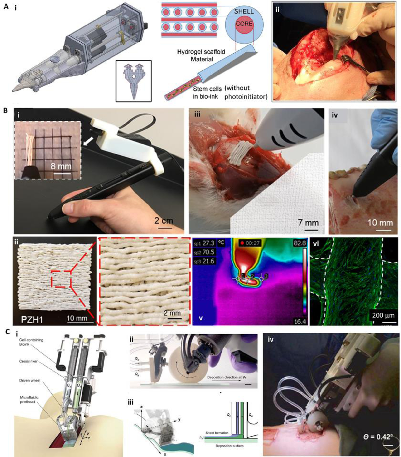
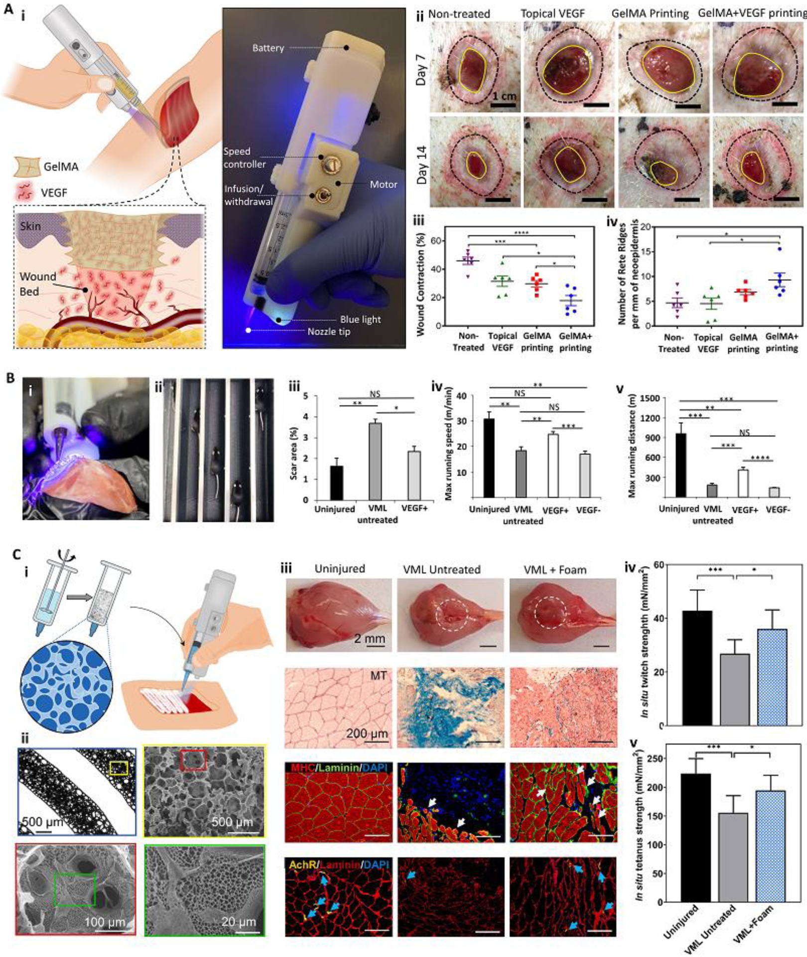
Bioprinting has emerged as a strong tool for devising regenerative therapies to address unmet medical needs. However, the translation of conventional in vitro bioprinting approaches is partially hindered due to challenges associated with the fabrication and implantation of irregularly shaped scaffolds and their limited accessibility for immediate treatment by healthcare providers. An alternative strategy that has recently drawn significant attention is to directly print the bioink into the patient's body, so-called 'in situ bioprinting'. The bioprinting strategy and the associated bioink need to be specifically designed for in situ bioprinting to meet the particular requirements of direct deposition in vivo. In this review, we discuss the developed in situ bioprinting strategies, their advantages, challenges, and possible future improvements.

Keywords: bioinks; bioprinting; handheld printers; in situ printing; robotic bioprinters.


Figures

Figure 1.
Figure 1.
Bioprinting integrated with scanning for minimizing errors during in situ bioprinting. (A) Error compensation before the main bioprinting procedure. Bioprinting was performed on a prototype defect model (i), the accumulative error of printing was detected with a scanner and compensated in the new printing process to reduce the error (ii). The corrected G-code was then used for in situ bioprinting of the bioink in a porcine long segmental bone defect (iii, iv). (B) Adaptive in situ printing using a closed-loop integrated scanning and printing system. The process of in situ printing on a breathing lung is shown schematically (i-iii), with the images of the actual setup (iv-vi). The surface of the lung was scanned (i, iv) and tracked in real-time using the fiducial guiding points (ii, v) while closed-loop feedback from the tracking module enabled printing on the moving lung (iii, vi). (C) Adaptive bioprinting for regeneration of the skin defects in live animals. Due to the movement of the body under anesthesia, in situ printing strategies can be improved by using adaptive bioprinting to compensate for movements during printing. A murine model (i) was used by the creation of a full-thickness wound and placement of fiducial markers (ii). The surface was scanned (iii, zoom-in inset) and a bioink was printed inside the defect. After 4 hr, the presence of live cells was confirmed using bioluminescence imaging (iv). Adapted with permission from Elsevier [28] (A), American Association for the Advancement of Science [27] (B), and Wiley [26] (C).
Figure 2.
Figure 2.
Minimally invasive internal in situ bioprinting. (A) Natural orifice transluminal endoscopic bioprinting strategy. Schematic representation (i) and practical model (ii) of in situ bioprinting for treatment of gastric wall injuries. The endoscopic robot could finely print multiple layers of cell-laden bioinks with high resolution (iii). (B) Laparoscopic bioprinting using a ferromagnetic soft nozzle. The bioprinting strategy was based on the insertion of the nozzle through a small incision, and its deformation in a programmable magnetic field while extruding the bioink to form the printing structure (i). The ferromagnetic nozzle was formed from a polymeric shell embedded with magnetic particles and reinforcing fibers (ii). Minimally invasive in situ printing on the liver of a living rat (iii-v). The process was consisted of CT scanning to reconstruct the liver surface (iii), definition the printing path on the upper liver surface (iii), and in situ printing (iv, v). The setup is shown in (iv) while a close-up view of the printing construct is shown in (v). Reproduced with permission from IOP Publishing [23] (A) and Nature Publishing Group [25] (B).
Figure 3.
Figure 3.
In situ bioprinting for the treatment of large and complex tissue defects. (A) In situ bioprinting for treatment of large burn wounds. The printing approach was based on integrated scanning and multimaterial inkjet printing (i). The scanner was first used to reconstruct the defect morphology, followed by deposition of fibroblast-laden dermal and keratinocyte-laden epidermal layers (ii, iii). Fibroblasts (green) and keratinocytes (red) layers formed in vitro (iii). An in situ bioprinting on porcine burn wounds demonstrated a rapid wound closure and reduced contraction when autologous cells were encapsulated in the bioink (iv, v). (B) The treatment of complex bone/skin composite defect with a hybrid in situ bioprinting approach. Scanning was used to reconstruct the defect geometry, while extrusion and inkjet printing methods were implemented for in situ printing of high viscosity acellular bone and low viscosity cellular skin bioinks, respectively (i, ii). Gross pictures of skin (iii) and bone (iv) tissue regeneration over 6 weeks post-surgery demonstrate major recovery of composite tissue. ST-ink: soft tissue ink consisting from collagen and fibrin; KGF: keratinocyte growth factor; rDF: rat primary dermal fibroblasts; HT-ink: hard tissue ink consisting from collagen, chitosan, nano-hydroxyapatite particles (nHAp), and β-Glycerophosphate disodium salt (β-GP); rhBMP2: recombinant human bone morphogenetic protein-2. Reproduced with permission from Nature Publishing Group [4] (A) and Wiley [38] (B).
Figure 4.
Figure 4.
Minimally invasive in situ SLA bioprinting. (A) The application of digital light processing for subcutaneous in situ bioprinting. A DMD chip was used to project NIR light through the intact skin and crosslink pre-injected cell-laden bioink (i). The in vitro (ii) and in vivo (iii) formation of an ear-like structure through intact skin. In this bioprinting strategy, the healthy tissue is scanned and mirrored to provide a representative model of the defected tissue (iia-iic). The model is then sliced and printed layer-by-layer. A fine structure with high cell viability could be achieved (iid, iie). The printed structure (iiia) was stable after 1 month (iiib), and demonstrated tissue integration by H&E staining (iiic) and immunostaining of collagen II (iiid) secreted by chondrocytes encapsulated in the printed structure. (B) Intramuscular bioprinting of scaffolds embedding muscle-derived stem cells. Elongated structures were printed to mimic the structure of native muscle (i). Results obtained after 7 days post-operation demonstrated that despite the injection of the bioink without subsequent selective crosslinking (ii), the injection of the bioink followed by selective crosslinking for the formation of elongated structures can induce organized muscle cell (green) architectures aligned with blood vessels (red). Reproduced with permission from the American Association for the Advancement of Science [37] (A) and Nature Publishing Group [36] (B).
Figure 5.
Figure 5.
Various handheld in situ printing strategies. (A) An extrusion-based handheld bioprinter for deposition of core-shell filaments for the treatment of osteochondral defects. The device (Bio Pen) could print a cell-laden core and a protective acellular shell (i). The Bio Pen was implemented for printing into a full-thickness osteochondral defect in the knee of a sheep (ii). (B) A melt-spinning handheld printer for treatment of bone defects. The handheld printer could be used to melt PCL-based material and deposit its filaments with a fine resolution, while a camera was integrated for better visibility (i, ii). The printer was used to fill ex vivo murine calvarial and porcine jaw defects (iii, iv). The temperature of the melt-extrusion filaments was shown to be lower than the threshold to induce cell death at the tissue filament interface (v). The printed scaffolds were reported to be osteoinductive after in vitro seeding (vi). Osteopontin (green) and nuclei (blue) were immunostained on human mesenchymal stem cells differentiated into osteoblasts for 28 days. (C) An extrusion-based handheld planar printer for treatment of large burn wounds. The device could co-deposit sheets of bioink and its crosslinker to fill and conform to clinically sized and shaped skin defects (i). The planar printer had a silicone wheel to control the relative velocity of the nozzle over the defect (ii). The printer enabled two degrees of spatial control to lay down the bioink and crosslinker along non-uniform edges of a skin wound (iii). The planar printer was used for the treatment of angled porcine full-thickness burn injuries (iv). Adapted with permission from Wiley [49] (A), Elsevier [46] (B), and IOP publishing [45] (C).
Figure 6.
Figure 6.
Handheld in situ printers used in vivo to induce tissue regeneration and functional recovery. (A) A custom handheld printer with an integrated photocrosslinking mechanism was used for in situ printing of VEGF-eluting GelMA scaffolds to treat full-thickness porcine wounds (i). The handheld printed VEGF-positive scaffolds improved wound healing quality (ii). Significant reduction in wound contraction (iii) and enhancement in the number of Rete Ridges (iv) was reported. (B) The handheld printer was implemented to deposit filaments of nano-engineered Muscle Ink for skeletal muscle regeneration. The printer enabled the deposition of filaments aligned with remnant muscle fascicles (i). The effect of Muscle Ink printed within a murine VML injury model on functional recovery was evaluated using a treadmill (ii). The use of VEGF-eluting Muscle Ink reduced scar area (iii) improved the maximum running speed (iv) and maximum running distance (v) over uninjured controls and VEGF-negative scaffolds. (C) The printer was used to deposit filaments with multiscale porosity for VML treatment. The porous bioink was developed by stirring a GelMA precursor (i). The printed foam resulted in a scaffold with hierarchical porous structures (ii). In situ printing of the porous bioink increased muscle volume, reduced fibrosis, improved myogenesis, and enhanced innervation after VML injury (iii). The foam scaffold induced functional recovery of the injured muscle, shown by in situ twitch (iv) and tetanus strength (v) results. Adapted with permission from Elsevier [12] (A), Wiley [53] (B), and AIP publishing [18] (C).

Similar articles

Cited by

References

    1. Cohen DL et al. (2010) Additive manufacturing for in situ repair of osteochondral defects. Biofabrication 2 (3), 035004. - PubMed
    1. Gatenholm B et al. (2020) Collagen 2A Type B Induction after 3D Bioprinting Chondrocytes In Situ into Osteoarthritic Chondral Tibial Lesion. Cartilage, 1947603520903788 - PMC - PubMed
    1. Li L et al. (2017) In situ repair of bone and cartilage defects using 3D scanning and 3D printing. Scientific reports 7 (1), 1–12. - PMC - PubMed
    1. Albanna M et al. (2019) In situ bioprinting of autologous skin cells accelerates wound healing of extensive excisional full-thickness wounds. Scientific reports 9 (1), 1–15. - PMC - PubMed
    1. Matai I et al. (2020) Progress in 3D bioprinting technology for tissue/organ regenerative engineering. Biomaterials 226, 119536. - PubMed

Publication types