. 2022 May;24(5):616-624.
doi: 10.1038/s41556-022-00898-9. Epub 2022 Apr 28.

Identification of a retinoic acid-dependent haemogenic endothelial progenitor from human pluripotent stem cells

Affiliations

Identification of a retinoic acid-dependent haemogenic endothelial progenitor from human pluripotent stem cells

Stephanie A Luff et al. Nat Cell Biol. 2022 May.

Abstract

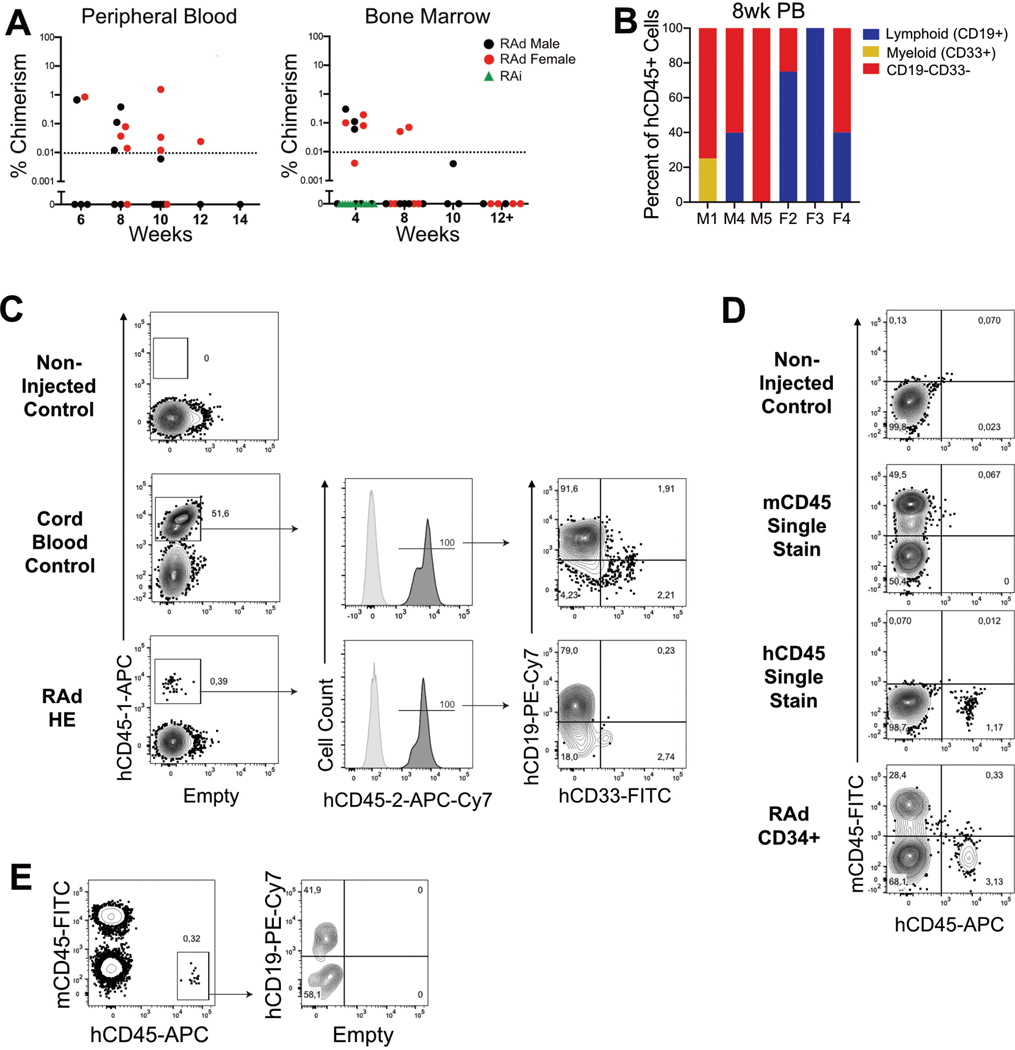
The generation of haematopoietic stem cells (HSCs) from human pluripotent stem cells (hPSCs) is a major goal for regenerative medicine. During embryonic development, HSCs derive from haemogenic endothelium (HE) in a NOTCH- and retinoic acid (RA)-dependent manner. Although a WNT-dependent (WNTd) patterning of nascent hPSC mesoderm specifies clonally multipotent intra-embryonic-like HOXA+ definitive HE, this HE is functionally unresponsive to RA. Here we show that WNTd mesoderm, before HE specification, is actually composed of two distinct KDR+ CD34neg populations. CXCR4negCYP26A1+ mesoderm gives rise to HOXA+ multilineage definitive HE in an RA-independent manner, whereas CXCR4+ ALDH1A2+ mesoderm gives rise to HOXA+ multilineage definitive HE in a stage-specific, RA-dependent manner. Furthermore, both RA-independent (RAi) and RA-dependent (RAd) HE harbour transcriptional similarity to distinct populations found in the early human embryo, including HSC-competent HE. This revised model of human haematopoietic development provides essential resolution to the regulation and origins of the multiple waves of haematopoiesis. These insights provide the basis for the generation of specific haematopoietic populations, including the de novo specification of HSCs.


Conflict of interest statement

Competing Interests

The methodology described in this publication is subject to patent PCT/US2020/014626 (Inventors: A.D. and C.M.S.).

Figures

Extended Data Fig. 1
Extended Data Fig. 1. Specification of HOXA+ HE from hPSCs in a WNT-dependent manner.
A, Schematic of hPSC directed differentiation towards hemogenic endothelium, as described in Sturgeon et. al. Definitive intra-embryonic-like hematopoietic potential is specified in a WNT-dependent (“WNTd”) manner, while extra-embryonic-like hematopoietic potential is WNT-independent (“WNTi”). B, Representative flow cytometric analyses of the T-lymphoid potential of WNTd CD34+ cells and WNTi CD34+CD43neg and CD43+ populations. T cell potential is positively identified by the presence of a CD4+CD8+ population following 21+ days of OP9-DL4 co-culture, while an absence of potential is identified by an absence of CD45+ lymphocytes,. C, Heatmaps visualizing the mean expression of HOXA genes across all biological replicates in mesoderm and hemogenic endothelium populations, as in (A). Grey indicates undetected gene. Scale bar: log10 FPKM. Biological replicates: WNTi/WNTd KDR+ (n=4); WNTi/WNTd HE (n=3). The expression of HOXA genes within WNTd-derived populations is suggestive of an intra-embryonic-like population, while a lack of HOXA expression in WNTi-derived populations is suggestive of an extra-embryonic-like population.
Extended Data Fig. 2
Extended Data Fig. 2. scRNA-seq analyses of day 3 WNTi and WNTd differentiation cultures.
A, Violin plots visualizing the number of genes per cell (left), the number of unique molecular identifiers (“UMIs”, middle), and the percent of expressed genes that are mitochondrial (right) following filtering of low quality cells from both WNTi and WNTd datasets combined (1 biological replicate each). B, UMAP visualizing before and after integration of WNTi (red) and WNTd (blue) datasets to account for batch effects between sequencing runs. C, UMAP visualizing quality control metrics, as in A, for the dataset following integration. Scale bar: values range as indicated in (A). D, Violin plots for CYP26A1 and ALDH1A2 expression within WNTi KDR+GYPA+ and WNTd KDR+GYPAneg cells, as indicated. E,F, Day 3 WNTd cultures are comprised of all germ layers. E, UMAP visualizing (i) clustering and (ii) KDR expression within WNTd differentiation cultures. Scale bar: gene expression scaled to WNTd subset. F, (i) UMAP plot with the projection of predicted germ layer type, where each label includes cells expressing the following genes: Pluripotent (SOX2, NANOS3, DND1, POU5F1, or TBXT), Ectoderm (TFAP2A, DLX5, or GATA3), Endoderm (FOXA2, APOA1, or APOA2), and Mesoderm (KDR, MEST, MESP1, TEK, or FLT1). 44 (0.64%) remaining cells were labeled based on clustering. (ii) Dot plot visualizing expression of germ layer-specific genes within each identified cell type, as in (i). Scale
Extended Data Fig. 3
Extended Data Fig. 3. Day 3 of differentiation WNTd KDR+CXCR4neg and KDR+CXCR4+ cells are transcriptionally distinct mesodermal subsets.
A, Heatmaps visualizing the mean expression across all biological replicates (log10 FPKM) of mesodermal, endothelial, hematopoietic, and RA-related genes within hPSC-derived day 3 WNTi KDR+CD235a+ cells and WNTd KDR+CXCR4+/neg cells, day 6 WNTi HE, and day 8 WNTd HE. Grey indicates undetected gene. Hierarchical clustering based on the expression of genes shown. Scale bar: log10 FPKM. Biological replicates: WNTi KDR+ (n=4); CXCR4+/neg KDR+, WNTi HE, WNTd HE (n=3). B, Representative flow cytometric analysis for endothelial markers CD34, CD144 (VE-Cadherin/CDH5), and TIE2 (TEK) within WNTd KDR+ cells. C, Heatmaps visualizing the mean expression of HOXA genes across all biological replicates in day 3 WNTi KDR+CD235a+ cells, or WNTd KDR+CXCR4+ or KDR+CXCR4neg cells, as in A. Scale bar: log10 FPKM. Biological replicates: WNTi KDR+ (n=4); CXCR4+/neg KDR+ (n=3). D, PCA plot for batch-corrected KDR+CXCR4+ and KDR+CXCR4neg replicates, as in A. E, Expression of CDX genes within WNTi and WNTd mesodermal populations, as in A, Two-way ANOVA with Tukey’s multiple comparison test comparing all biological replicates: WNTd CXCR4+/neg (n=3), WNTi CD235a+ (n=4). SEM, **p<0.01, ***p<0.001, ****p<0.0001, ns=not significant.
Extended Data Fig. 4
Extended Data Fig. 4. CXCR4neg mesoderm gives rise to HE in an RA-independent manner, while specification of HE from CXCR4+ mesoderm is RA-dependent.
A, Representative phase contrast microscopy of CD34+ cells from DMSO-treated CXCR4+/neg mesoderm (i) and retinol-treated CXCR4+ mesoderm (ii) following 1, 3, and 5 days after FACS isolation. 100X magnification, scale bar: 50um. B, Erythro-myeloid colony forming potential of HE specified
Extended Data Fig. 5
Extended Data Fig. 5. RA-dependent HE, similar to RAi HE, is a CD34+CD43negCD73negCXCR4neg population and undergoes the endothelial-to-hematopoietic transition (EHT) in a NOTCH-dependent manner.
A, Schematic for the differentiation of WNTd cultures towards either RAi and RAd CD34+ cells and their respective hematopoietic progenitor cells (HPCs) in the presence or absence of NOTCH inhibitor L-685458. B, Representative flow cytometric analyses and FACS isolation strategies within DEAB-treated (RAi) or retinol (“ROH”)-treated (RAd) cultures. Isolated populations were then assessed for hematopoietic potential. C(i), Representative flow cytometric analyses of T-lymphoid potential of populations, as in (B). (ii) Quantification of the erythro-myeloid CFC potential from the populations in (B), averaged across all biological replicates. Two-way ANOVA with Tukey’s multiple comparison test comparing all biological replicates: RAd HE plus NOTCH inhibitor (n=3), remaining samples (n=4), SEM, statistics shown for BFU-E (RAi HE vs. RAi HE+gSI (p=0.0059), RAi HE vs. RAi CXCR4+ (p=0.008), RAi HE vs. RAi CD73+ (p=0.0044), RAi HE vs. RAd CXCR4+ (p=0.0128), RAi HE vs. RAd CD73+ (p=0.0033), p<0.0001 for RAi HE vs. RAd HE, RAi HE+gSI vs. RAd HE, RAi CXCR4+ vs. RAd HE, RAi CD73+ vs. RAd HE, RAd HE vs. RAd HE+gSI, RAd HE vs. RAd CXCR4+, RAd HE vs. RAd CD73+), all comparisons not shown are not significant. All colony counts and statistical analyses are included in Source Extended Data Fig. 5. (iii) Representative flow cytometric analysis of CD34 and CD45 at 9 days after HE FACS isolation and percentage of CD34+CD45+ cells from each culture, averaged across all biological replicates (n=4). Two-tailed paired t-test, p=0.0458, SEM. D, Average expression across all biological replicates of embryonic (HBE), fetal (HBG), and adult (HBB) globin genes and BCL11A in BFU-E derived from WNTi CD34+ cells (purple), WNTd RAi HE (green), and RAd HE (blue). Ordinary one-way ANOVA comparing all biological replicates: HBE (n=3; WNTi vs. RAi, p=0.4636, WNTi vs. RAd, p=0.8811; RAi vs. RAd, p=0.3841), HBG (n=3; WNTi vs. RAi, p=0.234; WNTi vs. RAd, p=0.0018; RAi vs. RAd, p=0.007), HBB (n=3, WNTi vs. RAi, p=0.3253; WNTi vs. RAd, p=0.0662; RAi vs. RAd, p=0.286), BCL11A (n=6, p=0.002552), SEM, ns=not significant.
Extended Data Fig. 6
Extended Data Fig. 6. Xenograft analyses of hPSC-derived HE populations.
A, Transient xenograft persistence of RAd hematopoietic progenitors following injection in neonatal mice. Percent chimerism observed of hCD45 cells present in either the peripheral blood and bone marrow following injection with hPSC-derived CD34+ cells. B, Lineage distribution of hCD45+ cells present in the peripheral blood (PB) 8 weeks post-transplant. C-E, Detailed analysis of human engraftment in the bone marrow or peripheral blood of mice transplanted with RAd HE cells. C, Two independent CD45 antibodies (“CD45–1” and “CD45–2”) were used to label human cells. Representative flow cytometric 4-week analysis of the bone marrow of a non-injected control mouse (top), a recipient of 105 CD34+ cord blood cells (middle) and a recipient of 5×10⁴ RAd CD34+ cells. D-E, Alternative strategy for human chimerism analysis based on mCD45 and hCD45 exclusive staining. D, Representative flow cytometric analysis for single stains of both mCD45 and hCD45, from the bone marrow of an non-injected control mouse or a recipient of 2×10⁵ RAd CD34+ cells. E, Representative flow cytometric strategy for the detection of human chimerism in the peripheral blood, from a recipient of 105 RAd CD34+ cells.
Extended Data Fig. 7
Extended Data Fig. 7. Whole-transcriptome analysis of hPSC and human embryonic CD34+ populations.
A, Comparison of whole transcriptomes of hPSC-derived HE and HPCs (3 biological replicates each). (i) PCA plot demonstrating the similarity between batch-corrected biological replicates of WNTi, RAi, and RAd HE, along with CD34+CD45+ HPCs derived from RAi or RAd HE. (ii) Heatmap and hierarchical clustering showing the Euclidean distance between all biological replicates of WNTd RAi and RAd HE, as in (i). (iii) Selection of significant normalized enrichment scores (NES) from preranked GSEA between RAi HE (green) and RAd HE (blue), as in Extended Data Table 5C. RAi was enriched in embryonic development (NES=−1.80, FDR=0.23), endothelium development (NES=−1.85, FDR=0.23), cellular adhesion (NES=−1.78, FDR=0.24), the epithelial-to-mesenchymal transition (NES=−1.95, FDR 0.14), and response to mechanical stimuli (NES=−1.71, FDR=0.23). In contrast, RAd HE was enriched for RA signaling (NES=1.99, FDR=0.22) and several histone modification pathways (NES=2.12, FDR=0.14). B, (i) Mean expression (log10 FPKM) of hemato-endothelial genes for all biological replicates within hPSC-derived WNTi HE, RAi HE, RAd HE, RAi HPC, and RAd HPC, as in (A), with CD34+CD90+CD43neg cells (“AGM CD34+”), CD34+CD90+CD43+ hematopoietic stem/progenitors (“AGM HSPC”), and CD34+CD90negCD43+ hematopoietic progenitors (“AGM PR”) isolated from the aorta-gonad mesonephros (AGM) region of 5th-week of gestation human embryos. Grey indicates undetected gene. Scale bar: log10 FPKM. Biological replicates: WNTi HE, RAi HE, RAd HE, RAi HPC, RAd HPC (n=4); AGM CD34+, AGM HSPC, AGM PR (n=1, GEO SuperSeries GSE81102). (ii) Selection of significant normalized enrichment scores (NES) from preranked GSEA comparing RAi HE to AGM 34+ cells (green) and RAd HE to AGM CD34+ cells (blue), as in Extended Data Table 5E,F. RAi and RAd HE was enriched for hematopoietic stem and progenitor differentiation (NES≥1.63, FDR≤0.015), while AGM CD34+ cells were enriched for aorta and vascular development (NES≥1.82, FDR≤0.01), and BMP and VEGF signaling pathways (NES≥1.89, FDR≤0.004).
Extended Data Fig. 8
Extended Data Fig. 8. Establishment of human embryonic dataset for comparative analysis with hPSC-derived hemogenic and arterial endothelial populations.
A, UMAPs visualizing (i) Carnegie Stages and (ii) cell type labels from the complete dataset of Zeng, et al. Biological replicates: CS10, CS11, CS12, CS14, CS15 (n=1); CS13 (n=2, 1 each for CS13X (10X genomics) and CS13D (Modified STRT-Seq). B, UMAP visualizing the cells categorized as arterial endothelial cells (“AEC”, defined as CDH5+CXCR4+GJA5+DLL4+HEY2+SPNnegPTPRCneg) and hemogenic endothelial cells (“HEC”, defined as CDH5+RUNX1+HOXA+ITGA2BnegSPNnegPTPRCneg), as in Fig. 3B. C, UMAP visualizing the numbered groups for embryonic cells, as in Fig. 3B, within the categorized AEC and HEC. Di, Heatmap visualizing the expression of select broadly and arterial endothelial genes and RUNX1 in human CS10–14 AEC and HEC. Clusters of AEC and HEC are segregated by their relative similarity to hPSC-derived RAi or RAd HE, as in Fig. 3B. Scale bar: gene expression scaled to subset. (ii) Violin plot for scaled expression of select arterial endothelial genes across 6 AEC/HEC clusters, as in (i).
Extended Data Fig. 9
Extended Data Fig. 9. Gating strategy and controls for flow cytometric analyses.
A, Universal gating strategy for all hPSC-derived flow cytometric analyses. B, Single stain controls for markers assessed at the mesodermal stage (day 3 of differentiation). C, Single stain controls for markers assessed at the HE stage (day 8 of differentiation). D, Gating strategy and single stain controls for T cell assay (day 21 of OP9-DL4 co-culture). E, Gating strategy for assessment of xenograft persistence established using human cord blood CD34+ cells.
Figure 1:
Figure 1:. scRNAseq reveals distinct mesoderm populations.
A-B, UMAP plots of (A) sample origin or (B) transcriptionally distinct clusters within WNTi or WNTd day 3 of differentiation cultures. C, Expression of KDR, GYPA, or CDX genes within each differentiation culture. Scale bar: relative expression scaled for KDR and GYPA, and calculated module score for expression of CDX1/2/4. D, Clusters of WNTd KDR+ cells. UMAP plots visualizing (i) clustering of KDR+ WNTd cells, (ii) ALDH1A2 and (iii) CXCR4 expression. Scale bar: scaled expression within KDR+ mesodermal cells. E, Mesodermal CXCR4 expression under WNTd or WNTi differentiation conditions. (i) Representative flow cytometric analysis of KDR and CXCR4 expression on day 3 of differentiation, following control, WNTi, or WNTd differentiation conditions. (ii) Quantification of CXCR4+ cells within each KDR+ fraction, on day 3 of differentiation, across various hPSC lines. Two-way ANOVA with Tukey’s test comparing all biological replicates: H1 (n=12; Control vs. WNTi, p=0.0112; Control vs. WNTd, WNTi vs. WNTd, p<0.0001), H9 (n=5; Control vs. WNTi, p=0.1388; Control vs. WNTd, p=0.0008; WNTi vs. WNTd, p<0.0001), iPSC-1 (Control n=5, WNTi/WNTd n=7; Control vs. WNTi, p=0.0003; Control vs. WNTd, p=0.1109; WNTi vs. WNTd, p<0.0001). F, Expression of CYP26A1, ALDH1A1, and ALDH1A2 in day 3 KDR+ cells, as in (E). SEM, Two-way ANOVA with Tukey’s test comparing all biological replicates (n=3), CD235a+ vs. CXCR4+ (CYP26A1 p=0.152225, ALDH1A1 p=0.068067, ALDH1A2 p=0.003529), CD235a+ vs. CXCR4neg (CYP26A1 p=0.140911, ALDH1A1 p=0.103219, ALDH1A2 p=0.010088), CXCR4+ vs CXCR4neg (CYP26A1 p=0.000833, ALDH1A1 p=0.429912, ALDH1A2 p=0.035653). G, WNTd KDR+ cells with ALDEFLUOR (AF) activity have enriched expression of ALDH1A2. (i) Representative ALDEFLUOR (AF) flow cytometric analysis. (ii) ALDH1A2 and CYP26A1 expression within CXCR4+/negALDF+/neg KDR+ cells. One-way ANOVA with Tukey’s test comparing all biological replicates (n=3). SEM, ns=not significant, ALDH1A2 (CXCR4–ALDF– vs. CXCR4–ALDF+, p=0.0904; CXCR4–ALDF– vs. CXCR4+ALDF–, p=0.0818; CXCR4–ALDF– vs. CXCR4+ALDF+, p<0.0001; CXCR4–ALDF+ vs. CXCR4+ALDF–, p=0.9999; CXCR4–ALDF+ vs. CXCR4+ALDF+, p=0.0006; CXCR4+ALDF– vs. CXCR4+ALDF+, p=0.0006), CYP26A1 (CXCR4–ALDF– vs. CXCR4–ALDF+, p=0.0128; CXCR4–ALDF– vs. CXCR4+ALDF–, p=0.0033; CXCR4–ALDF– vs. CXCR4+ALDF+, p=0.0025; CXCR4–ALDF+ vs. CXCR4+ALDF–, p=0.7111; CXCR4–ALDF+ vs. CXCR4+ALDF+, p=0.5751; CXCR4+ALDF– vs. CXCR4+ALDF+, p=0.9945).
Figure 2:
Figure 2:. CXCR4neg and CXCR4+ mesoderm gives rise to hemogenic endothelium in a RA-independent and RA-dependent manner, respectively.
A, Separation of mesodermal progenitors of HE, based on CXCR4 cell surface expression. (i) Representative FACS gating scheme of KDR+ mesoderm for CXCR4 expression, within day 3 WNTd differentiation cultures. (ii) Representative FACS gating scheme of CD34 and CD43 expression, following 5 days of culture after mesoderm isolation. (iii) Representative flow cytometric analyses of CD4+CD8+T-lymphoid potential of CD34+CD43neg populations. B, Quantification of erythro-myeloid CFC potential from different HE populations, as in (Aii). Two-way ANOVA with Tukey’s test for all biological replicates (n=3), SEM, statistics shown for BFU-E (CXCR4– Control vs. CXCR4+ Control, p=0.0008; CXCR4+ Control vs. CXCR4+ ROH, p=0.009, ns=not significant), remaining statistics included in Source Data Fig. 2. Bar: mean count by colony type across all biological replicates, 〇: total CFCs for each biological replicate. C, Quantification of erythro-myeloid CFC potential of CD34+CD43neg populations, as in (Ai,ii), following ROH treatment initiating on either day 3, 4, or 5. Two-way ANOVA with Tukey’s test for all biological replicates (n=3), SEM, statistics shown for BFU-E (CXCR4+ Control v. D3 p=0.0005, D3 vs. D4 and D3 v. D5 p<0.0001, ns=not significant), remaining statistics included in Source Data Fig. 2. Bar: mean count by colony type across all biological replicates, 〇: total CFCs for each biological replicate. D, Quantification of erythro-myeloid CFC potential of CD34+CD43neg cells, following ATRA treatment on day 3, from isolated (i) WNTd KDR+CXCR4+, (ii) WNTd KDR+CXCR4neg, or (iii) WNTi KDR+CD235a+ cells, as in (A). Two-way ANOVA compared to DMSO with Dunnett’s test for all biological replicates, statistics shown for RAi/RAd BFU-E and WNTi Ery-P, remaining statistics included in Source Data Fig. 2. Bar: mean count by colony type across all biological replicates, 〇: total CFCs for each biological replicate. SEM, **p<0.01, ***p<0.001, ****p<0.0001. WNTi CD235a+: DMSO control, 0.1nM, 1nM (n=6); 0.01nM, 100nM, 1000nM (n=3); 10nM (n=5); WNTd CXCR4neg: DMSO control, 1nM (n=5); 0.01nM, 0.1nM, 10nM (n=2); 100nM (n=3). WNTd CXCR4+: DMSO control (n=5); 0.01nM, 0.1nM, 10nM (n=3); 1nM, 100nM (n=4).
Figure 3:
Figure 3:. In vivo correlates of hPSC-derived populations within the early human embryo.
A-B, Cells from hPSC WNTd differentiation cultures and early gastrulating human embryos cluster together following integration of the datasets. A, UMAP visualizing the contribution of embryonic and hPSCs to the integrated dataset (1 biological replicate each). B, UMAP visualizing (i) cell types in the human embryo, as defined by Tyser et al, and (ii) the cell type labels transferred from embryonic cells to hPSCs. C, UMAPs visualizing the calculated module score for the expression of CDX1/2/4, CXCR4, or ALDH1A2 within each dataset. Scale bar: expression scaled to each dataset and gene. D, UMAP of primitive streak and nascent mesoderm CS7 cells visualizing the (i) clusters and (ii) cell labels. E, Violin plot for scaled expression of ALDH1A2 and CXCR4 across clusters, as in D. F, Simultaneous expression of CDX1/2/4 or ALDH1A2/CXCR4 across the subset dataset, as in D. Scale bar: module score calculated for each gene combination. G, GSEA for enrichment of WNTd mesodermal gene signatures and RA-related processes within each subset cluster. (i) Normalized enrichment scores (NES) for each cluster using genes upregulated in KDR+CXCR4+ and KDR+CXCR4neg cells, based on all cells within each cluster in Di. Gene signatures were defined as in Extended Data Table 4D. *FDR<0.25, ns=not significant. Clusters 1, 2 and 3 were enriched for the hPSC-derived KDR+CXCR4neg transcriptional signature (Cluster 1: NES=1.689, FDR=0; Cluster 2: NES=1.541, FDR=0.001; Cluster 3 CXCR4neg: NES=1.530, FDR=0), while only cluster 4 harbored a statistically significant KDR+CXCR4+ gene signature (NES=1.972, FDR = 0). Additionally, cluster 2 was negatively correlated with the gene signature from KDR+CXCR4+ cells (NES=−1.234, FDR=0.051), while cluster 4 negatively correlated with the KDR+CXCR4neg gene signature (NES=−1.350, FDR=0.017). Other clusters include: Cluster 1 CXCR4+ (NES=1.511, FDR=0); Cluster 3 CXCR4+ (NES=1.530, FDR=0); Cluster 5 CXCR4+ (NES=0.800, FDR=0.880) and CXCR4neg (NES=−1.675, FDR=0); Cluster 6 CXCR4+ (NES=−0.716, FDR=0.980) and CXCR4neg (NES=−1.241, FDR=0.123); Cluster 7 CXCR4+ (NES=0.676, FDR=0.983) and CXCR4neg (NES=1.081, FDR=0.603). (ii) Enrichment plots for RA-related metabolic processing GO terms with opposite trends in cluster 2 (NES=−1.60, p=0.007, FDR=0.2147) and cluster 4 (NES=1.45, p=0.047, FDR=0.2498).
Figure 4:
Figure 4:. Distinct HOXA+ intra-embryonic-like HE from different ontogenic origins can be specified from hPSCs.
A, Heatmap visualizing the relative mean expression of HOXA genes within indicated hPSC and embryonic populations, averaged across all biological replicates (WNTi HE, RAi HE, RAd HE, RAi HPC, and RAd HPC: n=4; AGM CD34+, AGM HSPC, and AGM PR: n=1). Left: hPSC-derived CD34+CD43negCD73negCXCR4neg HE cells and primary embryonic CD34+CD90+CD43- cells from 5th week of gestation AGM (“AGM CD34+”). Right: hPSC-derived CD34+CD45+ cells (“HPC”) and primary embryonic CD34+CD90+CD43+ hematopoietic stem/progenitor (“HSPC”) or CD34+CD90negCD43+ committed progenitor (“AGM PR”) from 5th week of gestation AGM. Scale bar: robust z-score B, RAi and RAd HE exhibit transcriptional similarity to distinct subsets of intra-embryonic HECs in the human embryo. (i) Heatmap visualizing the similarity of individual early (CS10–11) and late (CS12–14) human embryonic arterial endothelial cells (“AEC”, CDH5+CXCR4+GJA5+DLL4+HEY2+SPNneg PTPRCneg) or HE cells (“HEC”, CDH5+RUNX1+HOXA+ITGA2BnegSPNnegPTPRCneg) compared to hPSC-derived CD34+CXCR4+ arterial endothelium (“CD34+ EC”), WNTi, WNTd RAi or RAd HE. Scale bar: relative Spearman coefficients. Each column is representative of a single embryonic cell scored across each hPSC-derived population indicated by the row name. (ii) Average similarity scores for each group of human embryonic cells. Two-way ANOVA with Tukey’s multiple comparison test comparing all single cells within each group illustrated in Bi (group 1: n=117, group 2: n=44, group 3: n=12, group 4: n=93, group 5: n=45, group 6: n=54), SEM, **p<0.01, ***p<0.001, ****p<0.0001, ns=not significant. C, Heatmap of relative mean expression for arterial genes within hPSC-derived RAi HE, RAd HE, and CD34+CXCR4+ arterial endothelial cells, averaged across all biological replicates (RAi HE, RAd HE, CD34+ EC: n=3). Scale bar: robust z-score D, Schematic of hPSC-derived HE from different ontogenic origins. KDR+CD235a+CYP26A1+ mesoderm (red) is obtained in a WNT-independent (WNTi) manner, and this population subsequently gives rise to HOXAlow/neg extra-embryonic-like HE and HPCs. Conversely, two distinct KDR+ populations are obtained under WNTd differentiation conditions, each of which gives rise to HOXA+ intra-embryonic-like HE. KDR+CXCR4negCYP26A1+ mesoderm (green) gives rise to multilineage, definitive HE in an RA-independent manner, while KDR+CXCR4+ALDH1A2+ mesoderm (blue) gives rise to multilineage, definitive HE, in a stage-specific RA-dependent manner.

References

    1. Chanda B, Ditadi A, Iscove NN & Keller G. Retinoic acid signaling is essential for embryonic hematopoietic stem cell development. Cell 155, 215–227 (2013). - PubMed
    1. Sturgeon CM, Ditadi A, Awong G, Kennedy M. & Keller G. Wnt Signaling Controls the Specification of Definitive and Primitive Hematopoiesis From Human Pluripotent Stem Cells. Nat Biotechnol 32, 554–561, doi:10.1038/nbt.2915 (2014). - DOI - PMC - PubMed
    1. Kennedy M. et al. T lymphocyte potential marks the emergence of definitive hematopoietic progenitors in human pluripotent stem cell differentiation cultures. Cell Rep 2, 1722–1735, doi:10.1016/j.celrep.2012.11.003. (2012). - DOI - PubMed
    1. Dege C. et al. Potently Cytotoxic Natural Killer Cells Initially Emerge from Erythro-Myeloid Progenitors during Mammalian Development. Dev Cell 53, 229–239 e227, doi:10.1016/j.devcel.2020.02.016 (2020). - DOI - PMC - PubMed
    1. Ditadi A. et al. Human Definitive Haemogenic Endothelium and Arterial Vascular Endothelium Represent Distinct Lineages. Nat Cell Biol 17, 580–591, doi:10.1038/ncb3161 (2015). - DOI - PMC - PubMed

Publication types