Review
. 2022 May;18(5):470-481.
doi: 10.1038/s41589-022-01017-3. Epub 2022 Apr 28.

Lipid metabolism in T cell signaling and function

Affiliations
Review

Lipid metabolism in T cell signaling and function

Seon Ah Lim et al. Nat Chem Biol. 2022 May.

Abstract

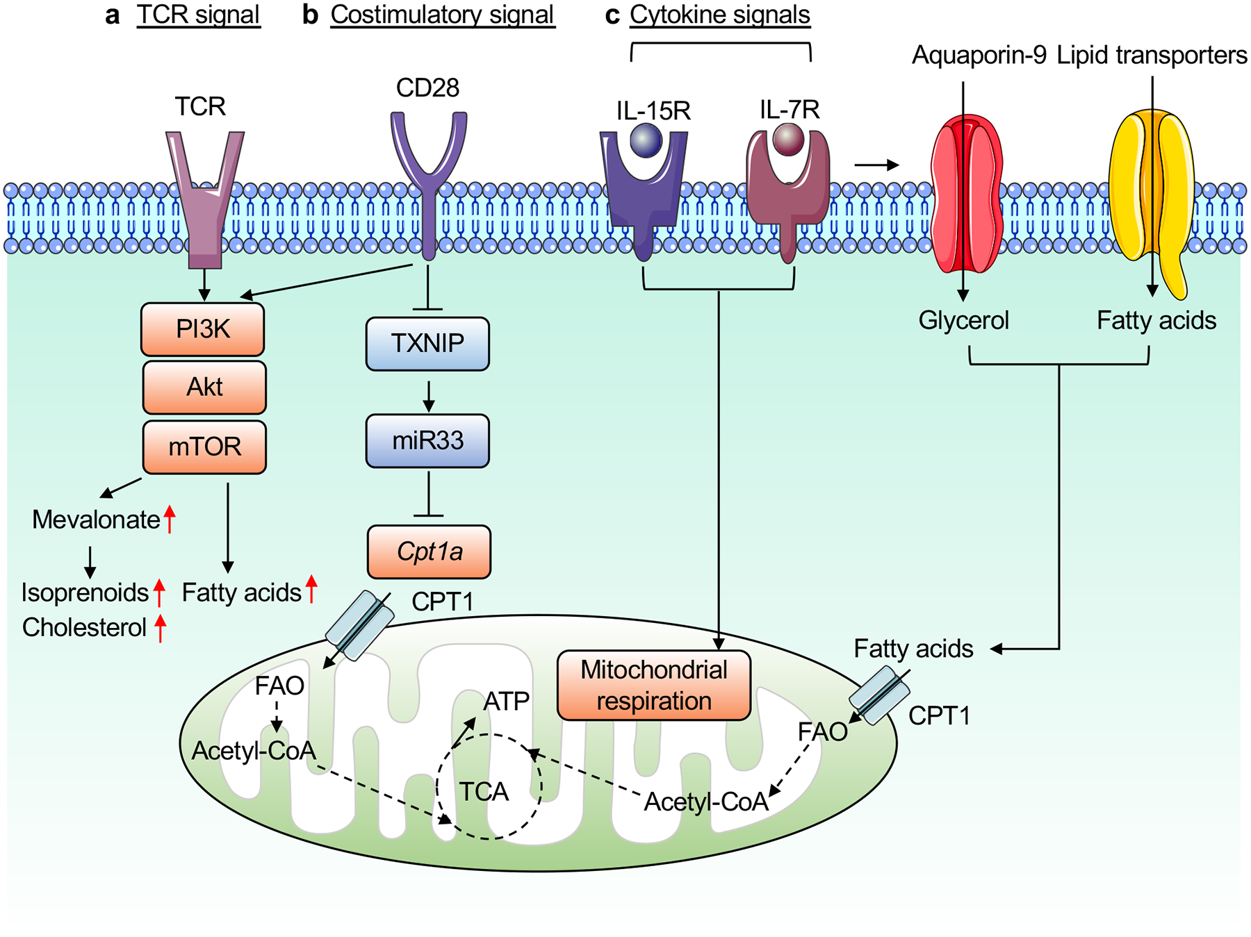
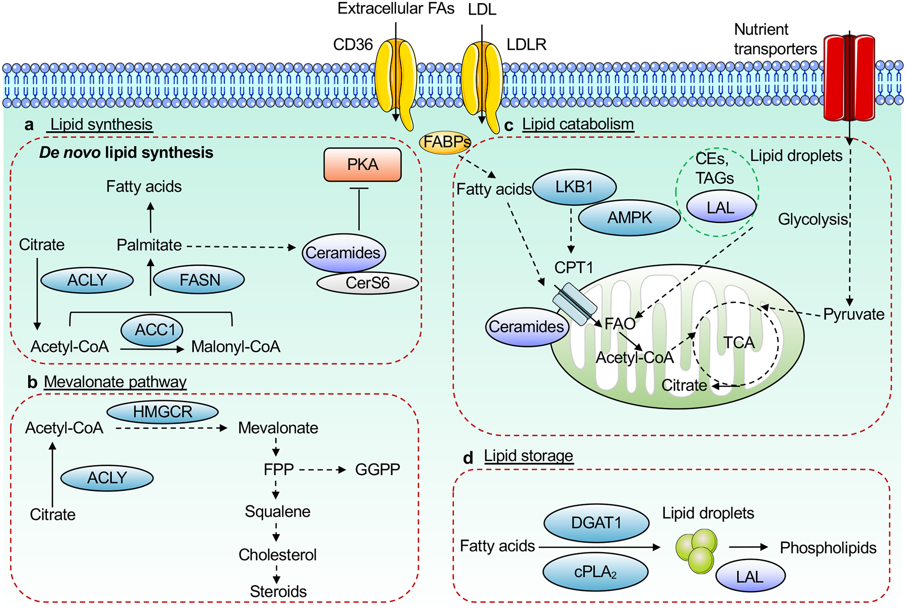
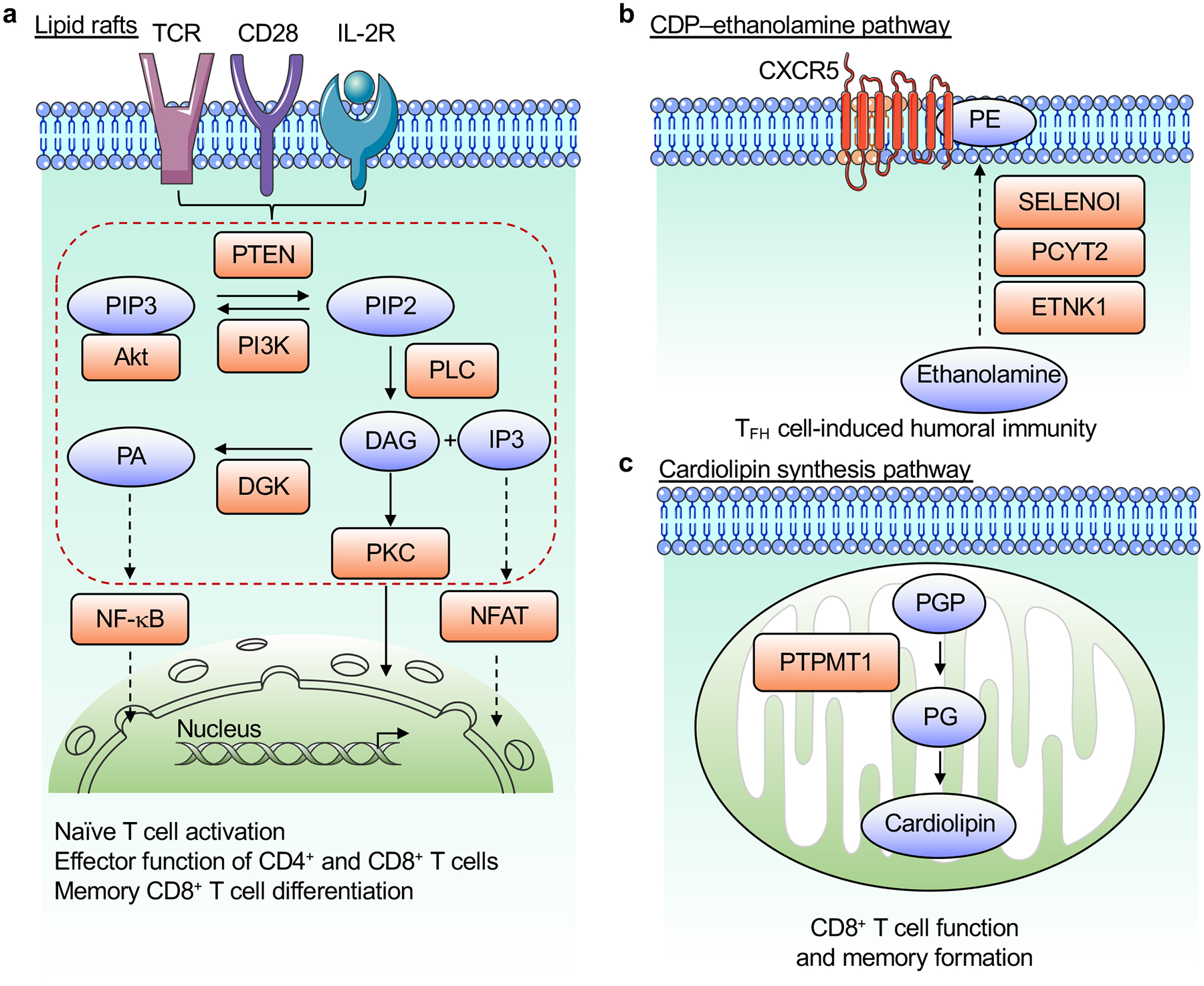
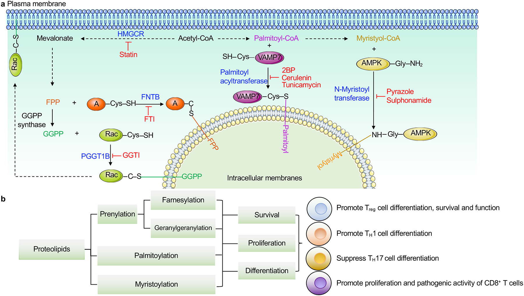
T cells orchestrate adaptive immunity against pathogens and other immune challenges, but their dysfunction can also mediate the pathogenesis of cancer and autoimmunity. Metabolic adaptation in response to immunological and microenvironmental signals contributes to T cell function and fate decision. Lipid metabolism has emerged as a key regulator of T cell responses, with selective lipid metabolites serving as metabolic rheostats to integrate environmental cues and interplay with intracellular signaling processes. Here, we discuss how extracellular, de novo synthesized and membrane lipids orchestrate T cell biology. We also describe the roles of lipids as regulators of intracellular signaling at the levels of transcriptional, epigenetic and post-translational regulation in T cells. Finally, we summarize therapeutic targeting of lipid metabolism and signaling, and conclude with a discussion of important future directions. Understanding the molecular and functional interplay between lipid metabolism and T cell biology will ultimately inform therapeutic intervention for human disease.


Conflict of interest statement

Competing interests

H.C. is a consultant for Kumquat Biosciences.

Figures

Figure 1.
Figure 1.. Extracellular lipids in T cell differentiation and functional adaptation.
Extracellular lipids, such as FAs, cholesterol or cholesterol-derived metabolites, and bile acids regulate multiple aspects of T cell biology. a, SCFAs, which are derived from dietary fiber via microbiota-dependent fermentation, regulate T cell accumulation and function. Exogenous LCFAs such as oleic acid are taken up via CD36 from the TME and impair the function of CD8+ T cells but enhance the function of Treg cells. b, T cells take up cholesterol from the TME, which inhibits the function of CD8+ T cells. Cholesterol accumulation also impedes Tc9 cell generation. Oxysterols signal via EBI2 to promote migration of T cells to the T cell–B cell border and contribute to TFH cell generation. c, Intestinal bile acids (lithocholic acid (LCA) and deoxycholic acid (DCA)) and their derivatives (3-oxoLCA, isoalloLCA, and isoDCA) modulate T cell differentiation and maintenance. 3-oxoLCA inhibits TH17 and promotes pTreg (marked by RORγt expression) cell differentiation, by binding to RORγt and vitamin D receptor (VDR), respectively. IsoalloLCA promotes pTreg cell differentiation, likely in an NR4A1-dependent manner. IsoDCA promotes pTreg cell differentiation by suppressing FXR activity in dendritic cells (DCs). Intestinal bile acids are detoxified by constitutive androstane receptor (CAR) to prevent activation of proinflammatory CD4+ T cell response. FAs, fatty acids; SCFAs, short-chain FAs; LCFAs, long-chain FAs; TME, tumor microenvironment. Receptors, mitochondrial, and nuclear structures used in this and other figures were obtained from Servier Medical Art website (http://smart.servier.com).
Figure 2.
Figure 2.. Regulation of lipid metabolism by immunological signals.
A summary of immunological signals for the regulation of lipid metabolism in T cells. a, Upon TCR engagement, T cells activates PI3K–Akt and mTOR signaling to induce FA synthesis and mevalonate metabolism. b, CD28 costimulation induces transient expression of CPT1a through thioredoxin-interacting protein (TXNIP) and miR33 (before first cell division), and may enhance mevalonate and lipid synthesis by boosting TCR-dependent mTORC1 activation. c, The cytokines IL-15 and IL-7 induce FAO, in part by enhancing expression of CPT1a and mediating glycerol uptake via aquaporin-9, respectively, to maintain mitochondrial respiration.
Figure 3.
Figure 3.. Intracellular lipid metabolism in T cells.
Intracellular lipid homeostasis is regulated by de novo lipid synthesis via the fatty acid (FA) and mevalonate pathways, catabolism, storage, and uptake (via specific transporters such as CD36 and LDLR). a, b, For de novo synthesis to occur, nutrients are catabolized into acetyl-CoA, which is used for synthesis of various FAs (via the enzymatic actions of ACLY, ACC1 and FASN, among others), ceramide (via CerS6), which suppresses PKA signaling (a), or mevalonate and downstream metabolites such as cholesterol and isoprenoids (via the mevalonate pathway including the rate-limiting enzyme HMGCR) (b). c, Lipid catabolism is initiated by FAO in the mitochondria, which is used to generate energy in certain T cell subsets. FAO can be mediated by exogenous lipids, whose intracellular levels are regulated by surface transporters (such as CD36 and LDLR) or intracellular FABPs, or lipid droplet-derived TAGs, which is mediated by LAL-dependent lipolysis. d, Lipid droplets form via cPLA2 or DGAT1-dependent mechanisms to store excess levels of triacylglycerides (TAGs) and cholesterol esters (CEs). TAGs and CEs stored in these lipid droplets undergo hydrolysis via LAL to generate phospholipids. CerS6, ceramide synthase 6; ACC1, acetyl-CoA carboxylase 1; FASN, FA synthase; PKA, protein kinase A; FABPs, FA binding proteins; LAL, lysosomal acid lipase; FAO, FA oxidation; HMGCR, HMG-CoA reductase; DGAT1, diacylglycerol acyltransferase 1; cPLA2, group IVA phospholipase A2.
Figure 4.
Figure 4.. Membrane lipids coordinate signaling in T cells.
a, Antigens, costimulatory signals and IL-2 stimulation induce PI3K activation that generates PIP3 from PIP2, which is opposed by PTEN. Immunological signals induce PLC activation to produce DAG and IP3 from PIP2. These lipid molecules activate several signaling cascades, including those downstream of PKC, to promote activation of NFAT and NF-κB in the nucleus. DGK opposes DAG-dependent signaling by converting DAG to PA. These lipid-coordinated signaling events play multiple roles in T cell biology. b, De novo PE synthesis via the CDP–ethanolamine pathway, mediated by the enzymes ETNK1, PCYT2, and SELENOI, selectively regulates PE localization to the outer layer of the TFH cell membrane and promotes humoral immunity. c, De novo cardiolipin synthesis in the mitochondria, which depends upon PTPMT1, promotes the function and memory differentiation of CD8+ T cells. DAG, diacylglycerol; IP3, inositol trisphosphate; DGK, diacylglycerol kinase; PA, phosphatidic acid; PIP2, phospholipid phosphatidylinositol 4,5-bisphosphate; PIP3, phosphatidylinositol 3,4,5-triphosphate; PKC, protein kinase C; PLC, phospholipase C; PE, phosphatidylethanolamine; PGP, phosphatidylglycerophosphate; PG, phosphatidylglycerol; PTPMT1, protein tyrosine phosphatase mitochondrial 1.
Figure 5.
Figure 5.. Lipid-dependent post-translational modifications orchestrate T cell responses.
a, A summary of lipid-dependent post-translational modifications in T cells. Acetyl-CoA is used for synthesis of either mevalonate or FAs. Mevalonate-derived farnesyl pyrophosphate (FPP) and geranylgeranyl pyrophosphate (GGPP) are covalently linked to specific cysteine residues in small G proteins, such as via Fntb-dependent protein farnesylation that attaches FPP to target proteins (denotated as A in the figure), and Pggt1b-dependent protein geranylgeranylation that conjugates GGPP to Rac. FA-derived palmitoyl-CoA and myristoyl-CoA are conjugated to glycine resides of certain proteins important for T cell biology. For example, palmitoyl acyltransferase DHHC18 promotes protein palmitoylation of VAMP7, while N-myristoyltransferase NMT leads to protein myristoylation of AMPK. These modifications serve important roles in establishing the localization of target proteins for the propagation of intracellular signaling. Inhibitors for the different lipid transferases are shown in red. b, Lipid-mediated post-translational modifications play critical roles in the survival, proliferation and differentiation of T cell subsets, including Treg, TH1, TH17, memory and effector T cells, as summarized in more detail in the figure. AMPK; AMP-activated protein kinase, GGTI; geranylgeranyl transferase type-1 inhibitor, FTI; farnesyltransferase inhibitor, HMGCR; HMG-CoA reductase, 2BP; 2-brompalmitate.

References

    1. Chapman NM & Chi H Metabolic adaptation of lymphocytes in immunity and disease. Immunity 55, 14–30, doi:10.1016/j.immuni.2021.12.012 (2022). - DOI - PMC - PubMed
    1. Maseda D, Ricciotti E & Crofford LJ Prostaglandin regulation of T cell biology. Pharmacol Res 149, 104456, doi:10.1016/j.phrs.2019.104456 (2019). - DOI - PubMed
    1. Trompette A et al. Dietary Fiber Confers Protection against Flu by Shaping Ly6c(−) Patrolling Monocyte Hematopoiesis and CD8(+) T Cell Metabolism. Immunity 48, 992–1005 e1008, doi:10.1016/j.immuni.2018.04.022 (2018). - DOI - PubMed
    1. Bachem A et al. Microbiota-Derived Short-Chain Fatty Acids Promote the Memory Potential of Antigen-Activated CD8(+) T Cells. Immunity 51, 285–297 e285, doi:10.1016/j.immuni.2019.06.002 (2019). - DOI - PubMed
    1. Dudek M et al. Auto-aggressive CXCR6(+) CD8 T cells cause liver immune pathology in NASH. Nature 592, 444–449, doi:10.1038/s41586-021-03233-8 (2021). - DOI - PubMed
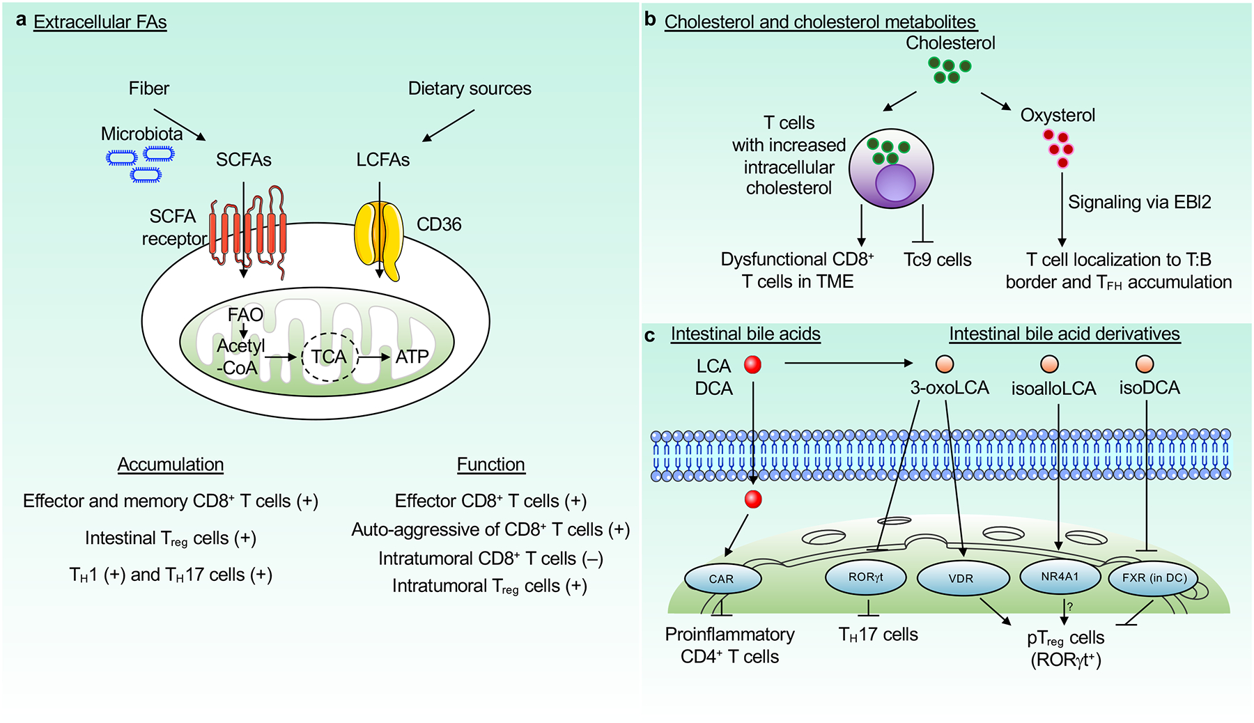
    2. This paper shows a pathogenic role for the SCFA acetate in CD8+ T cell-mediated autoinflammation and tissue damage in the liver, which is in contrast to the protective roles for SCFAs in tissue homeostasis or anti-pathogen immunity.

Publication types

Substances