

Comprehensive three-dimensional positional and morphological assessment of the temporomandibular joint in skeletal Class II patients with mandibular retrognathism in different vertical skeletal patterns
- PMID: 35484618
- PMCID: PMC9052647
- DOI: 10.1186/s12903-022-02174-6
Comprehensive three-dimensional positional and morphological assessment of the temporomandibular joint in skeletal Class II patients with mandibular retrognathism in different vertical skeletal patterns
Abstract
Background: Only a few studies have used 3D cone-beam computed tomography (CBCT) analysis to evaluate the positional and morphological characteristics of the temporomandibular joint (TMJ) in adults with skeletal Class II. No studies have focused on the case of skeletal Class II with mandibular retrognathism in different vertical skeletal patterns. As a result, this study aimed to evaluate and compare the position and morphology of TMJ in adults with skeletal Class II with mandibular retrognathism in different vertical skeletal patterns to the position and morphology of TMJ in the normal Chinese adult population in three dimensions.
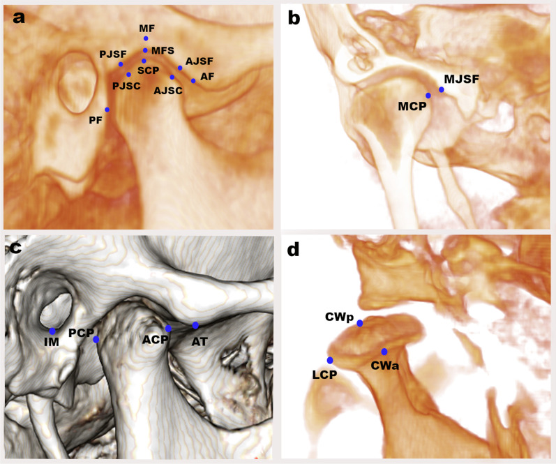
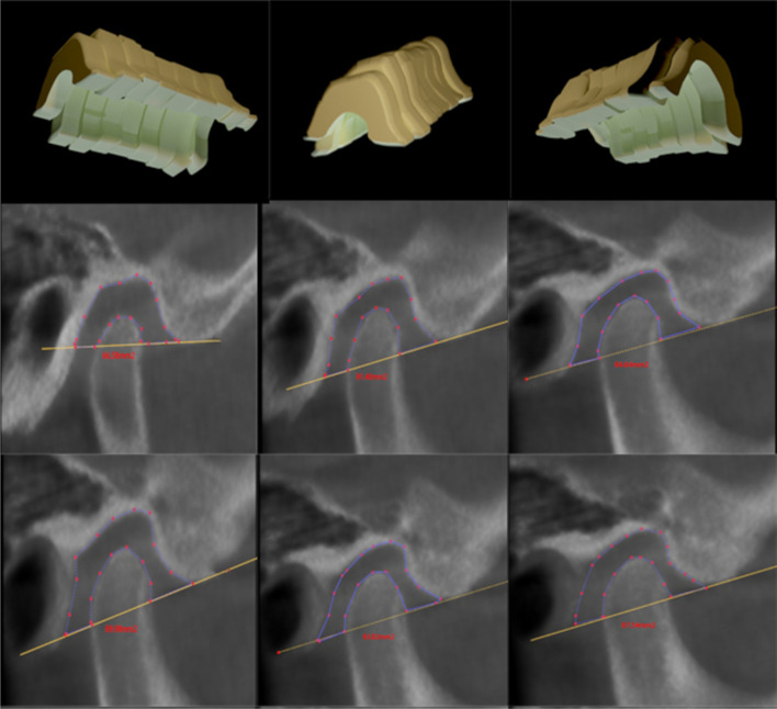
Methods: This retrospective study analyzed CBCT images of 80 adult patients. Subjects with skeletal Class II with a normal sagittal position of the maxilla and mandibular retrognathism were classified according to the mandibular angle and facial height ratio into three groups of 20 subjects each: hypodivergent, normodivergent, and hyperdivergent groups, as well as a control group of 20 subjects. The following 3D measurements of TMJ were evaluated: (1) position, parameters, and inclination of the mandibular fossa; (2) position, parameters, and inclination of the mandibular condyle; (3) condyle centralization in their respective mandibular fossae; (4) anterior, posterior, superior, and medial joint spaces; and (5) 3D volumetric measurements of the TMJ spaces. Measurements were statistically analyzed by one-way ANOVA test, followed by Tukey's post hoc test.
Results: Significant differences were found in the hyperdivergent and hypodivergent groups compared with the normal group in the vertical and anteroposterior mandibular fossa position, vertical condylar inclination, and condylar width and length. The hyperdivergent group showed the significantly highest condylar inclination with the midsagittal plane; anterior and superior positioning of the condyle; smallest anterior, superior, and medial joint spaces; and largest volumetric total joint space relative to the two other groups.
Conclusions: The condyle-fossa position and morphology differ with various vertical facial patterns in individuals with skeletal Class II mandibular retrognathism. These differences could be considered during TMD diagnosis and orthodontic treatment.
Keywords: CBCT; Class II; Retrognathism; Temporomandibular joint; Three-dimensional; Vertical skeletal patterns.
© 2022. The Author(s).
Conflict of interest statement
The authors declare that they have no competing interests.
Figures

References
-
- Krisjane Z, Urtane I, Krumina G, Bieza A, Zepa K, Rogovska I. Condylar and mandibular morphological criteria in the 2D and 3D MSCT imaging for patients with Class II division 1 subdivision malocclusion. Stomatologija. 2007;9:67–71. - PubMed
-
- Proffit WR, Fields H, Jr, Moray L. Prevalence of malocclusion and orthodontic treatment need in the United States: estimates from the NHANES III survey. Int J Adult Orthodon Orthognath Surg. 1998;13:97–106. - PubMed
Publication types
MeSH terms
LinkOut - more resources
Full Text Sources
Research Materials
