. 2023 Mar 1;108(3):717-731.
doi: 10.3324/haematol.2021.280557.

Molecular characterization and clinical outcome of B-cell precursor acute lymphoblastic leukemia with IG-MYC rearrangement

Affiliations

Molecular characterization and clinical outcome of B-cell precursor acute lymphoblastic leukemia with IG-MYC rearrangement

Simon Bomken et al. Haematologica. .

Abstract

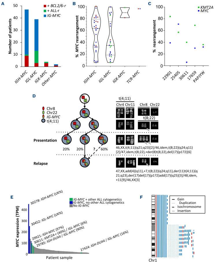
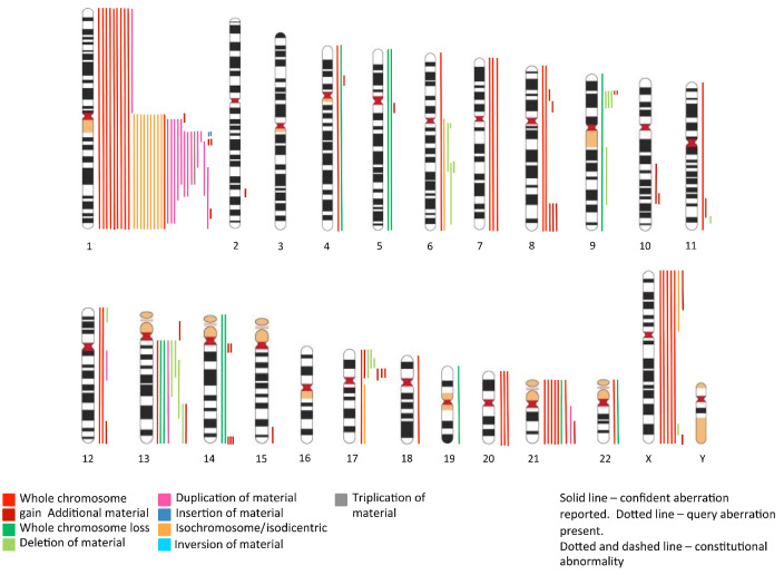
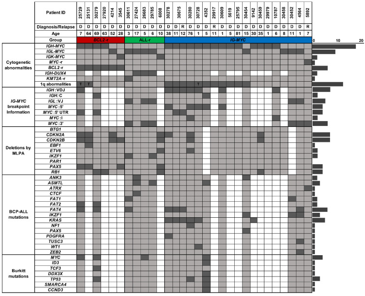
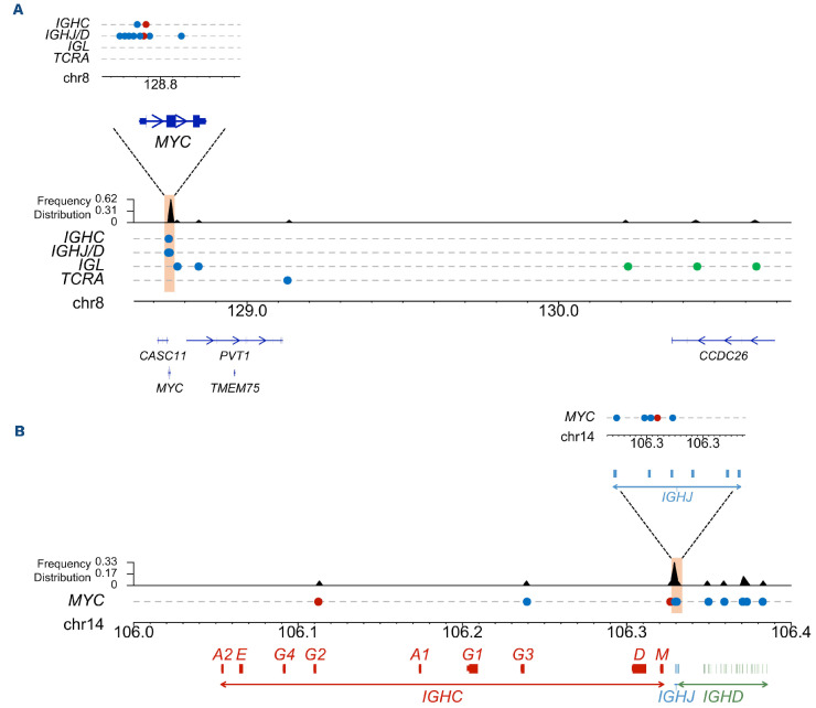
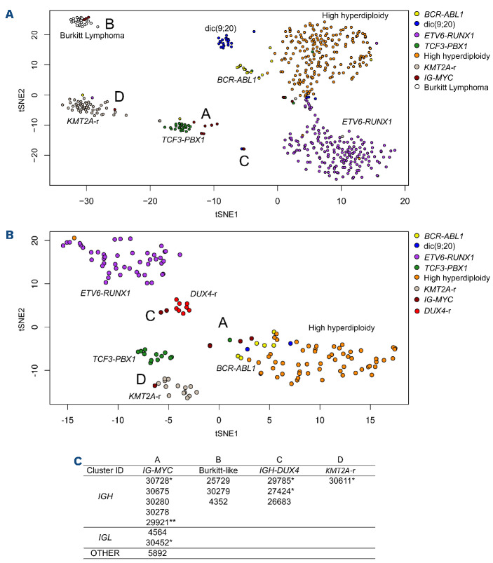
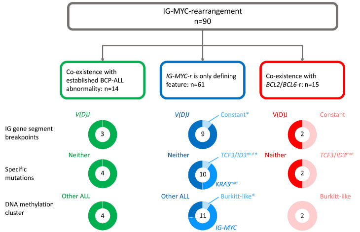
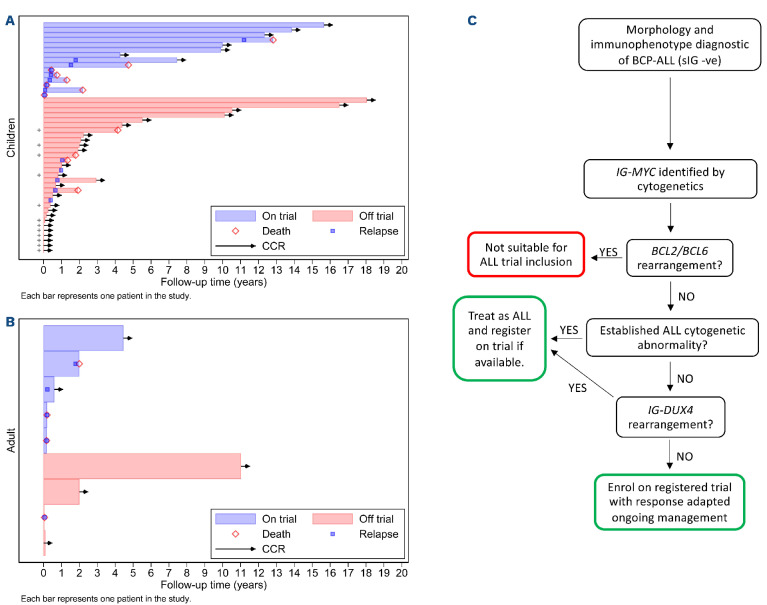
Rarely, immunophenotypically immature B-cell precursor acute lymphoblastic leukemia (BCP-ALL) carries an immunoglobulin- MYC rearrangement (IG-MYC-r). This can result in diagnostic confusion with Burkitt lymphoma/leukemia and use of individualized treatment schedules of unproven efficacy. Here we compare the molecular characteristics of these conditions and investigate historic clinical outcome data. We identified 90 cases registered in a national BCP-ALL clinical trial/registry. When present, diagnostic material underwent cytogenetic, exome, methylome and transcriptome analyses. The outcomes analyzed were 3-year event-free survival and overall survival. IG-MYC-r was identified in diverse cytogenetic backgrounds, co-existing with either established BCP-ALL-specific abnormalities (high hyperdiploidy, n=3; KMT2A-rearrangement, n=6; iAMP21, n=1; BCR-ABL1, n=1); BCL2/BCL6-rearrangements (n=15); or, most commonly, as the only defining feature (n=64). Within this final group, precursor-like V(D)J breakpoints predominated (8/9) and KRAS mutations were common (5/11). DNA methylation identified a cluster of V(D)J-rearranged cases, clearly distinct from Burkitt leukemia/lymphoma. Children with IG-MYC-r within that subgroup had a 3-year event-free survival of 47% and overall survival of 60%, representing a high-risk BCP-ALL. To develop effective management strategies this group of patients must be allowed access to contemporary, minimal residual disease-adapted, prospective clinical trial protocols.


Figures

Figure 1.
Figure 1.
Cytogenetic characterization of IG-MYC-rearranged patients. (A) Karyotype reveals the distribution of immunoglobulin chain involvement in MYC translocations (fluorescence in situ hybridization [FISH] data were used for one patient who presented with a normal karyotype). +BCL2/6-r: concomitant BCL2/BCL6-rearrangement; +ALL-r, concomitant established acute lymphoblastic leukemia rearrangement; IG-MYC: IG-MYC rearrangement as the defining cytogenetic abnormality. (B) Percentage of nuclei carrying IG-MYC-r grouped by immunoglobulin chain involvement. Blue dots, IG-MYC-r alone. Green dots, IG-MYC-r and established ALL rearrangement. Red dots, IG-MYC-r and BCL2/BCL6-r. Dotted and dashed lines, interquartile ranges. (C) Percentage rearrangement of MYC (blue dots) and KMT2A (green dots) using FISH. (D) Evolution of t(4;11) and subsequent gain of t(8;22) in case 17659. Percentages at diagnosis represent the proportion of metaphases seen with each abnormality (left panel). Representative chromosomes taken from diagnostic and relapse metaphases (right panel). Gray arrows mark the portion of chromosome 22 translocated to chromosome 8. (E) RNA sequencing comparing expression of MYC among (labeled) cases from the IG-MYC-r cohort with other in-house B-cell precursor ALL cases. TPM: transcripts per million reads. (F) Chromosomal abnormalities observed for chromosome 1 in the karyotypes of patients with IG-MYC-r. Red line: patients with concomitant BCL2-r.
Figure 2.
Figure 2.
Ideogram of cytogenetic rearrangements reported in the karyotypes of 70 patients with IG-MYC-rearrangements.
Figure 3.
Figure 3.
Oncoplot depicting the incidence of selected genomic features of IG-MYC-rearranged patients with material available for genomic studies. D: diagnosis; R: relapse; dark-shaded square: positive result; light-shaded square: negative result; white square: not tested; T: 1q translocation.
Figure 4.
Figure 4.
Targeted IG and MYC sequencing identified heterogeneous breakpoints. (A) Distribution of breakpoints within the MYC locus. (B) Distribution of breakpoints within the IGH locus. Each line shows the breakpoints for patients with translocations involving individual genomic loci. Frequency distribution defines a region of increased frequency of breaks (peach shaded area). Upper panels provide an expanded view of the breakpoint hotspots. Each dot represents an individual breakpoint. Blue dots: IGMYC-rearrangement alone. Green dots, IG-MYC-rearrangement and established acute lymphoblastic leukemia rearrangement. Red dots, IG-MYC-rearrangement and BCL2/BCL6-rearrangement.
Figure 5.
Figure 5.
Methylation and transcriptome data identified five clusters within the cohort of patients with IG-MYC-rearrangements. (A) Consensus clustering of methylation data. Common t-distributed stochastic neighbor embedding (t-SNE) visualization of IGMYC-rearranged patients combined with publicly available data for subtypes of B-acute lymphoblastic leukemia (GSE49031, GSE69229, GSE76585), a patient with Burkitt lymphoma (GSE114210) and cell line samples (GSE92378). Each dot represents an individual patient. (B) Consensus clustering of RNA-sequencing data. Common t-SNE visualization of IG-MYC-rearranged patients combined with publicly available RNA-sequencing data for subtypes of B-acute lymphoblastic leukemia (EGAS00001001795). Each dot represents an individual patient. (C) Patients’ identities assigned to clusters. *Methylation and RNA-sequencing data, **RNA-sequencing only.
Figure 6.
Figure 6.
Molecular characteristics of cases of IG-MYC-rearranged acute lymphoblastic leukemia. Summary of three groups defined within the molecular analysis. Cases with a co-existing acute lymphoblastic leukemia (ALL)-specific cytogenetic abnormality are shown in green in the left panel. Cases with an IG-MYC-rearrangement (IG-MYC-r) as the only recurrent cytogenetic feature are shown in blue in the center panel and cases with a co-existing rearrangement of BCL2 and/or BCL6 are shown in red in the right panel. Each donut plot shows the proportion of cases studied which have a specific molecular feature. The total number of cases in each study is shown within each donut. The upper donut plots show the proportion with variable V(D)J gene segment breakpoints (dark segments) and constant segment breakpoints (light segments). The middle donut plots show the proportion of cases with either Burkitt-like TCF3/ID3 mutations (light segments), KRAS mutations (medium segments) or neither such mutation (dark segments). The lower donut plots show the proportion of cases within each of three DNA methylation clusters, either Burkitt-like cluster B (light segments), the IG-MYC-r cluster A (medium segments) or one of the other ALL specific clusters (dark segments). *The single case in the center panel harboring both a constant region breakpoint and an ID3 mutation (case 4352) clustered with Burkitt lymphoma/leukemia cases in our DNA methylation analysis.
Figure 7.
Figure 7.
Swimmer plots displaying event-free survival and overall survival. (A) Children and (B) adults with IG-MYC-rearrangements but no other acute lymphoblastic leukemia-specific cytogenetic abnormality. Suggested approach for the assignment of patients to a treatment strategy (C). + For patients without data on relapse or second tumor, death was assumed to be the first event. ALL: acute lymphoblastic leukemia; BCP: B-cell precursor; CCR: continuous complete remission; sIG: surface immunoglobulin.

Comment in

References

    1. Klapper W, Stoecklein H, Zeynalova S, et al. . Structural aberrations affecting the MYC locus indicate a poor prognosis independent of clinical risk factors in diffuse large B-cell lymphomas treated within randomized trials of the German High-Grade Non-Hodgkin's Lymphoma Study Group (DSHNHL). Leukemia. 2008;22(12):2226-2229. - PubMed
    1. Savage KJ, Johnson NA, Ben-Neriah S, et al. . MYC gene rearrangements are associated with a poor prognosis in diffuse large B-cell lymphoma patients treated with R-CHOP chemotherapy. Blood. 2009;114(17):3533-3537. - PubMed
    1. Ott G, Rosenwald A, Campo E. Understanding MYC-driven aggressive B-cell lymphomas: pathogenesis and classification. Blood. 2013;122(24):3884-3891. - PubMed
    1. Mikulasova A, Ashby C, Tytarenko RG, et al. . Microhomology-mediated end joining drives complex rearrangements and overexpression of MYC and PVT1 in multiple myeloma. Haematologica. 2020;105(4):1055-1066. - PMC - PubMed
    1. Swerdlow SH, Campo E, Harris NL, et al. . WHO Classification of Tumours of Haematopoietic and Lymphoid Tissues (Revised 4th edition): International Agency for Research on Cancer (IARC). 2017

Publication types

MeSH terms

Substances