

Computational fluid dynamics (CFD) analysis in a ruptured vertebral artery dissecting aneurysm implanted by Pipeline when recurrent after LVIS-assisted coiling treatment: Case report and review of the literatures
- PMID: 35484808
- PMCID: PMC10399494
- DOI: 10.1177/15910199221097766
Computational fluid dynamics (CFD) analysis in a ruptured vertebral artery dissecting aneurysm implanted by Pipeline when recurrent after LVIS-assisted coiling treatment: Case report and review of the literatures
Abstract
Backgrounds: Hemodynamics plays an important role in the natural history of the process of rupture and recurrence of intracranial aneurysms. This study aimed to investigate the role of hemodynamics for recurrence in a vertebral artery dissecting aneurysm (VADA).
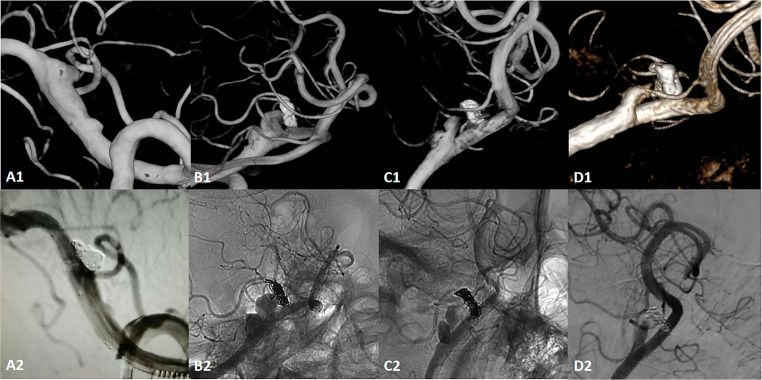
Methods: A patient with a ruptured VADA firstly treated by low-profile visualized intraluminal support (LVIS)-assisted coiling, and was implanted with a Pipeline Embolization Device (PED) after aneurysm recurrence. Finite element analysis and computational fluid dynamics simulations were conducted in 6 serial imaging procedures, and the calculated hemodynamics was correlated with aneurysm recurrence.
Results: Wall shear stress (WSS) was not effectively suppressed, resulting in aneurysm recurrence with initial entry tear to occur above the protuberance after 7 months of LVIS stent-assisted coiling. With the implantation of PED, WSS, inflow stream and velocity at the aneurysm neck significantly decreased. During the 3-month follow-up after PED deployment, there was significant shrinkage of the sac and the blood flow in the sac was reduced considerably. The 27-month follow-up after PED deployment indicated the aneurysm was stable.
Conclusions: The present case study suggests that insufficient suppression of high WSS and high inflow velocity at the neck of the parent artery, especially near the posterior inferior cerebellar artery, might be associated with aneurysm recurrence.
Keywords: aneurysm recurrence; compuational fluid dynamics; finite element analysis; vertebral artery dissecting aneurysm.
Conflict of interest statement
The author(s) declared no potential conflicts of interest with respect to the research, authorship, and/or publication of this article.
Figures


References
-
- Yamaura A, Ono J, Hirai S. Clinical picture of intracranial non-traumatic dissecting aneurysm. Neuropathology 2000; 20: 85–90. - PubMed
-
- Takagi T, Takayasu M, Suzuki Y, et al. Prediction of rebleeding from angiographic features in vertebral artery dissecting aneurysms. Neurosurg Rev 2007; 30: 32–38; discussion -9. - PubMed
-
- Sluzewski M, van Rooij W, Rinkel G, et al. Endovascular treatment of ruptured intracranial aneurysms with detachable coils: long-term clinical and serial angiographic results. Radiology 2003; 227: 720–724. - PubMed
-
- Campi A, Ramzi N, Molyneux AJ, et al. Retreatment of ruptured cerebral aneurysms in patients randomized by coiling or clipping in the international subarachnoid aneurysm trial (ISAT). Stroke 2007; 38: 1538–1544. - PubMed
Publication types
MeSH terms
LinkOut - more resources
Full Text Sources
Medical
Research Materials
Miscellaneous
