

The meningeal lymphatic vessels and the glymphatic system: Potential therapeutic targets in neurological disorders
- PMID: 35484910
- PMCID: PMC9274866
- DOI: 10.1177/0271678X221098145
The meningeal lymphatic vessels and the glymphatic system: Potential therapeutic targets in neurological disorders
Abstract
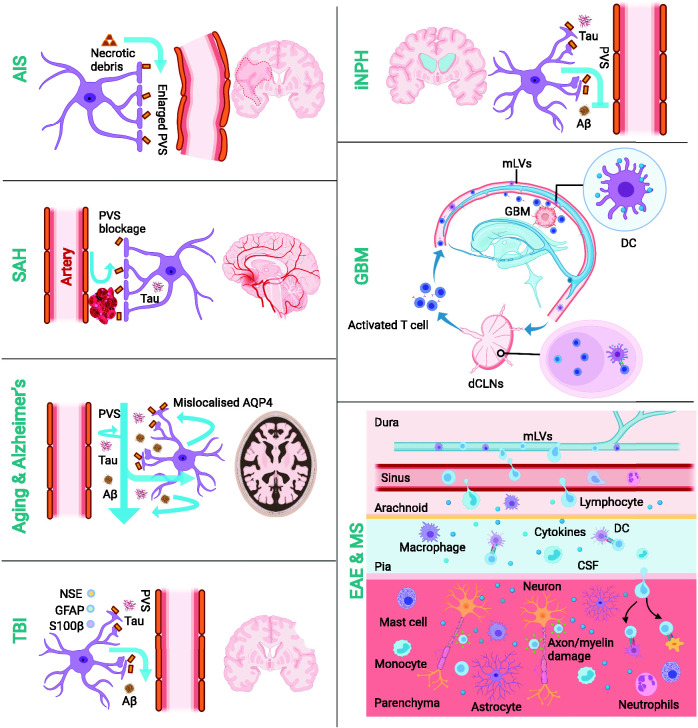
The recent discovery of the meningeal lymphatic vessels (mLVs) and glymphatic pathways has challenged the long-lasting dogma that the central nervous system (CNS) lacks a lymphatic system and therefore does not interact with peripheral immunity. This discovery has reshaped our understanding of mechanisms underlying CNS drainage. Under normal conditions, a close connection between mLVs and the glymphatic system enables metabolic waste removal, immune cell trafficking, and CNS immune surveillance. Dysfunction of the glymphatic-mLV system can lead to toxic protein accumulation in the brain, and it contributes to the development of a series of neurodegenerative disorders, such as Alzheimer's and Parkinson's diseases. The identification of precise cerebral transport routes is based mainly on indirect, invasive imaging of animals, and the results cannot always be applied to humans. Here we review the functions of the glymphatic-mLV system and evidence for its involvement in some CNS diseases. We focus on emerging noninvasive imaging techniques to evaluate the human glymphatic-mLV system and their potential for preclinical diagnosis and prevention of neurodegenerative diseases. Potential strategies that target the glymphatic-mLV system in order to treat and prevent neurological disorders are also discussed.
Keywords: CNS diseases; brain clearance system; glymphatic pathways; mLVs; meningeal immunity.
Conflict of interest statement
Figures
Similar articles
-
Therapeutic approaches to CNS diseases via the meningeal lymphatic and glymphatic system: prospects and challenges.Front Cell Dev Biol. 2024 Sep 6;12:1467085. doi: 10.3389/fcell.2024.1467085. eCollection 2024. Front Cell Dev Biol. 2024. PMID: 39310229 Free PMC article. Review.
-
Current Understanding of Central Nervous System Drainage Systems: Implications in the Context of Neurodegenerative Diseases.Curr Neuropharmacol. 2020;18(11):1054-1063. doi: 10.2174/1570159X17666191113103850. Curr Neuropharmacol. 2020. PMID: 31729299 Free PMC article. Review.
-
Photobiomodulation Therapy and the Glymphatic System: Promising Applications for Augmenting the Brain Lymphatic Drainage System.Int J Mol Sci. 2022 Mar 10;23(6):2975. doi: 10.3390/ijms23062975. Int J Mol Sci. 2022. PMID: 35328396 Free PMC article. Review.
-
Emerging Roles of Meningeal Lymphatic Vessels in Alzheimer's Disease.J Alzheimers Dis. 2023;94(s1):S355-S366. doi: 10.3233/JAD-221016. J Alzheimers Dis. 2023. PMID: 36683509 Free PMC article. Review.
-
The glymphatic system and meningeal lymphatics of the brain: new understanding of brain clearance.Rev Neurosci. 2021 Feb 23;32(7):693-705. doi: 10.1515/revneuro-2020-0106. Print 2021 Nov 25. Rev Neurosci. 2021. PMID: 33618444 Review.
Cited by
-
Therapeutic approaches to CNS diseases via the meningeal lymphatic and glymphatic system: prospects and challenges.Front Cell Dev Biol. 2024 Sep 6;12:1467085. doi: 10.3389/fcell.2024.1467085. eCollection 2024. Front Cell Dev Biol. 2024. PMID: 39310229 Free PMC article. Review.
-
Bridging the brain and gut: neuroimmune mechanisms of neuroinflammation and therapeutic insights.Front Cell Neurosci. 2025 Jun 13;19:1590002. doi: 10.3389/fncel.2025.1590002. eCollection 2025. Front Cell Neurosci. 2025. PMID: 40584220 Free PMC article. Review.
-
The role of astrocytes in the glymphatic network: a narrative review.Metab Brain Dis. 2024 Mar;39(3):453-465. doi: 10.1007/s11011-023-01327-y. Epub 2023 Nov 27. Metab Brain Dis. 2024. PMID: 38008886 Review.
-
Image analysis techniques for in vivo quantification of cerebrospinal fluid flow.Exp Fluids. 2023 Nov;64(11):181. doi: 10.1007/s00348-023-03719-3. Epub 2023 Oct 30. Exp Fluids. 2023. PMID: 39691852 Free PMC article.
-
Glymphatic system dysfunction in cerebral infarction: advances and perspectives based on DTI-derived ALPS measures.Am J Transl Res. 2025 Mar 15;17(3):1630-1642. doi: 10.62347/OQRE2088. eCollection 2025. Am J Transl Res. 2025. PMID: 40226042 Free PMC article. Review.
References
-
- Stritt S, Koltowska K, Mäkinen T. Homeostatic maintenance of the lymphatic vasculature. Trends Mol Med 2021; 27: 955–970. - PubMed
-
- Sandrone S, Moreno-Zambrano D, Kipnis J, et al.. A (delayed) history of the brain lymphatic system. Nat Med 2019; 25: 538–540. - PubMed
-
- Li J, Zhou J, Shi Y. Scanning electron microscopy of human cerebral meningeal stomata. Ann Anat 1996; 178: 259–261. - PubMed
-
- Kida S, Pantazis A, Weller RO. CSF drains directly from the subarachnoid space into nasal lymphatics in the rat. Anatomy, histology and immunological significance. Neuropathol Appl Neurobiol 1993; 19: 480–488. - PubMed
Publication types
MeSH terms
LinkOut - more resources
Full Text Sources
Medical
