Neonatal Myocardial Infarction: A Proposed Algorithm for Coronary Arterial Thrombus Management
- PMID: 35485231
- PMCID: PMC11225359
- DOI: 10.1161/CIRCINTERVENTIONS.121.011664
Neonatal Myocardial Infarction: A Proposed Algorithm for Coronary Arterial Thrombus Management
Abstract
Background: Neonatal myocardial infarction is rare and is associated with a high mortality of 40% to 50%. We report our experience with neonatal myocardial infarction, including presentation, management, outcomes, and our current patient management algorithm.
Methods: We reviewed all infants admitted with a diagnosis of coronary artery thrombosis, coronary ischemia, or myocardial infarction between January 2015 and May 2021.
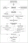
Results: We identified 21 patients (median age, 1 [interquartile range (IQR), 0.25-9.00] day; weight, 3.2 [IQR, 2.9-3.7] kg). Presentation included respiratory distress (16), shock (3), and murmur (2). Regional wall motion abnormalities by echocardiogram were a key criterion for diagnosis and were present in all 21 with varying degrees of depressed left ventricular function (severe [8], moderate [6], mild [2], and low normal [5]). Ejection fraction ranged from 20% to 54% (median, 43% [IQR, 34%-51%]). Mitral regurgitation was present in 19 (90%), left atrial dilation in 15 (71%), and pulmonary hypertension in 18 (86%). ECG was abnormal in 19 (90%). Median troponin I was 0.18 (IQR, 0.12-0.56) ng/mL. Median BNP (B-type natriuretic peptide) was 2100 (IQR, 924-2325) pg/mL. Seventeen had documented coronary thrombosis by cardiac catheterization. Seventeen (81%) were treated with intracoronary tPA (tissue-type plasminogen activator) followed by systemic heparin, AT (antithrombin), and intravenous nitroglycerin, and 4 (19%) were treated with systemic heparin, AT, and intravenous nitroglycerin alone. Nineteen of 21 recovered. One died (also had infradiaphragmatic total anomalous pulmonary venous return). One patient required a ventricular assist device and later underwent heart transplant; this patient was diagnosed late at 5 weeks of age and did not respond to tPA. Nineteen of 21 (90%) regained normal left ventricular function (ejection fraction, 60%-74%; mean, 65% [IQR, 61%-67%]) at latest follow-up (median, 6.8 [IQR, 3.58-14.72] months). Two of 21 (10%) had residual trivial mitral regurgitation. After analysis of these results, we present our current algorithm, which developed and matured over time, to manage neonatal myocardial infarction.
Conclusions: We experienced a lower mortality rate for infants with neonatal infarction than that reported in the literature. We propose a post hoc algorithm that may lead to improvement in patient outcomes following coronary artery thrombus.
Keywords: dilatation; infant; stroke volume; troponin I; umbilical cord.
Figures


References
-
- Saliba E, Debillon T, Auvin S, Baud O, Biran V, Chabernaud JL, Chabrier S, Cneude F, Cordier AG, Darmency-Stamboul V, et al.; Recommandations Accident Vasculaire Cérébral (AVC) Néonatal. [Neonatal arterial ischemic stroke: review of the current guidelines]. Arch Pediatr. 2017;24:180–188. doi: 10.1016/j.arcped.2016.11.005 - DOI - PubMed

