. 2023 Mar 1;146(3):1006-1020.
doi: 10.1093/brain/awac157.

Recovered grasping performance after stroke depends on interhemispheric frontoparietal connectivity

Affiliations

Recovered grasping performance after stroke depends on interhemispheric frontoparietal connectivity

Lukas Hensel et al. Brain. .

Abstract

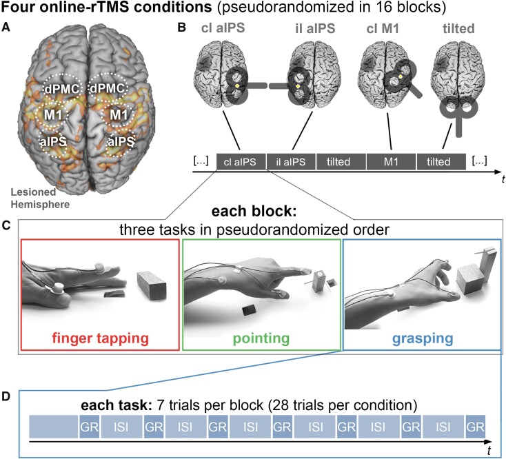
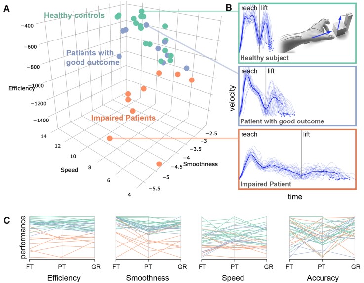
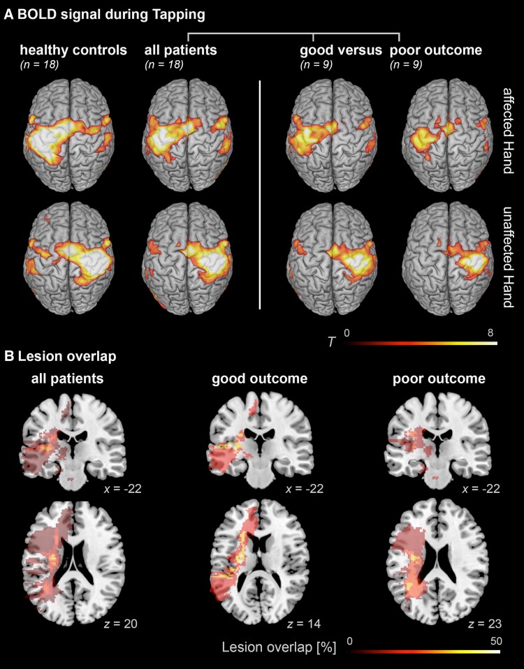
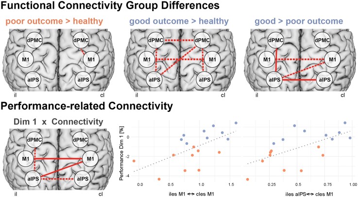
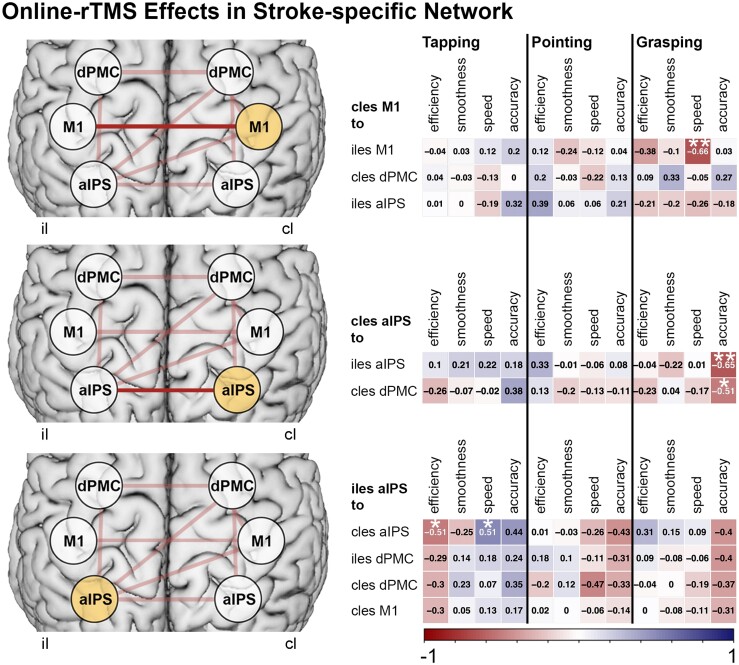
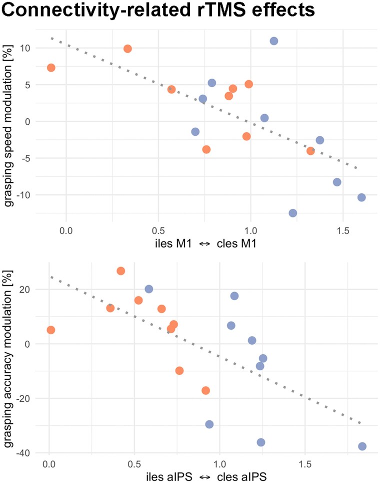
Activity changes in the ipsi- and contralesional parietal cortex and abnormal interhemispheric connectivity between these regions are commonly observed after stroke, however, their significance for motor recovery remains poorly understood. We here assessed the contribution of ipsilesional and contralesional anterior intraparietal cortex (aIPS) for hand motor function in 18 recovered chronic stroke patients and 18 healthy control subjects using a multimodal assessment consisting of resting-state functional MRI, motor task functional MRI, online-repetitive transcranial magnetic stimulation (rTMS) interference, and 3D movement kinematics. Effects were compared against two control stimulation sites, i.e. contralesional M1 and a sham stimulation condition. We found that patients with good motor outcome compared to patients with more substantial residual deficits featured increased resting-state connectivity between ipsilesional aIPS and contralesional aIPS as well as between ipsilesional aIPS and dorsal premotor cortex. Moreover, interhemispheric connectivity between ipsilesional M1 and contralesional M1 as well as ipsilesional aIPS and contralesional M1 correlated with better motor performance across tasks. TMS interference at individual aIPS and M1 coordinates led to differential effects depending on the motor task that was tested, i.e. index finger-tapping, rapid pointing movements, or a reach-grasp-lift task. Interfering with contralesional aIPS deteriorated the accuracy of grasping, especially in patients featuring higher connectivity between ipsi- and contralesional aIPS. In contrast, interference with the contralesional M1 led to impaired grasping speed in patients featuring higher connectivity between bilateral M1. These findings suggest differential roles of contralesional M1 and aIPS for distinct aspects of recovered hand motor function, depending on the reorganization of interhemispheric connectivity.

Keywords: intraparietal sulcus; kinematics; motor recovery; rTMS; resting-state fMRI.


Figures

Figure 1
Figure 1
Experimental setup. (A) Regions of interest for the connectivity analysis, drawn on an activation map of an example patient during paretic hand movements. (B) The online-rTMS session consisted of four rTMS conditions, each occurring in four blocks in a pseudo-randomized order. (C) In each block, participants performed three tasks: finger-tapping, pointing, and reach-grasp-lift an object. (D) Each task was performed in seven trials while rTMS was applied for 1.5 s after the onset of each trial. In total, each task was performed in 112 trials (28 trials per condition). Cl = contralesional; il = ipsilesional; GR = reach-grasp-lift condition; ISI = intertrial interval (5.5 s). Lesioned hemispheres are schematically marked by darkened lesion areas.
Figure 2
Figure 2
Kinematic hand motor phenotypes. K-means clustering of hand kinematics yielded a two-cluster solution, (A) grouping healthy controls and patients with good motor outcome into the same cluster, in contrast to impaired patients. Participants are plotted based on reach-grasp-lift kinematics. (B) Examples of velocity profiles of one healthy participant (top), a patient with near-to-normal performance (middle), and one impaired patient (bottom). The dark blue lines indicate the mean velocity profile. (C) Parallel plot of all kinematic input variables entered into the clustering analysis, showing each participant’s performance for each scaled variable. FT = finger-tapping; PT = pointing; GR = grasping.
Figure 3
Figure 3
Activation and lesion maps. (A) Group effects of BOLD activation indicating reduced activation in the ipsilesional M1 in impaired patients when tapping with the affected hand, whereas patients with good outcome show more activation of contralesional frontoparietal regions. Results were FWE cluster-level corrected at a threshold of P < 0.05 (cluster-forming threshold at the voxel level: P < 0.001). (B) Lesion overlap of stroke patients, based on T2 images. Lesion maps showed maximal overlap in the internal capsule in all patients as well as the two patient subgroups.
Figure 4
Figure 4
Stroke-related connectivity alterations. Top: Higher frontoparietal and interhemispheric connectivity was found for patients with good motor outcome, compared to healthy controls and compared to patients with poor outcome. Bottom: Correlating connectivity with motor performance across tasks (dimension 1 of the multiple factor analysis) revealed similar connections related to motor performance after stroke. Solid lines indicate FDR-corrected results P < 0.05. Dotted lines indicate uncorrected results P < 0.05. Orange = impaired patients; blue = patients with good motor outcome.
Figure 5
Figure 5
Online-rTMS effects. Stroke-specific connections (top row, connections in light red) with each stimulation target were tested for correlations with patients’ performance changes of kinematics during rTMS. This analysis revealed interhemispheric connections (top row, connections in dark red) to be related to the modulation of grasping kinematics during rTMS over contralesional M1 and aIPS. **FDR-corrected correlations P < 0.05. *Uncorrected correlations P < 0.05.
Figure 6
Figure 6
Connectivity-related rTMS effects. Top: Grasping speed was reduced during rTMS of contralesional M1 in participants with higher interhemispheric M1-connectivity. Bottom: Accuracy in the grasp-to-lift movement was reduced during rTMS of contralesional aIPS in participants with higher interhemispheric aIPS-connectivity. Orange = impaired patients; blue = patients with good motor outcome.

Comment in

References

    1. GBD 2016 Neurology Collaborators . Global, regional, and national burden of neurological disorders, 1990-2016: a systematic analysis for the Global Burden of Disease Study 2016. Lancet Neurol. 2019;18:459–480. - PMC - PubMed
    1. Carod-Artal J, Egido JA, González JL, de Seijas E V. Quality of life among stroke survivors evaluated 1 year after stroke: experience of a stroke unit. Stroke. 2000;31:2995–3000. - PubMed
    1. Grefkes C, Fink GR. Recovery from stroke: current concepts and future perspectives. Neurol Res Pract. 2020;2:17. - PMC - PubMed
    1. Hensel L, Grefkes C, Tscherpel C, et al. . Intermittent theta burst stimulation applied during early rehabilitation after stroke: study protocol for a randomised controlled trial. BMJ Open. 2019;9:e034088. - PMC - PubMed
    1. Rehme AK, Eickhoff SB, Rottschy C, Fink GR, Grefkes C. Activation likelihood estimation meta-analysis of motor-related neural activity after stroke. NeuroImage. 2012;59:2771–2782. - PubMed

Publication types