. 2022 Jun;36(6):e22334.
doi: 10.1096/fj.202101800R.

IL-20 promotes cutaneous inflammation and peripheral itch sensation in atopic dermatitis

Affiliations

IL-20 promotes cutaneous inflammation and peripheral itch sensation in atopic dermatitis

Zhiping Lu et al. FASEB J. 2022 Jun.

Abstract

Atopic dermatitis (AD) is a chronic skin disease, which is associated with intense itch, skin barrier dysfunction and eczematous lesions. Aberrant IL-20 expression has been implicated in numerous inflammatory diseases, including psoriasis. However, the role of IL-20 in AD remains unknown. Here, RNA-seq, Q-PCR, and immunocytochemistry were utilized to examine disease-driven changes of IL-20 and its cognate receptor subunits in skin from healthy human subjects, AD patients and murine AD-models. Calcium imaging, knockdown and cytokine array were used to investigate IL-20-evoked responses in keratinocytes and sensory neurons. The murine cheek model and behavioral scoring were employed to evaluate IL-20-elicited sensations in vivo. We found that transcripts and protein of IL-20 were upregulated in skin from human AD and murine AD-like models. Topical MC903 treatment in mice ear enhanced IL-20R1 expression in the trigeminal sensory ganglia, suggesting a lesion-associated and epidermal-driven mechanism for sensitization of sensory IL-20 signaling. IL-20 triggered calcium influx in both keratinocytes and sensory neurons, and promoted their AD-related molecule release and transcription of itch-related genes. In sensory neurons, IL-20 application increased TLR2 transcripts, implicating a link between innate immune response and IL-20. In a murine cheek model of acute itch, intradermal injection IL-20 and IL-13 elicited significant itch-like behavior, though only when co-injected. Our findings provide novel insights into IL-20 function in peripheral (skin-derived) itch and clinically relevant intercellular neuron-epidermal communication, highlighting a role of IL-20 signaling in the pathophysiology of AD, thus forming a new basis for the development of a novel antipruritic strategy via interrupting IL-20 epidermal pathways.

Keywords: IL-13; IL-13Rα1; IL-13Rα2; IL-20; IL-20R1; IL-20R2; atopic dermatitis; cytokine; toll-like receptor.


Figures

FIGURE 1
FIGURE 1
In AD skin, transcription, and expression levels of IL‐20, but not its receptors, are correlated with disease severity. (A) Transcriptional analysis by RNA‐seq of IL‐20, IL‐20R1, and IL‐20R2 in human LAD versus HC, in LAD versus NLAD, and in NLAD versus HC (n = 5 donors/group). (B) Transcriptome analysis of IL‐20 and its receptors in skin from flaky‐tail and ova‐induced mice models. Full dataset and methods are available from a previous report. (C) Representative immuno‐staining of IL‐20, IL‐20R1 and IL‐20R2 in skin samples from human LAD and HC (n = 3 donors/group). Scales = 50 µm. (D) Analysis of fluorescence intensity of randomly selected keratinocytes in epidermis in panel C. For (A), DEG threshold: 2‐fold up‐ or down‐regulation, ***FDR < 0.001, *0.01 < FDR < 0.05, nsFDR > 0.05. For (C), data are means ± SEMs (n = 3); ***p < .001, a two‐tailed Student's t‐test followed by Welch's correction was used
FIGURE 2
FIGURE 2
IL‐20 triggers calcium influx, upregulates transcription levels of AD‐related gene transcripts, and induces release of itch mediators in cultured phKCs. (A–C) IL‐20‐induced calcium mobilization (A), percentage of responders (B), and subpopulation analysis with regard to Timemax versus (F/F0)max. (C); n = 396 phKCs were recorded. (D, E) IL‐20‐induced upregulation in gene transcription (D) and cytokine release (E). For data in (D), DEG threshold: 2‐fold up‐ or down‐regulation, ***FDR < 0.001, **0.001 < FDR < 0.01, nsFDR > 0.05. For (E), data are means ± SEMs (n = 3); ns p > .05, *p < .05, **p < .01, and ***p < .001, a two‐tailed Student's t‐test followed by Welch's correction was used
FIGURE 3
FIGURE 3
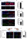
IL‐20 is upregulated in MC903‐treated mouse ear skin when compared to control, whereas IL‐20 receptors are downregulated. In contrast, IL‐20R1 transcription levels are upregulated in sensory trigeminal ganglia (TG) ipsilateral to MC903‐treated ear. Representative immunofluorescence staining of IL‐20 (A), and IL‐20R1 plus IL‐20R2 (C) in MC903‐ and vehicle‐treated mouse ear, and their fluorescence intensity analysis (B, D); n = 5 slides/mice (n = 3 mice). (E) Q‐PCR analysis of IL‐20R1 and IL‐20R2. (F, G) Immunofluorescence staining of IL‐20R1 in ipsi. and contra. TG to MC903‐treated mice ear as depicted above (F), and IL‐20R1 fluorescence intensity analysis (G); n = 5 slides/mice (n = 3 mice). For (B, D, E and G), means ± SEMs (n = 3); ns p > .05, *p < .05, and ***p < .001, a two‐tailed Student's t‐test followed by Welch's correction was used. For (A, C and F), scales = 50 µm
FIGURE 4
FIGURE 4
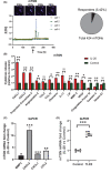
IL‐20 triggers calcium transients in mTGNs, induces release of AD‐related cytokines, and upregulates itch‐related gene transcripts. (A) Calcium transients induced by IL‐20 in mTGNs and analysis of responders; n = 424 mTGNs; representative micrographs for calcium spikes are shown on the top panel. (B) Cytokine release induced by IL‐20 versus vehicle. (C, D) Q‐PCR analysis of IL‐20‐induced gene transcripts (C) and TLR2 mRNA (D) relative to vehicle control; n = 6. For (B–D), means ± SEMs (n = 3); ns p > .05, *p < .05, **p < .01, and ***p < .001, a two‐tailed Student's t‐test followed by Welch's correction was used
FIGURE 5
FIGURE 5
In phKCs, KD of IL‐13 receptors results in increased transcription levels of IL‐20 and reduced levels of IL‐20 receptors. Injection of IL‐20 and IL‐13 in mouse cheek induces itch‐like behavior, though only when co‐injected. (A) Lentiviral shRNA‐mediated KD of IL‐13Rα1 and IL‐13Rα2 in phKCs. SNAP‐23 serves as the internal control in Western‐blotting. Percentage (%) KD relative to scrambled controls from n = 3 independent experiments. (B) Transcriptome analysis of IL‐20 and its receptors in phKCs after KD of IL‐13 receptors. KD cells and scrambled control‐treated control cells were incubated with vehicle (left panel) or IL‐13 (right panel) before RNA‐seq. (C) Scratching bouts in mouse cheek model after injection of vehicle, or IL‐13, or IL‐20, or following co‐injection of IL‐13 and IL‐20 (2‐way ANOVA followed by Bonferroni's post hoc analysis; data are presented as mean ± SEM; n = 8 mice/group.). For (A) and (B) ns p > .05, *p < .05, **p < .01, and ***p < .001, a two‐tailed Student's t‐test followed by Welch's correction was used
FIGURE 6
FIGURE 6
Schematic showing pathway of IL‐20 in AD. IL‐20 is upregulated in skin from AD, particularly in lesional AD. Skin lesion induces upregulation of IL‐20 receptors in sensory ganglia. IL‐20 triggers calcium influx in both keratinocytes and sensory neurons, promoting their AD‐related molecule release, and transcription of itch‐related genes. In sensory neurons, IL‐20 increases TLR2 transcripts, implicating a link between innate immune response and IL‐20. Intradermal co‐injection of IL‐13 and IL‐20 induces itch‐like behaviors in mice, but this is not observed in mice injected with IL‐13 or IL‐20 alone. Our findings provide novel insights into IL‐20 function in peripheral (skin‐derived) itch and clinically relevant intercellular neuron‐epidermal communication and highlight IL‐20 is functionally linked with lesional AD in human, thus forming a new basis for the development of a novel antipruritic strategy via interrupting IL‐20 signaling pathways

Similar articles

Cited by

References

    1. Langan SM, Irvine AD, Weidinger S. Atopic dermatitis. Lancet. 2020;396:345‐360. - PubMed
    1. Bieber T. Atopic dermatitis. N Engl J Med. 2008;358:1483‐1494. - PubMed
    1. Weidinger S, Beck LA, Bieber T, Kabashima K, Irvine AD. Atopic dermatitis. Nat Rev Dis Primers. 2018;4:1. - PubMed
    1. Yosipovitch G. Introduction for understanding and treating itch. Dermatol Ther. 2013;26:83. - PubMed
    1. Ikoma A. [Therapeutic agents of today and the future for atopic dermatitis]. Nihon Yakurigaku Zasshi. 2006;128:411‐415. - PubMed

Publication types