Practice Guideline

Idiopathic Pulmonary Fibrosis (an Update) and Progressive Pulmonary Fibrosis in Adults: An Official ATS/ERS/JRS/ALAT Clinical Practice Guideline

Ganesh Raghu et al. Am J Respir Crit Care Med. .

Abstract

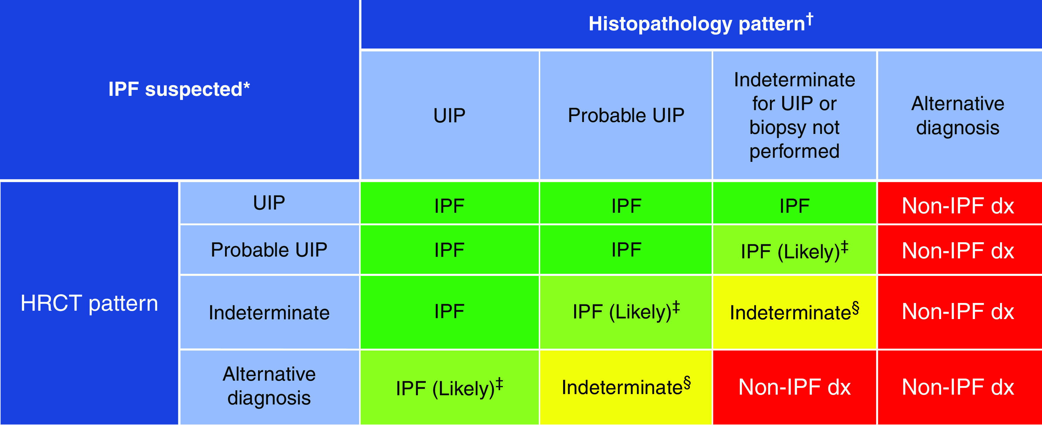
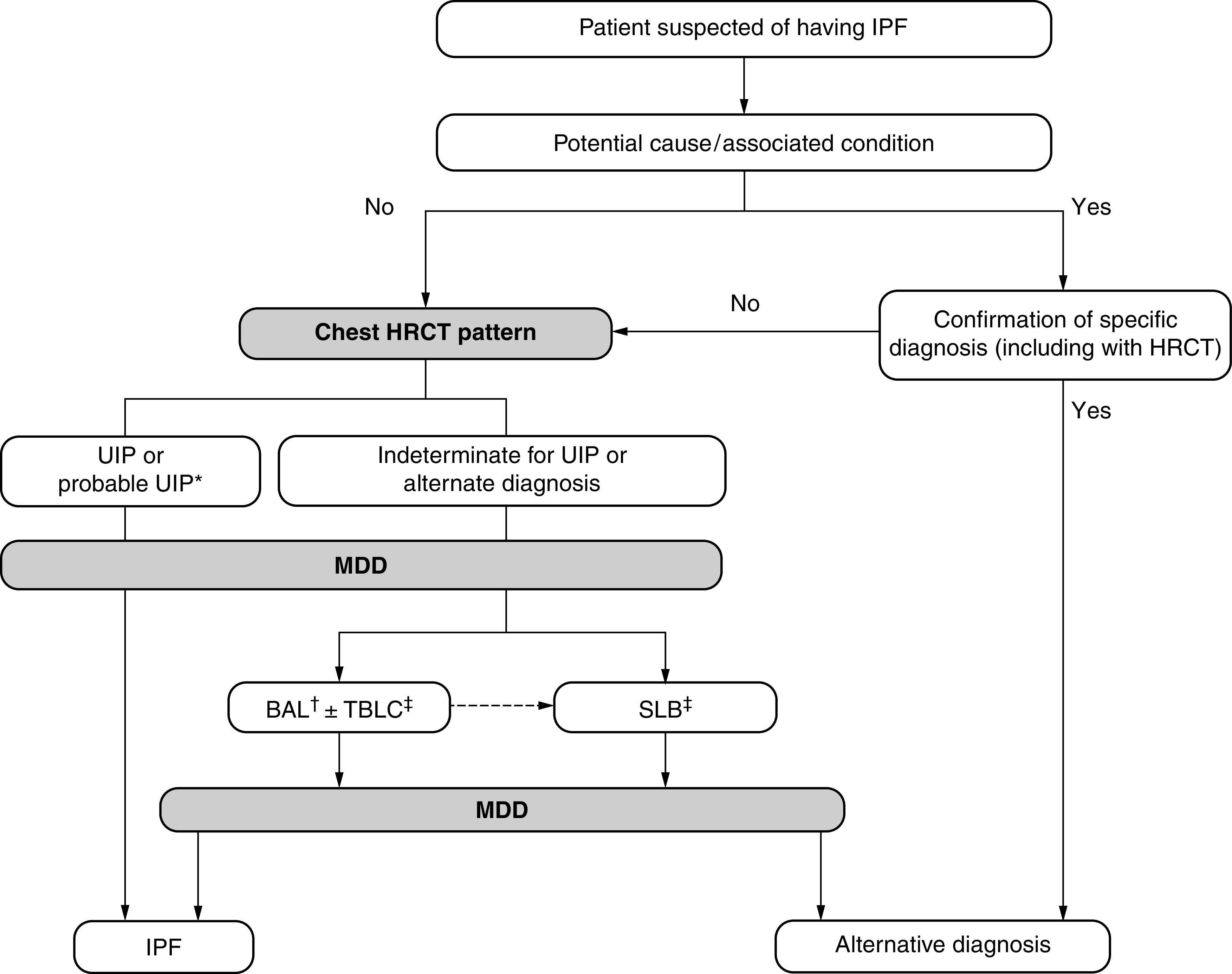
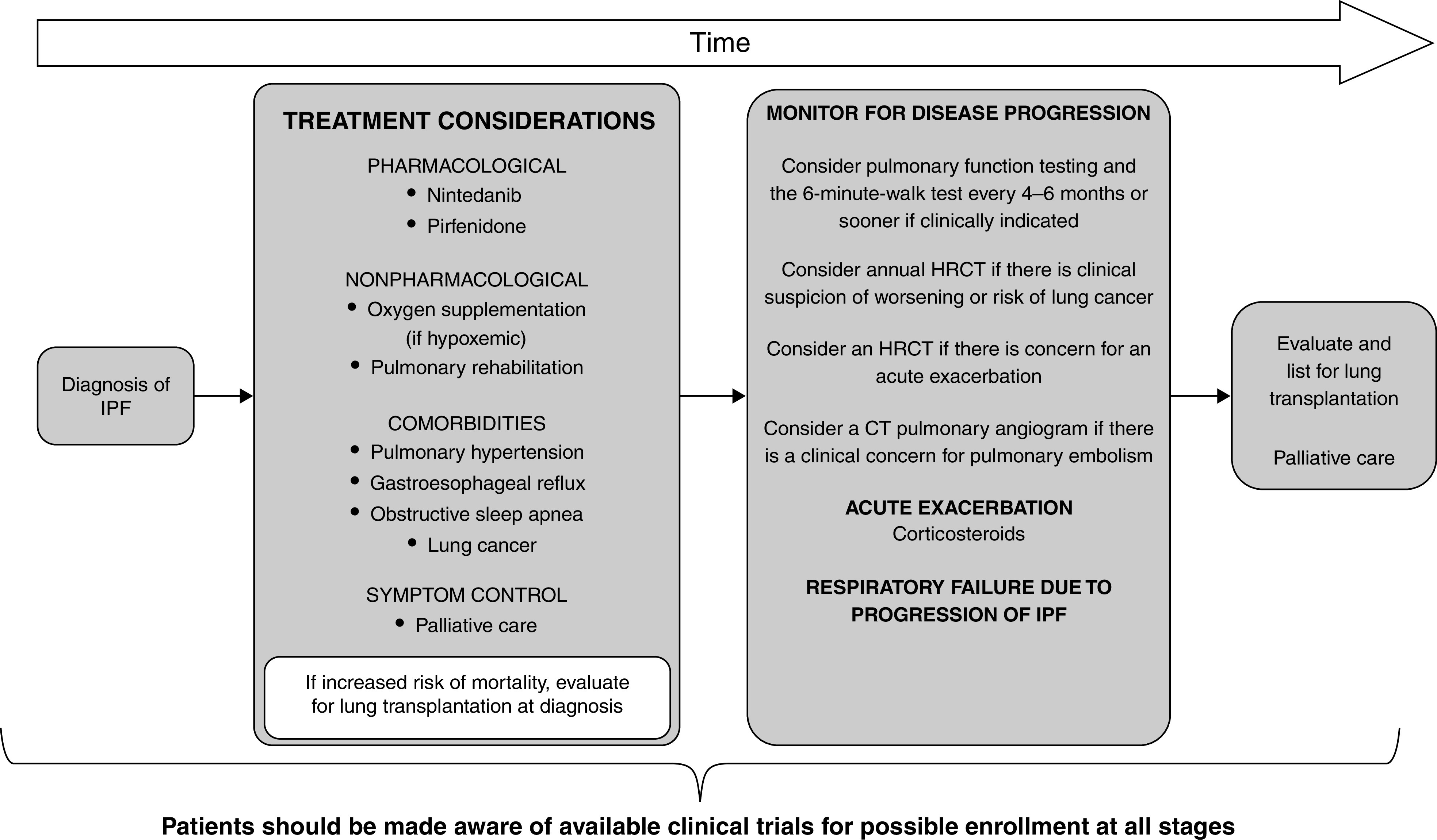
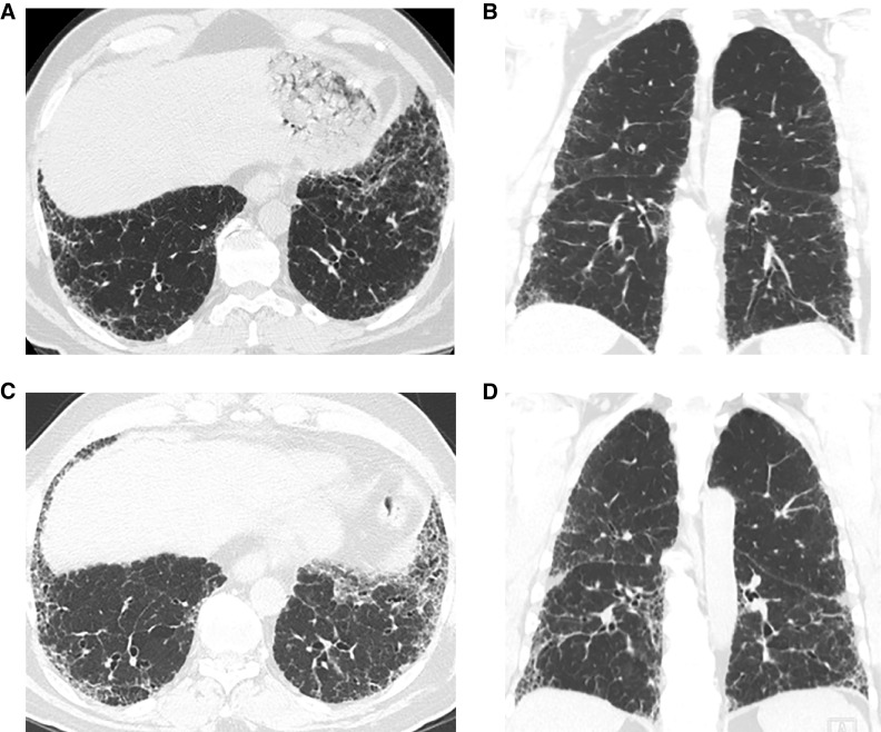
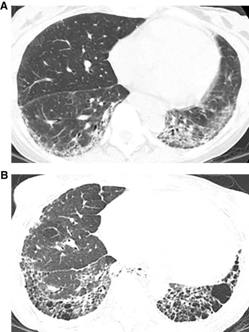
Background: This American Thoracic Society, European Respiratory Society, Japanese Respiratory Society, and Asociación Latinoamericana de Tórax guideline updates prior idiopathic pulmonary fibrosis (IPF) guidelines and addresses the progression of pulmonary fibrosis in patients with interstitial lung diseases (ILDs) other than IPF. Methods: A committee was composed of multidisciplinary experts in ILD, methodologists, and patient representatives. 1) Update of IPF: Radiological and histopathological criteria for IPF were updated by consensus. Questions about transbronchial lung cryobiopsy, genomic classifier testing, antacid medication, and antireflux surgery were informed by systematic reviews and answered with evidence-based recommendations using the Grading of Recommendations, Assessment, Development and Evaluation (GRADE) approach. 2) Progressive pulmonary fibrosis (PPF): PPF was defined, and then radiological and physiological criteria for PPF were determined by consensus. Questions about pirfenidone and nintedanib were informed by systematic reviews and answered with evidence-based recommendations using the GRADE approach. Results:1) Update of IPF: A conditional recommendation was made to regard transbronchial lung cryobiopsy as an acceptable alternative to surgical lung biopsy in centers with appropriate expertise. No recommendation was made for or against genomic classifier testing. Conditional recommendations were made against antacid medication and antireflux surgery for the treatment of IPF. 2) PPF: PPF was defined as at least two of three criteria (worsening symptoms, radiological progression, and physiological progression) occurring within the past year with no alternative explanation in a patient with an ILD other than IPF. A conditional recommendation was made for nintedanib, and additional research into pirfenidone was recommended. Conclusions: The conditional recommendations in this guideline are intended to provide the basis for rational, informed decisions by clinicians.

Keywords: histopathology; idiopathic pulmonary fibrosis; progressive pulmonary fibrosis; radiology.


Figures

Figure 1.
Figure 1.
Traction bronchiectasis/bronchiolectasis. Axial, sagittal, and coronal computed tomography images show subpleural-predominant, lower lung–predominant reticular abnormality with traction bronchiectasis (arrows). Traction bronchiectasis/bronchiolectasis represents irregular bronchial and/or bronchiolar dilatation caused by surrounding retractile fibrosis; distorted airways are thus identified in a background of reticulation and/or ground-glass attenuation. On contiguous high-resolution computed tomography sections, the dilated bronchi or bronchioles can be tracked back toward more central bronchi. The pattern in this patient represents the probable usual interstitial pneumonia pattern.
Figure 2.
Figure 2.
Honeycombing. Axial, sagittal, and coronal computed tomography images show subpleural-predominant, lower lung–predominant reticular abnormality with honeycombing (arrowheads). Honeycombing is defined by clustered, thick-walled, cystic spaces of similar diameters, measuring between 3 and 10 mm but up to 2.5 cm in size. The size and number of cysts often increase as the disease progresses. Often described in the literature as being layered, a single layer of subpleural cysts is also a manifestation of honeycombing. Honeycombing is an essential computed tomography criterion for typical (“definite”) usual interstitial pneumonia–idiopathic pulmonary fibrosis pattern when seen with a basal and peripheral predominance. In this pattern, honeycombing is usually associated with traction bronchiolectasis and a varying degree of ground-glass attenuation.
Figure 3.
Figure 3.
Paraseptal emphysema. Axial and coronal computed tomography images show relatively large subpleural cysts of paraseptal emphysema (arrows), mainly in the upper lobes. Centrilobular emphysema is also present. The subpleural cysts of paraseptal emphysema usually occur in a single layer and are larger than honeycomb cysts (typically >1 cm); they are not associated with other features of fibrosis such as reticular abnormality or traction bronchiectasis.
Figure 4.
Figure 4.
Airspace enlargement with fibrosis (AEF), also called smoking-related interstitial fibrosis, in a cigarette smoker. Axial and sagittal images show clustered asymmetric cysts that are larger and more irregular than typical honeycomb cysts, without traction bronchiectasis or other signs of fibrosis (arrows). Emphysema is also present. AEF is not regarded as a distinct form of idiopathic interstitial pneumonia but results from the presence of a greater amount of fibrosis than usually described in the classic definition of emphysema.
Figure 5.
Figure 5.
Spectrum of computed tomography (CT) appearances in usual interstitial pneumonia (UIP) pattern due to hypersensitivity pneumonitis (HP). (A) Coronal CT section obtained at deep inspiration showing honeycombing with traction bronchiolectasis in the peripheral part of the right lower lobe (short arrows) and numerous hyperlucent lobules in the left lower lobe (long arrows). (B) Lobular air trapping was confirmed on expiratory CT. HP-UIP should be considered when fibrosis and honeycomb cysts predominate in the upper or mid lungs, when mosaic attenuation or three-density sign is present, or when the fibrosis appears diffuse in the axial plane.
Figure 6.
Figure 6.
Usual interstitial pneumonia (UIP) pattern due to connective tissue disease (CTD-UIP) in a patient with dermatomyositis/scleroderma overlap. Axial and coronal images show sharply demarcated fibrosis with exuberant honeycombing in the lower lobes and in the anterior upper lobes. CTD-UIP should be considered when honeycomb cysts are extensive, occupying >70% of the fibrotic portions of the lung (exuberant honeycombing sign); when fibrotic abnormality is sharply demarcated on coronal images from the relatively normal upper lungs (straight-edge sign); and when there is relative increase in fibrosis in the anterior upper lobes (anterior upper lobe sign).
Figure 7.
Figure 7.
Combined pleuroparenchymal fibroelastosis and usual interstitial pneumonia patterns. Coronal computed tomography image shows dense subpleural fibrosis at the lung apices with traction bronchiectasis and upper lobe volume loss. There is subpleural reticular abnormality and honeycombing in both lower lobes.
Figure 8.
Figure 8.
Three of the four high-resolution computed tomography patterns of usual interstitial pneumonia (UIP): (A) UIP pattern (associated with some paraseptal and centrilobular emphysema in the upper lobes), (B) probable UIP pattern with fibrotic features in the lung periphery (and some centrilobular emphysema in the upper lobes), and (C) indeterminate for UIP pattern (peribronchovascular and subpleural ground-glass opacities, intermingled with fine reticulation but no honeycombing or traction bronchiectasis). The fourth category, alternative diagnosis, is widely variable, depending on the specific alternative diagnosis, and is not shown.
Figure 9.
Figure 9.
Idiopathic pulmonary fibrosis (IPF) diagnosis on the basis of high-resolution computed tomography (HRCT) and biopsy patterns, developed using consensus by discussion. *“Clinically suspected of having IPF” is defined as unexplained patterns of bilateral pulmonary fibrosis on chest radiography or chest computed tomography, bibasilar inspiratory crackles, and age > 60 years. Middle-aged adults (>40 and <60 yr old) can rarely present with otherwise similar clinical features, especially in patients with features suggesting familial pulmonary fibrosis. Diagnostic confidence may need to be downgraded if histopathological assessment is based on transbronchial lung cryobiopsy given the smaller biopsy size and greater potential for sampling error compared with surgical lung biopsy. IPF is the likely diagnosis when any of the following features are present: 1) moderate to severe traction bronchiectasis and/or bronchiolectasis (defined as mild traction bronchiectasis and/or bronchiolectasis in four or more lobes, including the lingula as a lobe, or moderate to severe traction bronchiectasis in two or more lobes) in a man >50 years old or in a woman >60 yr old, 2) extensive (>30%) reticulation on HRCT and age > 70 yr, 3) increased neutrophils and/or absence of lymphocytosis in BAL fluid, and 4) multidisciplinary discussion produces a confident diagnosis of IPF. §Indeterminate for IPF 1) without an adequate biopsy remains indeterminate and 2) with an adequate biopsy may be reclassified to a more specific diagnosis after multidisciplinary discussion and/or additional consultation. Adapted from Reference . dx = diagnosis; UIP = usual interstitial pneumonia.
Figure 10.
Figure 10.
Diagnostic algorithm for idiopathic pulmonary fibrosis (IPF), developed using consensus by discussion. *Patients with a radiological pattern of probable usual interstitial pneumonia (UIP) can receive a diagnosis of IPF after multidisciplinary discussion (MDD) without confirmation by lung biopsy in the appropriate clinical setting (e.g., 60 yr old, male, smoker). BAL may be appropriate in some patients with a probable UIP pattern. BAL may be performed before MDD in some patients evaluated in experienced centers. Transbronchial lung cryobiopsy (TBLC) may be preferred to surgical lung biopsy (SLB) in centers with appropriate expertise and/or in some patient populations, as described in the text. A subsequent SLB may be justified in some patients with nondiagnostic findings on TBLC. Adapted from Reference . HRCT = high-resolution computed tomography.
Figure 11.
Figure 11.
Schematic pathway for clinical management of patients with idiopathic pulmonary fibrosis (IPF), developed using consensus by discussion. Treatment considerations should include both pharmacological (nintedanib and pirfenidone) and nonpharmacological (oxygen supplementation and/or pulmonary rehabilitation) therapies. Patients should be evaluated and treated for existing comorbidities, including pulmonary hypertension, gastroesophageal reflux, obstructive sleep apnea, and lung cancer. Patients may benefit from involvement of palliative care to help with symptom management (cough, dyspnea, and/or anxiety). Patient values and preferences should be explored. Patients at increased risk of mortality should be referred for lung transplantation at diagnosis. Patients should be evaluated every 3–6 months or more often for disease progression. Acute exacerbations may be treated with corticosteroids. Mechanical ventilation is not recommended for the majority of patients with respiratory failure. Adapted from Reference . CT = computed tomography; HRCT = high-resolution computed tomography.
Figure 12.
Figure 12.
Interstitial lung diseases (ILDs) manifesting progressive pulmonary fibrosis (PPF), developed using consensus by discussion. The shaded area represents the estimated proportion of patients with various types of ILD who manifest PPF. Note that idiopathic pulmonary fibrosis (IPF) is not included in the figure, because it is excluded from the definition of PPF. While virtually all patients with IPF will manifest disease progression similar to PPF, the proportion of patients with ILDs other than IPF who manifest PPF is based on the consensus of opinions and the perception of the international committee. There are no data to provide the exact or estimated proportion of patients manifesting PPF in ILDs, other than IPF. *The committee acknowledges that eosinophilic pneumonia of unknown cause was not included in the IIP classification. Myositis includes PM/DM/antisynthetase syndrome, which may be amyopathic. Although respiratory bronchiolitis interstitial lung disease (RBILD) is acknowledged to be a consequence of exposure to cigarette smoke in virtually all patients with RBILD, RBILD and desquamative interstitial pneumonia (DIP) often coexist. Although DIP is also related to exposure to cigarette smoke in a majority of patients, DIP is also seen in some patients with connective tissue disease, without exposure to cigarette smoke, and without a known cause. Antifibrotic treatment is indicated for patients diagnosed with IPF (3). Antifibrotic treatment of the other types of ILD upon manifesting PPF is as suggested/recommended in this guideline. AFOP = acute fibrinous and organizing pneumonia; AIP = acute interstitial pneumonia; COP = cryptogenic organizing pneumonia; DM = dermatomyositis; HP = hypersensitivity pneumonitis; iDIP = idiopathic DIP; IIP = idiopathic interstitial pneumonia; iLIP = idiopathic lymphoid interstitial pneumonia; iNSIP = idiopathic nonspecific interstitial pneumonia; iPPFE = idiopathic pleuroparenchymal fibroelastosis; LAM = lymphangioleiomyomatosis; LCH = Langerhans cell histiocytosis; MCTD = mixed connective tissue disease; PAP = pulmonary alveolar proteinosis; PM = polymyositis; RA = rheumatoid arthritis; SLE = systemic lupus erythematosus; SSc = systemic sclerosis.
Figure 13.
Figure 13.
Progressive pulmonary fibrosis in a patient with idiopathic pulmonary fibrosis (probable usual interstitial pneumonia pattern). (A and B) Baseline axial and coronal images show moderately extensive reticular abnormality with traction bronchiectasis, with predominance in the subpleural lower lung. (C and D) Matched images 4 years later show substantial increase in extent of abnormality and increased traction bronchiectasis.
Figure 14.
Figure 14.
Progressive pulmonary fibrosis due to fibrotic nonspecific interstitial pneumonia (NSIP). (A) Computed tomography in a 45-year-old woman with scleroderma shows lower lung–predominant reticular and ground-glass abnormality with subpleural sparing, typical for NSIP. (B) Nine years later, the fibrosis has progressed with increased extent of reticular abnormality, increased traction bronchiectasis, and evolution of reticular abnormality to honeycombing. Small bilateral pleural effusions are present.

Comment in

References

    1. Raghu G, Collard HR, Egan JJ, Martinez FJ, Behr J, Brown KK, et al. ATS/ERS/JRS/ALAT Committee on Idiopathic Pulmonary Fibrosis An official ATS/ERS/JRS/ALAT statement: idiopathic pulmonary fibrosis: evidence-based guidelines for diagnosis and management. Am J Respir Crit Care Med . 2011;183:788–824. - PMC - PubMed
    1. Raghu G, Remy-Jardin M, Myers JL, Richeldi L, Ryerson CJ, Lederer DJ, et al. American Thoracic Society, European Respiratory Society, Japanese Respiratory Society, and Latin American Thoracic Society Diagnosis of idiopathic pulmonary fibrosis: an official ATS/ERS/JRS/ALAT clinical practice guideline. Am J Respir Crit Care Med . 2018;198:e44–e68. - PubMed
    1. Raghu G, Rochwerg B, Zhang Y, Garcia CAC, Azuma A, Behr J, et al. American Thoracic Society European Respiratory society; Japanese Respiratory Society; Latin American Thoracic Association. An official ATS/ERS/JRS/ALAT clinical practice guideline: treatment of idiopathic pulmonary fibrosis. An update of the 2011 clinical practice guideline. Am J Respir Crit Care Med . 2015;192:e3–e19. - PubMed
    1. Flaherty KR, Wells AU, Cottin V, Devaraj A, Walsh SLF, Inoue Y, et al. INBUILD Trial Investigators Nintedanib in progressive fibrosing interstitial lung diseases. N Engl J Med . 2019;381:1718–1727. - PubMed
    1. Wells AU, Brown KK, Flaherty KR, Kolb M, Thannickal VJ, IPF Consensus Working Group What’s in a name? That which we call IPF, by any other name would act the same. Eur Respir J . 2018;51:1800692. - PubMed

Publication types