

Fabrication of Customizable Intraplaque Hemorrhage Phantoms for Magnetic Resonance Imaging
- PMID: 35486294
- PMCID: PMC9581813
- DOI: 10.1007/s11307-022-01722-4
Fabrication of Customizable Intraplaque Hemorrhage Phantoms for Magnetic Resonance Imaging
Abstract
Purpose: Magnetic resonance (MR) imaging detection of methemoglobin, a molecular marker of intraplaque hemorrhage (IPH), in atherosclerotic plaque is a promising method of assessing stroke risk. However, the multicenter imaging studies required to further validate this technique necessitate the development of IPH phantoms to standardize images acquired across different scanners. This study developed a set of phantoms that modeled methemoglobin-laden IPH for use in MR image standardization.
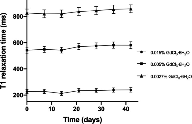
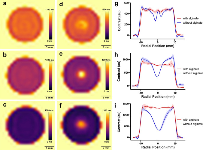
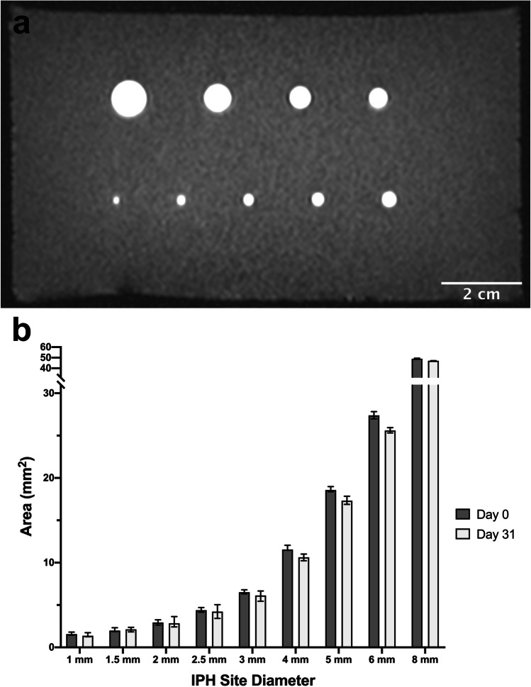
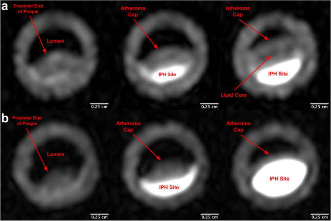
Procedures: A time-stable material mimicking the MR properties of methemoglobin in IPH was created by doping agarose hydrogel with gadolinium and sodium alginate. This material was used to create a phantom that consisted of 9 cylindrical IPH sites (with sizes from 1 to 8 mm). Anatomical replicas of IPH-positive atherosclerosis were also created using 3D printed molds. These plaque replicas also modeled other common plaque components including a lipid core and atheroma cap. T1 mapping and a magnetization-prepared rapid acquisition gradient echo (MPRAGE) carotid imaging protocol were used to assess phantom realism and long-term stability.
Results: Cylindrical phantom IPH sites possessed a T1 time of 335 ± 51 ms and exhibited little change in size or MPRAGE signal intensity over 31 days; the mean (SD) magnitude of changes in size and signal were 6.4 % (2.7 %) and 7.3 % (6.7 %), respectively. IPH sites incorporated into complex anatomical plaque phantoms exhibited contrast comparable to clinical images.
Conclusions: The cylindrical IPH phantom accurately modeled the short T1 time characteristic of methemoglobin-laden IPH, with the IPH sites exhibiting little variation in imaging properties over 31 days. Furthermore, MPRAGE images of the anatomical atherosclerosis replicas closely matched those of clinical plaques. In combination, these phantoms will allow for IPH imaging protocol standardization and thus facilitate future multicenter IPH imaging.
Keywords: Carotid plaque; Intraplaque hemorrhage; MR imaging; MRI phantoms; Magnetic resonance angiography; Methemoglobin.
© 2022. The Author(s).
Conflict of interest statement
NM is a senior editor (Nanomaterials and Delivery Platforms) of MIB. MAB, ARM, and JMD declare that they have no conflict of interest.
Figures
References
Publication types
MeSH terms
Substances
Grants and funding
LinkOut - more resources
Full Text Sources
Medical
Miscellaneous
