

Association of Elevated Maternal Psychological Distress, Altered Fetal Brain, and Offspring Cognitive and Social-Emotional Outcomes at 18 Months
- PMID: 35486403
- PMCID: PMC9055453
- DOI: 10.1001/jamanetworkopen.2022.9244
Association of Elevated Maternal Psychological Distress, Altered Fetal Brain, and Offspring Cognitive and Social-Emotional Outcomes at 18 Months
Abstract
Importance: Prenatal maternal psychological distress is associated with disturbances in fetal brain development. However, the association between altered fetal brain development, prenatal maternal psychological distress, and long-term neurodevelopmental outcomes is unknown.
Objective: To determine the association of fetal brain development using 3-dimensional magnetic resonance imaging (MRI) volumes, cortical folding, and metabolites in the setting of maternal psychological distress with infant 18-month neurodevelopment.
Design, setting, and participants: Healthy mother-infant dyads were prospectively recruited into a longitudinal observational cohort study from January 2016 to October 2020 at Children's National Hospital in Washington, DC. Data analysis was performed from January 2016 to July 2021.
Exposures: Prenatal maternal stress, anxiety, and depression.
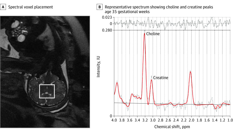
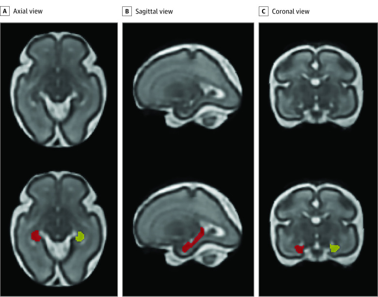
Main outcomes and measures: Prenatal maternal stress, anxiety, and depression were measured using validated self-report questionnaires. Fetal brain volumes and cortical folding were measured from 3-dimensional, reconstructed T2-weighted MRI scans. Fetal brain creatine and choline were quantified using proton magnetic resonance spectroscopy. Infant neurodevelopment at 18 months was measured using Bayley Scales of Infant and Toddler Development III and Infant-Toddler Social and Emotional Assessment. The parenting stress in the parent-child dyad was measured using the Parenting Stress Index-Short Form at 18-month testing.
Results: The cohort consisted of 97 mother-infant dyads (mean [SD] maternal age, 34.79 [5.64] years) who underwent 184 fetal MRI visits (87 participants with 2 fetal studies each) with maternal psychological distress measures between 24 and 40 gestational weeks and completed follow-up infant neurodevelopmental testing. Prenatal maternal stress was negatively associated with infant cognitive performance (β = -0.51; 95% CI, -0.92 to -0.09; P = .01), and this association was mediated by fetal left hippocampal volume. In addition, prenatal maternal anxiety, stress, and depression were positively associated with all parenting stress measures at 18-month testing. Finally, fetal cortical local gyrification index and sulcal depth were negatively associated with infant social-emotional performance (local gyrification index: β = -54.62; 95% CI, -85.05 to -24.19; P < .001; sulcal depth: β = -14.22; 95% CI, -23.59 to -4.85; P = .002) and competence scores (local gyrification index: β = -24.01; 95% CI, -40.34 to -7.69; P = .003; sulcal depth: β = -7.53; 95% CI, -11.73 to -3.32; P < .001).
Conclusions and relevance: In this cohort study of 97 mother-infant dyads, fetal cortical local gyrification index and sulcal depth were associated with infant 18-month social-emotional and competence outcomes, and fetal left hippocampal volume mediated the association between prenatal maternal stress and infant cognitive outcome. These findings suggest that altered prenatal brain development in the setting of elevated maternal distress has adverse infant sociocognitive outcomes, and identifying early biomarkers associated with long-term neurodevelopment may assist in early targeted interventions.
Conflict of interest statement
Figures
Comment in
References
-
- McKee KA. The Effects of Prenatal Maternal Stress and Early Life Maternal Stress on Adolescent Hippocampal Morphology: Project Ice Storm. McGill University; 2018.
Publication types
MeSH terms
Grants and funding
LinkOut - more resources
Full Text Sources
Medical
