Review
. 2022 Nov 16;33(11):2018-2034.
doi: 10.1021/acs.bioconjchem.2c00030. Epub 2022 Apr 29.

Engineering Self-Assembling Protein Nanoparticles for Therapeutic Delivery

Affiliations
Review

Engineering Self-Assembling Protein Nanoparticles for Therapeutic Delivery

Audrey Olshefsky et al. Bioconjug Chem. .

Abstract

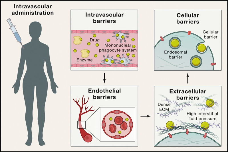
Despite remarkable advances over the past several decades, many therapeutic nanomaterials fail to overcome major in vivo delivery barriers. Controlling immunogenicity, optimizing biodistribution, and engineering environmental responsiveness are key outstanding delivery problems for most nanotherapeutics. However, notable exceptions exist including some lipid and polymeric nanoparticles, some virus-based nanoparticles, and nanoparticle vaccines where immunogenicity is desired. Self-assembling protein nanoparticles offer a powerful blend of modularity and precise designability to the field, and have the potential to solve many of the major barriers to delivery. In this review, we provide a brief overview of key designable features of protein nanoparticles and their implications for therapeutic delivery applications. We anticipate that protein nanoparticles will rapidly grow in their prevalence and impact as clinically relevant delivery platforms.


Conflict of interest statement

The authors declare the following competing financial interest(s): N.P.K. is a co-founder, shareholder, paid consultant, and chair of the scientific advisory board of Icosavax, Inc. The King lab has received an unrelated sponsored research agreement from Pfizer.

Figures

Figure 1
Figure 1
Barriers to in vivo nanoparticle delivery following intravascular administration. For nanoparticles to successfully reach their targets, they must pass several barriers. This example uses intravascular administration as a case study; other routes of administration are reviewed elsewhere. Unless immune responses are specifically desired, the nanoparticles must evade recognition by the innate and adaptive immune system during circulation (e.g., the complement system, the mononuclear phagocytic system (MPS), and B cells). Once near the target organ, they must travel past endothelial cells and tight junctions, which line blood vessels. Then the nanoparticles must travel through extracellular matrices within the target tissue, and once they reach the target cells, must access their intended subcellular compartment for therapeutic payload delivery. Reprinted with permission.(8)
Figure 2
Figure 2
Designable features of nanoparticle scaffolds. Two main considerations when designing protein nanoparticle delivery systems are scaffold source and scaffold modifications. (A–C) Protein nanoparticle geometries commonly used for delivery applications have icosahedral, octahedral, tetrahedral, or dihedral symmetry.,, Notably, the octahedral nanoparticle in (B) is composed of eight identical trimer subunits, which are colored differently to help visually distinguish individual subunits in the context of the global structure. (D–F) Additional functional elements are designed into the nanoparticle through subunit interface, interior, and exterior modifications. (D) Interfaces between the trimer subunits (slate) and pentamer subunits (gray) were computationally designed. (E) Interior residues were mutated to hold a net positive charge, leading to electrostatic association of mRNA. (F) Additional protein domains can be displayed on the surface of existing nanoparticles. A, D: Reprinted with permission.(18) B: Reprinted with permission.(19) C: Reprinted with permission.(71)
Figure 3
Figure 3
Qualitative impacts of nanoparticle size, shape, and surface charge on biodistribution. The general effects of surface physicochemical properties on biodistribution were comprehensively reviewed by Blanco, Shen, & Ferrari and qualitatively graphed as relative accumulation in major mouse organs. Data were included from gold nanoparticles, liposomes, polymer micelles, zwitterionic nanoparticles, hydrogel nanoparticles, and more. (A) Nanoparticles greater than about 150 nm in diameter show increased accumulation in the lungs, liver, and spleen, while nanoparticles less than 5 nm in diameter show rapid renal clearance. (B) Spherical nanoparticles tend to have the least uptake by major clearance organs compared to cylindrical and discoidal nanoparticles. (C) Nanoparticles with positively charged surfaces show much higher nonspecific uptake than nanoparticles with negatively charged or neutral surfaces. Reprinted with permission.(10)
Figure 4
Figure 4
The DS-Cav1-I53–50 nanoparticle immunogen. (A) A vaccine against respiratory syncytial virus (RSV) was engineered by outwardly displaying a trimeric RSV antigen (DS-Cav1) on the trimeric subunit of a computationally designed nanoparticle (I53–50). (B) Negative stain electron microscopy of I53–50 and DS-Cav1-I53–50 nanoparticles. Left: representative micrographs. Right: averages of nanoparticle micrographs. (C–D) Antibody binding titers ofDS-Cav-1-specific antibodies (C) or serum neutralizing antibodies (D) from mice immunized with bare nanoparticle (I53–50), free immunogen (DS-Cav1), or nanoparticle immunogens (DS-Cav1-I53–50) at valencies of 33%, 67%, or 100%. Each point represents and individual animal, geometric means are represented by horizontal lines and indicated at the bottom of the plots, and statistical significance is indicated: *p < 0.05; **p < 0.01; ***p < 0.001; ****p < 0.0001. See original publication for more details. Reprinted with permission.(98)
Figure 5
Figure 5
Coadministration of liposomal doxorubicin (Doxil) with ferritin codisplaying clot-targeting and fibrinolytic domains. (A) The ferritin monomer was engineered to display a clot-targeting peptide (CLT) and fibrinolytic domain (μP), resulting in surface display on ferritin nanoparticles. (B) Fibrinolytic nanocages (FNC) coadministered with Doxil show increased tumor cell accumulation and decreased fibrin signal compared to saline or Doxil controls. (C–D) When delivered to tumor-bearing mice at the indicated dosing schedule (C), Doxil-FNC coadministration show increased tumor growth inhibition (purple) compared to mice treated with Doxil alone (red) and other controls (D). Reprinted with permission.(33)
Figure 6
Figure 6
Computationally designed antibody cages (AbCs) activate apoptosis and angiogenic signaling pathways. (A and B) Caspase-3/7 is activated by AbCs formed with α-DR5 antibody (A), but not the free antibody, in RCC4 renal cancer cells (B). (C and D) α-DR5 AbCs (C), but not Fc AbC controls (D), reduce cell viability 4 days after treatment. (E) α-DR5 AbCs reduce viability 6 days after treatment. (F and G) o42.1 α-DR5 AbCs enhance PARP cleavage, a marker of apoptotic signaling; (G) is a quantification of (F) relative to PBS control. (H) The F-domain from angiopoietin-1 was fused to Fc (A1F-Fc) and assembled into octahedral (o42.1) and icosahedral (i52.3) AbCs. (I) Representative Western blots show that A1F-Fc AbCs, but not controls, increase pAKT and pERK1/2 signals. (J) Quantification of (I): pAKT quantification is normalized to o42.1 A1F-Fc signaling (no pAKT signal in the PBS control); pERK1/2 is normalized to PBS. (K) A1F-Fc AbCs increase vascular stability after 72 h. (Left) Quantification of vascular stability compared with PBS. (Right) Representative images; scale bars, 100 μm. All error bars represent means ± SEM; means were compared using analysis of variance and Dunnett posthoc tests (tables S8 and S9). *P ≤ 0.05; **P ≤ 0.01; ***P ≤ 0.001; ****P ≤ 0.0001. Reprinted with permission.(21)
Figure 7
Figure 7
Computationally designed nanoparticle O3–33 was redesigned for siRNA delivery. The computationally designed octahedral protein nanoparticle O3–33 (porous protein cage) containing 144 hexahistidine tags on its surface was redesigned to have a positively charged interior to electrostatically associate with nucleic acid in vitro (positively charged capsule loaded with nucleic acid)., The authors reported nuclease protection, uptake of the nanoparticles by HeLa cells, and subsequent cargo release leading to knockdown of intracellular GFP mRNA. The authors attributed successful endosomal escape to the hexahistidine tags. Reprinted with permission.(166)
Figure 8
Figure 8
Example of ferritin engineered as a theranostic by displaying a fluorescent protein and encapsulating a cytotoxic peptide. (A) Schematic for genetically fusing a cytotoxic peptide (chamber 1) and a fluorescent protein (chamber 2) to ferritin. (B) Electron micrographs of ferritin, ferritin displaying GFP, and ferritin fused to both GFP and a cytotoxic peptide (“KLAK” repeats). (C) Tumor cryosections from mice treated with a saline control, cytotoxic peptide (KLAK), ferritin-GFP and cytotoxic peptide (sFt-GFP + KLAK), or ferritin fused to both cytotoxic peptide and GFP (KLAK-sFt-GFP). Scale bars are 40 μm. Reprinted with permission.(83)

References

    1. Mitchell M. J.; Billingsley M. M.; Haley R. M.; Wechsler M. E.; Peppas N. A.; Langer R. Engineering Precision Nanoparticles for Drug Delivery. Nat. Rev. Drug Discov 2021, 20 (2), 101–124. 10.1038/s41573-020-0090-8. - DOI - PMC - PubMed
    1. Davis M. E.; Chen Z. G.; Shin D. M. Nanoparticle Therapeutics: An Emerging Treatment Modality for Cancer. Nat. Rev. Drug Discov 2008, 7 (9), 771–782. 10.1038/nrd2614. - DOI - PubMed
    1. Waterhouse D. N.; Tardi P. G.; Mayer L. D.; Bally M. B. A Comparison of Liposomal Formulations of Doxorubicin with Drug Administered in Free Form: Changing Toxicity Profiles. Drug Saf 2001, 24 (12), 903–920. 10.2165/00002018-200124120-00004. - DOI - PubMed
    1. Barenholz Y. Doxil®–the First FDA-Approved Nano-Drug: Lessons Learned. J. Controlled Release 2012, 160 (2), 117–134. 10.1016/j.jconrel.2012.03.020. - DOI - PubMed
    1. Office of the Commissioner. FDA approves first COVID-19 vaccine https://www.fda.gov/news-events/press-announcements/fda-approves-first-c... (accessed 2021 −11 −02).

Publication types