. 2022 Apr 29;13(1):2349.
doi: 10.1038/s41467-022-29829-w.

Molecular basis of antibiotic self-resistance in a bee larvae pathogen

Affiliations

Molecular basis of antibiotic self-resistance in a bee larvae pathogen

Tam Dang et al. Nat Commun. .

Abstract

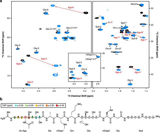
Paenibacillus larvae, the causative agent of the devastating honey-bee disease American Foulbrood, produces the cationic polyketide-peptide hybrid paenilamicin that displays antibacterial and antifungal activity. Its biosynthetic gene cluster contains a gene coding for the N-acetyltransferase PamZ. We show that PamZ acts as self-resistance factor in Paenibacillus larvae by deactivation of paenilamicin. Using tandem mass spectrometry, nuclear magnetic resonance spectroscopy and synthetic diastereomers, we identified the N-terminal amino group of the agmatinamic acid as the N-acetylation site. These findings highlight the pharmacophore region of paenilamicin, which we very recently identified as a ribosome inhibitor. Here, we further determined the crystal structure of PamZ:acetyl-CoA complex at 1.34 Å resolution. An unusual tandem-domain architecture provides a well-defined substrate-binding groove decorated with negatively-charged residues to specifically attract the cationic paenilamicin. Our results will help to understand the mode of action of paenilamicin and its role in pathogenicity of Paenibacillus larvae to fight American Foulbrood.


Conflict of interest statement

The authors declare no competing interests.

Figures

Fig. 1
Fig. 1. Biosynthetic gene cluster and structure of paenilamicin variants.
The paenilamicin (pam) gene cluster contains core biosynthetic (red), auxiliary biosynthetic (orange), resistance (pamZ and pamJ; blue) and other (gray) genes and expresses the nonribosomal peptide synthetase-polyketide synthase (NRPS-PKS) hybrid biosynthetic machinery for the production of paenilamicin A1 (Glm, Lys), A2 (Glm, Orn), B1 (Aga, Lys), and B2 (Aga, Orn). Glm - galantinamic acid, Aga - agmatinamic acid, Lys lysine, Orn - ornithine, Ala - alanine, mDap - N-methyldiaminopropionic acid, Gla - galantinic acid, Gly - glycine, Spd - 4,3-spermidine.
Fig. 2
Fig. 2. Deactivation of paenilamicins through PamZ-mediated N-acetylation tested by agar diffusion assay against B. megaterium as the indicator strain.
Paenilamicin variants (PamA1, A2, B1, B2) isolated from P. larvae and synthetic paenilamicin B2 (PamB2_3) were incubated in vitro with both acetyl-CoA and PamZ (1), acetyl-CoA only (2), or PamZ only (3). Samples 2 and 3 are negative controls and indicate the lack of bacterial growth.
Fig. 3
Fig. 3. Identification of the N-acetylation site through 2D NMR spectroscopy.
a Overlay of relevant 1H-13C HSQC sections of paenilamicin B2 (black) and N-acetylpaenilamicin B2 (blue). Strongly perturbed cross-peaks are highlighted with red labels. Known impurities are labeled with one, two and three asterisks arising from glycerol, acetic acid and residual purification traces of paenilamicin B1, respectively. The numbers (in backbone) and Greek letters (in amino acid residues) refer to atom positions in the corresponding building blocks as labeled in b. b Significant chemical shift perturbations (CSPs) and corresponding positions are indicated as circles in the chemical structure of N-acetylpaenilamicin B2 (see legend for color code). Aga - agmatinamic acid, Ala - alanine, mDap - N-methyldiaminopropionic acid, Orn - ornithine, Gla - galantinic acid, Gly - glycine, Spd - 4,3-spermidine, Ac - acetyl group.
Fig. 4
Fig. 4. Substrate specificity and stereoselectivity of PamZ.
The natural product (PamB2), synthetic paenilamicin B2 (PamB2_3) and synthetic diastereomers of paenilamicin B2 (PamB2_2, PamB2_1) were incubated with PamZ and acetyl-CoA in vitro and tested in an agar diffusion assay against Bacillus megaterium (insets). From the chemical structure of paenilamicins, only the agmatinamic and galantinic acid are depicted to emphasize the changes in stereoconfiguration highlighted in purple and circles. Each single reaction was verified by HPLC-ESI-MS. Dashed lines indicate the mass shift of 42 Da (4 × 10.5 Da) due to N-acetylation.
Fig. 5
Fig. 5. X-ray crystal structure of Gcn5-related N-acetyltransferase PamZ.
a Structural topology of PamZ with its characteristic tandem-GNAT fold. The protein structure is divided into an N-terminal (NTD) and a C-terminal (CTD) domain. Color coding of protein regions follows that of other bacterial GNATs, such as aminoglycoside N-acetyltransferases (AACs). b Cartoon representations of PamZ from two perspectives. The first perspective (left) follows the color code as in panel a. Acetyl-CoA (AcCoA) bound to the P-loop and β-bulge of the CTD is depicted as sticks. The β-bulge is formed by strands β4b and β5. The tandem-GNAT domains are highlighted (right) in blue (NTD) and purple (CTD). Secondary structure elements are labeled according to the protein topology. c Identical view as in panel b with the electrostatic potential mapped on the surface of PamZ, illustrating positive (blue) and negative (red) charges. The acetyl group attached to CoA (sticks) points into the active site highlighted by an asterisk. GNAT is an abbreviation of Gcn5-related N-acetyltransferase (Gcn5: general control non-repressed protein 5).
Fig. 6
Fig. 6. Active site of PamZ.
a Motifs A (β4–α3) and B (β5–α4) located in the C-terminal domain (CTD) interact with co-substrate acetyl-CoA. b Close-up view of the active site displaying the negatively charged groove (color code as in Fig. 5c). c Highlighted amino acid residues with hydrogen-donating and -accepting groups form the groove and are well-positioned to potentially interact with the substrate paenilamicin.
Fig. 7
Fig. 7. Self-resistance of P. larvae against paenilamicin.
a Deactivation of a paenilamicin mixture A1/B1 (left) was tested by an agar diffusion assay against P. larvae WT (top) and P. larvaepamZ (bottom). The negative control (right) contained water only. b HPLC-ESI-MS spectra of cell lysates of P. larvae WT (top) and P. larvaepamZ (bottom) are depicted. Relevant peaks for paenilamicin (A1/B1) and N-acetylpaenilamicin (Ac-A1/Ac-B1) species are labeled with corresponding m/z ratios (z = 4). WT - wild-type.

References

    1. Klein AM, et al. Importance of pollinators in changing landscapes for world crops. Proc. R. Soc. B Biol. Sci. 2007;274:303–313. doi: 10.1098/rspb.2006.3721. - DOI - PMC - PubMed
    1. Henry M, et al. A common pesticide decreases foraging success and survival in honey bees. Science. 2012;336:348–350. doi: 10.1126/science.1215039. - DOI - PubMed
    1. Cornman RS, et al. Pathogen webs in collapsing honey bee colonies. PLoS ONE. 2012;7:e43562. doi: 10.1371/journal.pone.0043562. - DOI - PMC - PubMed
    1. Genersch E, et al. Reclassification of Paenibacillus larvae subsp. pulvifaciens and Paenibacillus larvae subsp. larvae as Paenibacillus larvae without subspecies differentiation. Int. J. Syst. Evol. Microbiol. 2006;56:501–511. doi: 10.1099/ijs.0.63928-0. - DOI - PubMed
    1. Lindström A, Korpela S, Fries I. Horizontal transmission of Paenibacillus larvae spores between honey bee (Apis mellifera) colonies through robbing. Apidologie. 2008;39:515–522. doi: 10.1051/apido:2008032. - DOI

Publication types

LinkOut - more resources