

Parathyroid venous sampling for the preoperative localisation of parathyroid adenoma in patients with primary hyperparathyroidism
- PMID: 35487946
- PMCID: PMC9054741
- DOI: 10.1038/s41598-022-11238-0
Parathyroid venous sampling for the preoperative localisation of parathyroid adenoma in patients with primary hyperparathyroidism
Abstract
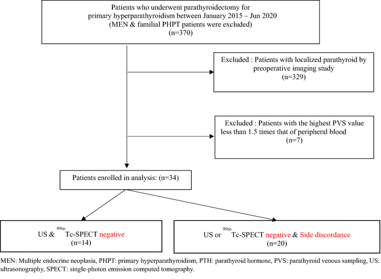
Preoperative localisation studies are essential for parathyroidectomy in patients with primary hyperparathyroidism. If the location of abnormal parathyroid glands cannot be identified through non-invasive studies, parathyroid venous sampling (PVS) may be employed. In this study, we evaluated the utility of preoperative PVS in parathyroid surgery. Patients with primary hyperparathyroidism who underwent preoperative PVS at Severance Hospital between January 2015 and June 2020 were identified. Patients for whom the results of non-invasive imaging studies were inconsistent or negative underwent PVS. The results of PVS were compared with operative findings and pathologic results. For 14 patients, the results of preoperative ultrasonography and 99mTc-sestamibi single-photon emission computed tomography (SPECT) were negative; for 20 patients, either the result of only one test was positive, or the results of the two tests were inconsistent. With respect to the lateralisation of diseased adenoma, the results of PVS and pathological examination were inconsistent only for one patient in either group (total: 2/34 patients). This study showed that PVS could be used effectively for preoperative localisation in patients with primary hyperparathyroidism in whom the location of diseased parathyroid glands cannot be determined through non-invasive image studies.
© 2022. The Author(s).
Conflict of interest statement
The authors declare no competing interests.
Figures
Similar articles
-
Update on Preoperative Parathyroid Localization in Primary Hyperparathyroidism.Endocrinol Metab (Seoul). 2022 Oct;37(5):744-755. doi: 10.3803/EnM.2022.1589. Epub 2022 Oct 25. Endocrinol Metab (Seoul). 2022. PMID: 36327985 Free PMC article. Review.
-
Role of four-dimensional computer tomography (4D-CT) in non-localising and discordant first-line imaging in primary hyperparathyroidism.Ann R Coll Surg Engl. 2023 Nov;105(8):739-746. doi: 10.1308/rcsann.2022.0126. Epub 2023 Feb 7. Ann R Coll Surg Engl. 2023. PMID: 36748800 Free PMC article.
-
Adenoma localization for recurrent or persistent primary hyperparathyroidism using dynamic four-dimensional CT and venous sampling.J Vasc Interv Radiol. 2015 Jan;26(1):79-86. doi: 10.1016/j.jvir.2014.09.019. Epub 2014 Nov 14. J Vasc Interv Radiol. 2015. PMID: 25454737
-
ROLE OF IMAGING TESTS FOR PREOPERATIVE LOCATION OF PATHOLOGIC PARATHYROID TISSUE IN PATIENTS WITH PRIMARY HYPERPARATHYROIDISM.Endocr Pract. 2016 Sep;22(9):1062-7. doi: 10.4158/EP151137.OR. Epub 2016 May 23. Endocr Pract. 2016. PMID: 27214298
-
Surgery for primary hyperparathyroidism.Cancer. 2014 Dec 1;120(23):3602-16. doi: 10.1002/cncr.28891. Epub 2014 Jul 9. Cancer. 2014. PMID: 25042934 Review.
Cited by
-
Update on Preoperative Parathyroid Localization in Primary Hyperparathyroidism.Endocrinol Metab (Seoul). 2022 Oct;37(5):744-755. doi: 10.3803/EnM.2022.1589. Epub 2022 Oct 25. Endocrinol Metab (Seoul). 2022. PMID: 36327985 Free PMC article. Review.
-
[Special features of the diagnostics and treatment of hereditary primary hyperparathyroidism].Chirurgie (Heidelb). 2023 Jul;94(7):586-594. doi: 10.1007/s00104-023-01897-8. Epub 2023 Jun 8. Chirurgie (Heidelb). 2023. PMID: 37291366 Review. German.
-
Complex Primary Hyperparathyroidism: Hereditary and Recurrent Disease.Surg Clin North Am. 2024 Aug;104(4):811-823. doi: 10.1016/j.suc.2024.02.010. Epub 2024 Mar 23. Surg Clin North Am. 2024. PMID: 38944501 Free PMC article. Review.
-
Imaging Recommendations for Diagnosis and Management of Primary Parathyroid Pathologies: A Comprehensive Review.Cancers (Basel). 2024 Jul 19;16(14):2593. doi: 10.3390/cancers16142593. Cancers (Basel). 2024. PMID: 39061231 Free PMC article. Review.
References
MeSH terms
Substances
LinkOut - more resources
Full Text Sources
Research Materials
