Review
. 2022 Sep;131(3):107-125.
doi: 10.1007/s00412-022-00773-4. Epub 2022 Apr 30.

Life of double minutes: generation, maintenance, and elimination

Affiliations
Review

Life of double minutes: generation, maintenance, and elimination

Mila Ilić et al. Chromosoma. 2022 Sep.

Abstract

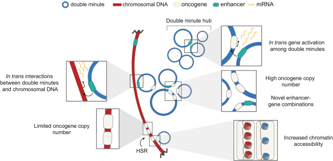
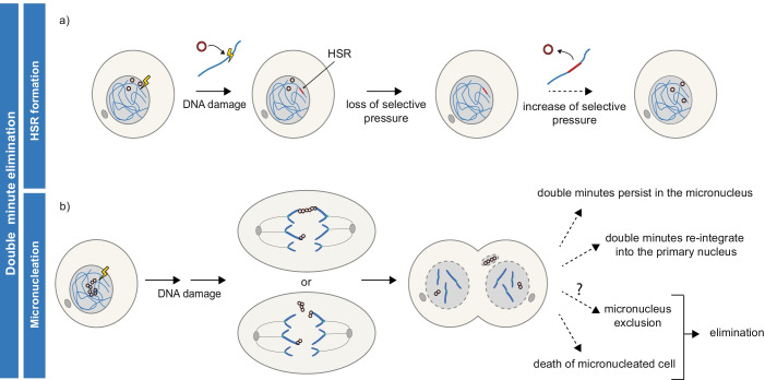
Advances in genome sequencing have revealed a type of extrachromosomal DNA, historically named double minutes (also referred to as ecDNA), to be common in a wide range of cancer types, but not in healthy tissues. These cancer-associated circular DNA molecules contain one or a few genes that are amplified when double minutes accumulate. Double minutes harbor oncogenes or drug resistance genes that contribute to tumor aggressiveness through copy number amplification in combination with favorable epigenetic properties. Unequal distribution of double minutes over daughter cells contributes to intratumoral heterogeneity, thereby increasing tumor adaptability. In this review, we discuss various models delineating the mechanism of generation of double minutes. Furthermore, we highlight how double minutes are maintained, how they evolve, and discuss possible mechanisms driving their elimination.

Keywords: Double minutes; Extrachromosomal DNA; Extrachromosomal oncogene amplification; Gene amplification; ecDNA.


Conflict of interest statement

The authors declare no competing interests.

Figures

Fig. 1
Fig. 1
Double minutes in HeLa cell line. Double minutes are derived through inducing drug resistance
Fig. 2
Fig. 2
Proposed models of “simple” double minute generation with no or limited chromosomal rearrangements. Top: Three models were proposed to explain non-deletion-associated double minute generation: a Re-replication caused by origin refiring is followed by excision of DNA fragments and their circularization (replication – re-replication – excision). b In a model summarized as replication – excision – continued replication, stalling and destabilization of the replication forks would lead to excision of a DNA fragment. Repair of the stalled replication forks could happen through breakage-induced repair mechanisms. For simplicity, we depict the continued replication simply as further advancement of replication forks. c DNA damage on one of the sister chromatids leads to excision of a DNA fragment, followed by repair through homologous recombination (post-replicative excision – homologous recombination). Excision has been depicted here as two DSBs. Bottom: d A model explaining the non-deletion- and deletion-associated generation by a single mechanism. Double minutes are generated in G2 phase by excision of a DNA fragment followed by circularization through non-homologous end joining. Upon cell division, the double minute can end up in the same daughter cell as the intact chromatid (non-deletion-associated generation) or as the chromatid with the deletion (deletion-associated generation). In case of the non-deletion-associated phenotype, negative selection of the cell harbouring the deletion can lead to it not being detected
Fig. 3
Fig. 3
Mechanisms of double minute generation through chromothripsis. a Chromothripsis is caused by lagging chromosome micronucleation. Often, a derivative chromosome is formed in this process. b Breakage-fusion-bridge cycles can result in generation of double minutes when a chromosome bridge is shattered. Here, there are two possible scenarios. A micronucleus-independent process, where the damage occurring at the site of the bridge breakage leads to double minute generation. Alternatively, micronucleation could precede double minute formation
Fig. 4
Fig. 4
Proposed model for the behaviour of double minutes throughout the cell cycle. Double minutes are replicated during S phase to form paired structures. In mitosis, double minutes tether to chromosomes at metaphase and are found in proximity of the chromosome tips in anaphase. Sister double minutes remain paired during G2 and mitosis, and their mitotic nondisjunction results in unequal distribution of double minutes over daughter cells
Fig. 5
Fig. 5
Unique modes of double minute-gene expression. Double minutes reach higher gene copy number when compared to intrachromosomal gene amplification in homogeneously staining regions (HSRs). Even when normalized to copy number, transcriptional output of double minutes could be higher as (1) double minute chromatin is more accessible; (2) double minute formation may lead to novel in cis regulation of oncogenes through incorporation of enhancer-gene pairs that normally localize to different topologically associated domains; (3) genes on double minutes may be activated in trans by enhancers on different double minutes within transcriptional hubs. Interestingly, gene activation in trans may also result in global increased expression of chromosomal genes
Fig. 6
Fig. 6
Routes of double minute elimination. a Double minutes are eliminated from cells when they integrate into a chromosome, thereby forming an HSR. Proposedly, double minute integration is a random event that is selected for when selective pressure is lost. Formed HSRs may serve as reservoirs of double minutes, generating new double minutes when selective pressures are in favour. b The fate of micronuclei containing double minutes are manifold. Double minutes may be lost through micronucleation, through cell death or other not fully characterized mechanisms

References

    1. Alitalo K, Schwab M, Lin CC, et al. Homogeneously staining chromosomal regions contain amplified copies of an abundantly expressed cellular oncogene (c-myc) in malignant neuroendocrine cells from a human colon carcinoma. Proc Natl Acad Sci U S A. 1983;80:1707–1711. doi: 10.1073/pnas.80.6.1707. - DOI - PMC - PubMed
    1. Baiker A, Maercker C, Piechaczek C, et al. Mitotic stability of an episomal vector containing a human scaffold/matrix-attached region is provided by association with nuclear matrix. Nat Cell Biol. 2000;2:182–184. doi: 10.1038/35004061. - DOI - PubMed
    1. Barker P, Drwinga H, Hittelman W, Maddox A. Double minutes replicate once during S phase of the cell cycle. Exp Cell Res. 1980;130:353–360. doi: 10.1016/0014-4827(80)90012-9. - DOI - PubMed
    1. Borrie MS, Campor JS, Joshi H, Gartenberg MR. Binding, sliding, and function of cohesin during transcriptional activation. Proc Natl Acad Sci U S A. 2017;114:E1062–E1071. doi: 10.1073/pnas.1617309114. - DOI - PMC - PubMed
    1. Cai M, Zhang H, Hou L, et al. Inhibiting homologous recombination decreases extrachromosomal amplification but has no effect on intrachromosomal amplification in methotrexate-resistant colon cancer cells. Int J Cancer. 2019;144:1037–1048. doi: 10.1002/ijc.31781. - DOI - PMC - PubMed

Publication types