. 2022 Apr 29;12(1):6998.
doi: 10.1038/s41598-022-10981-8.

Sensorimotor inhibition during emotional processing

Affiliations

Sensorimotor inhibition during emotional processing

Alessandro Botta et al. Sci Rep. .

Abstract

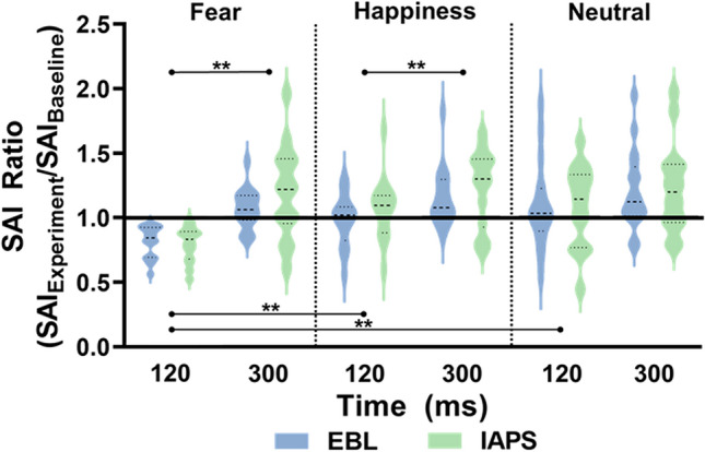
Visual processing of emotional stimuli has been shown to engage complex cortical and subcortical networks, but it is still unclear how it affects sensorimotor integration processes. To fill this gap, here, we used a TMS protocol named short-latency afferent inhibition (SAI), capturing sensorimotor interactions, while healthy participants were observing emotional body language (EBL) and International Affective Picture System (IAPS) stimuli. Participants were presented with emotional (fear- and happiness-related) or non-emotional (neutral) EBL and IAPS stimuli while SAI was tested at 120 ms and 300 ms after pictures presentation. At the earlier time point (120 ms), we found that fear-related EBL and IAPS stimuli selectively enhanced SAI as indexed by the greater inhibitory effect of somatosensory afferents on motor excitability. Larger early SAI enhancement was associated with lower scores at the Behavioural Inhibition Scale (BIS). At the later time point (300 ms), we found a generalized SAI decrease for all kind of stimuli (fear, happiness or neutral). Because the SAI index reflects integrative activity of cholinergic sensorimotor circuits, our findings suggest greater sensitivity of such circuits during early (120 ms) processing of threat-related information. Moreover, the correlation with BIS score may suggest increased attention and sensory vigilance in participants with greater anxiety-related dispositions. In conclusion, the results of this study show that sensorimotor inhibition is rapidly enhanced while processing threatening stimuli and that SAI protocol might be a valuable option in evaluating emotional-motor interactions in physiological and pathological conditions.


Conflict of interest statement

The authors declare no competing interests.

Figures

Figure 1
Figure 1
Experimental design. SAI was tested via an electrical stimulus (ES) delivered over of the median nerve (dashed blue arrow), followed 20 ms later by a transcranial magnetic stimulus (TMS) stimulus (red arrow) over the left first dorsal interosseus (FDI) cortical area at two timepoints, 120 and 300 ms after visual stimulus onset (black lightning). The blue dot indicates the somatosensory afferences to the sensorimotor cortex, while the yellow dot represents the FDI muscle. Each visual emotional stimulus had duration of 1000 ms, interspersed by a blank, white fixation screen of 4000 ms, for a total of 45 stimuli for each trial [emotional body language (EBL) and International Affective Picture System (IAPS)]. The same experimental design was followed also for the control experiment, with the emotional visual stimuli replaced by a black cross on a white screen.
Figure 2
Figure 2
SAI Ratio data. The picture shows the violin plots of all SAI Ratio data for EBL and IAPS. A value greater than 1 (black thick line) indicates a decreased inhibition (SAI reduction) in relation to baseline, whereas a value lower than 1 indicates an augmented inhibition (SAI increase) in relation to SAI recorded at baseline. As observable, the only increase of SAI was at 120 ms after stimulus onset for fearful emotional stimuli. All SAI Ratio data computed at 300 ms showed a decreased SAI across emotional and neutral stimuli in both EBL and IAPS. SAI Ratio data are reported on the y-axis, while the two timepoints 120 and 300 ms are reported on the x-axis, separately for every condition. **p < 0.01.
Figure 3
Figure 3
SAI data. All SAI data are graphically reported. In (A), the increase of SAI at 120 ms after stimulus onset for fearful stimuli is observable in both conditions (EBL and IAPS) in the comparison with SAI recorded at baseline (no visual stimuli). At 300 ms after stimulus onset, SAI was decreased compared to baseline for all emotional (A,B) and neutral stimuli (C) in both conditions (EBL and IAPS). SAI data (MEP conditioned/MEP test) are reported on the y-axis, while the three timepoints at which SAI was tested (Baseline, 120 ms and 300 ms) are reported on the x-axis. *p < 0.05, **p < 0.01, $p > 0.05 (trend).
Figure 4
Figure 4
Correlation analysis on SAI Ratio and BIS. Pearson correlation analysis for SAI Ratio computed for fearful stimuli at 120 ms after stimulus onset and BIS scores of participants from both EBL and IAPS. A positive correlation is observable between the two sets of data, meaning that at a higher BIS score corresponded a decreased SAI and vice versa.

References

    1. Gladwin TE, Hashemi MM, van Ast V, Roelofs K. Ready and waiting: Freezing as active action preparation under threat. Neurosci. Lett. 2016;619:182–188. doi: 10.1016/j.neulet.2016.03.027. - DOI - PubMed
    1. Smith NK, Cacioppo JT, Larsen JT, Chartrand TL. May I have your attention, please: Electrocortical responses to positive and negative stimuli. Neuropsychologia. 2003;41:171–183. doi: 10.1016/S0028-3932(02)00147-1. - DOI - PubMed
    1. Vuilleumier P. How brains beware: Neural mechanisms of emotional attention. Trends Cogn. Sci. 2005;9:585–594. doi: 10.1016/j.tics.2005.10.011. - DOI - PubMed
    1. van Heijnsbergen CCRJ, Meeren HKM, Grèzes J, de Gelder B. Rapid detection of fear in body expressions, an ERP study. Brain Res. 2007 doi: 10.1016/j.brainres.2007.09.093. - DOI - PubMed
    1. Roelofs K. Freeze for action: Neurobiological mechanisms in animal and human freezing. Philos. Trans. R. Soc. B Biol. Sci. 2017;372:20160206. doi: 10.1098/rstb.2016.0206. - DOI - PMC - PubMed

Publication types

MeSH terms