

Use of Imaging Technology to Assess the Effect of COVID-19 on Retinal Tissues: A Systematic Review
- PMID: 35488102
- PMCID: PMC9053559
- DOI: 10.1007/s40123-022-00509-8
Use of Imaging Technology to Assess the Effect of COVID-19 on Retinal Tissues: A Systematic Review
Abstract
Purpose: To evaluate the effect of COVID-19 on retinal tissues by conducting a systematic review and meta-analysis of the current literature.
Background: The novel coronavirus disease is not yet well understood. The orbit provides a window into the body's microvasculature, and as such, it is a non-invasive opportunity to analyse the systemic circulation in vivo. By analysing the current literature, we test the hypothesis that non-invasive imaging of the retina could provide insight into the effect of COVID-19 on the retinal microvasculature.
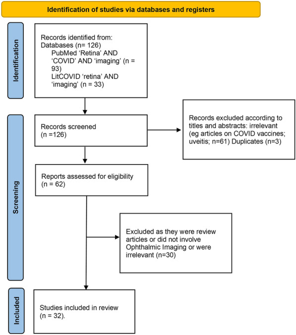
Methods: For this systematic review and meta-analysis, we screened PubMed databases and LitCOVID19 using the search criteria: (OCTA or Optical Coherence Tomography Angiography) AND (COVID-19 or corona or SARS-CoV-2) AND (retina or fundus). Databases were searched on 11 January 2022. The primary study outcomes were studies that utilised OCTA to analyse the retina; secondary outcomes involved studies that involved other imaging modalities such as OCT, fundus photography, and fundus autofluorescence.
Findings: The total number of studies included in this review was 32. Optical coherence tomography angiography scans show reduced central retinal vascular density, a thinner ganglion cell layer, a thicker retinal nerve fibre layer, and an enlarged foveal avascular zone. Optical coherence tomography scans demonstrate a thicker central macular thickness and other changes to the macula, ganglion cell, and inner nuclear layers. Many fundus photographs depicted cotton wool spots, microhaemorrhages, and vascular occlusions. Non-invasive imaging technology has demonstrated that COVID-19 can profoundly affect the retina. Therefore, there is a requirement for long-term follow-up of COVID-19 patients to assess whether the retinal damage caused by COVID-19 is reversible.
Keywords: COVID-19; Imaging; OCT; OCTA; Retina.
© 2022. The Author(s).
Figures
References
Publication types
LinkOut - more resources
Full Text Sources
Miscellaneous
