Case Reports
. 2022 Dec;35(6):763-767.
doi: 10.1177/19714009221096836. Epub 2022 Apr 29.

Multiple recurrences of unknown primary tumor in a meningioma: A case report

Affiliations
Case Reports

Multiple recurrences of unknown primary tumor in a meningioma: A case report

Muhammed Amir Essibayi et al. Neuroradiol J. 2022 Dec.

Abstract

Metastasis of a systemic tumor into a primary brain tumor has been rarely reported in the literature. These metastases could be seen mostly in meningiomas and in less frequently in the other intracranial tumors. Carcinoma of an unknown primary site (CUP) metastasizing into an intracranial meningioma is not a common occasion. According to the best of our knowledge, in this report, we present the first case with multiple recurrent CUP, which metastasized to intracranial meningioma, reported in the literature. The whole body was investigated with FDG PET/CT and biomarkers of the most common carcinomas for primary cancer. However, these tests showed no primary affected region. Despite all adjuvant therapies, the tumor had multiple recurrences. Such cases are still a challenge to offer optimal management.

Keywords: Brain metastasis; meningioma; primary unknown metastasis.


Conflict of interest statement

The author(s) declared no potential conflicts of interest with respect to the research, authorship, and/or publication of this article.

Figures
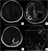
Figure 1.
Figure 1.
Contrast-enhancing MRI demonstrated a heterogeneously enhancing left parieto-occipital mass that had two components and measured 53 × 52 × 63 mm. The mass was surrounded by edema. The central component was hypointense on T2WIs and heterogeneously enhancing the contrast substance. The peripheral component was isointense on both T1- and T2-weighted sequences and homogenously surrounded the central component; (a) axial T1WI; (b) axial T2WI; (c) contrast-enhanced axial T1WI; and (d) contrast-enhanced sagittal T1WI demonstrated a dural tail—one of the most common radiological feature of meningiomas—and cerebrospinal fluid (CSF) cleft sign (i.e., a thin rim of CSF between a tumoral mass and brain parenchyma) that can be used to distinguish an extra-axial lesion from intra-axial lesions.
Figure 2.
Figure 2.
Serial axial postoperative MRI sequences; (a) the early postoperative T1WI; (b) contrast-enhanced T1WI, the comparison between both sequences demonstrated that the GTR was performed on the tumoral mass at the first surgical intervention; (c) the third-month postoperative contrast-enhanced T1WI revealed recurrent heterogeneously enhancing mass measured 31 × 29 × 25 mm; (d-e) the ninth-month postoperative contrast-enhanced T1WIs revealed no residual or recurrent masses; and (f) the 12th-month postoperative contrast-enhanced T1WI demonstrated the second recurrence mass that measured 18 × 24 × 19 mm. The patient was treated with GTR followed by standard WBRT and chemotherapy. (g) The 19th-month postoperative contrast-enhanced T1WI demonstrated a third recurrence mass that measured 25 ×18 × 10 mm. The patient was re-treated with GTR.
Figure 3.
Figure 3.
Photos of the histopathological examination for the pieces obtained from the tumoral mass in the first surgical intervention. The final histopathological diagnosis of the mass was confirmed to be atypical meningioma (WHO 2016 grade II) with the metastasis cells (poorly differentiated neuroendocrine carcinoma). (a) Fascicular architecture of spindle-shaped cells (meningioma cells) (showed by arrows) and solid-shaped carcinoma cells in the below-left side of the photo (star shows carcinoma component) (hematoxylin and eosin staining; magnification, ×100); (b) solid-shaped carcinoma cells with necrosis (showed by arrows) (hematoxylin and eosin staining; magnification, ×200); (c-f) immunohistochemistry for pancytokeratin (c); HMB-45 (d); synaptophysin (e); and CD 56 (f) (magnification, ×200).

Similar articles

References

    1. Han HJ, Chang WS, Jung HH, et al. Optimal treatment decision for brain metastases of unknown primary origin: the role and timing of radiosurgery. Brain Tumor Res Treat 2016; 4(2): 107–110. - PMC - PubMed
    1. Lanotte M, Benech F, Panciani PP, et al. Systemic cancer metastasis in a meningioma: report of two cases and review of the literature. Clin Neurol Neurosurg 2009; 111(1): 87–93. - PubMed
    1. Benedetto N, Perrini P, Scollato A, et al. Intracranial meningioma containing metastatic colon carcinoma. Acta Neurochirurgica 2007; 149(8): 799–803. - PubMed
    1. Chambers PW, Davis RL, Blanding JD, et al. Metastases to primary intracranial meningiomas and neurilemomas. Arch Pathol Lab Med 1980; 104: 350–354. - PubMed
    1. Lodrini S, Savoiardo M. Metastases of carcinoma to intracranial meningioma: report of two cases and review of the literature. Cancer 1981; 48: 2668–2673. - PubMed

Publication types

MeSH terms

LinkOut - more resources