Pedicled frontal periosteal rescue flap via eyebrow incision for skull base reconstruction (SevEN-002)
- PMID: 35488272
- PMCID: PMC9052618
- DOI: 10.1186/s12893-022-01590-3
Pedicled frontal periosteal rescue flap via eyebrow incision for skull base reconstruction (SevEN-002)
Abstract
Purpose: Cerebrospinal fluid (CSF) leakage is one of the major complications after endoscopic endonasal surgery. The reconstructive nasoseptal flap is widely used to repair CSF leakage. However, it could not be utilized in all cases; thus, there was a need for an alternative. We developed a pericranial rescue flap that could cover both sellar and anterior skull base defects via the endonasal approach. A modified surgical technique that did not violate the frontal sinus and cause cosmetic problems was designed using the pericranial rescue flap.
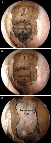
Methods: We performed 12 cadaveric dissections to investigate the applicability of the lateral pericranial rescue flap. An incision was made, extending from the middle to the lateral part of the eyebrow. The pericranium layer was dissected away from the galea layer, from the supraorbital region towards the frontoparietal region. With endoscopic assistance, the periosteal flap was raised, the flap base was the pericranium layer at the eyebrow incision. After a burr-hole was made in the supraorbital bone, the pericranial flap was inserted via the intradural or extradural pathway.
Results: The mean size of the pericranial flap was 11.5 cm × 3.2 cm. It was large enough to cross the midline and cover the dural defects of the anterior skull base, including the sellar region.
Conclusion: We demonstrated a modified endoscopic technique to repair the anterior skull base defects. This minimally invasive pericranial flap may resolve neurosurgical complications, such as CSF leakage.
Keywords: CSF leak; Endonasal approach; Endoscopic surgery; Pericranial flap; Skull base.
© 2022. The Author(s).
Conflict of interest statement
All authors certify that they have no affiliations with or involvement in any organization or entity with any financial interest (such as honoraria; educational grants; participation in speakers' bureaus; membership, employment, consultancies, stock ownership, or other equity interest; and expert testimony or patent-licensing arrangements), or non-financial interest (such as personal or professional relationships, affiliations, knowledge or beliefs) in the subject matter or materials discussed in this manuscript.
Figures


References
-
- Alobid I, Ensenat J, Marino-Sanchez F, de Notaris M, Centellas S, Mullol J, Bernal-Sprekelsen M. Impairment of olfaction and mucociliary clearance after expanded endonasal approach using vascularized septal flap reconstruction for skull base tumors. Neurosurgery. 2013;72:540–546. doi: 10.1227/NEU.0b013e318282a535. - DOI - PubMed
-
- Alqahtani A, Padoan G, Segnini G, Lepera D, Fortunato S, Dallan I, Pistochini A, Abdulrahman S, Abbate V, Hirt B, Castelnuovo P. Transorbital transnasal endoscopic combined approach to the anterior and middle skull base: a laboratory investigation. Acta Otorhinolaryngol Ital. 2015;35:173–179. - PMC - PubMed
-
- Cappelletti M, Ruggeri AG, Giovannetti F, Priore P, Pichierri A, Delfini R. Endoscopic applica’tion of autologous fibrin glue to treat postoperative CSF leak after expanded endonasal approach: Report of two cases. Interdiscip Neurosurg. 2018;14:72–75. doi: 10.1016/j.inat.2018.06.001. - DOI
MeSH terms
LinkOut - more resources
Full Text Sources
Medical

