. 2022 Apr 29;15(1):46.
doi: 10.1186/s13045-022-01266-8.

Universal immunotherapeutic strategy for hepatocellular carcinoma with exosome vaccines that engage adaptive and innate immune responses

Affiliations

Universal immunotherapeutic strategy for hepatocellular carcinoma with exosome vaccines that engage adaptive and innate immune responses

Bingfeng Zuo et al. J Hematol Oncol. .

Abstract

Background: Personalized immunotherapy utilizing cancer vaccines tailored to the tumors of individual patients holds promise for tumors with high genetic heterogeneity, potentially enabling eradication of the tumor in its entirety.

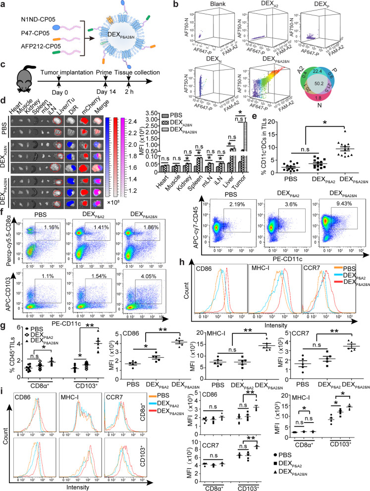
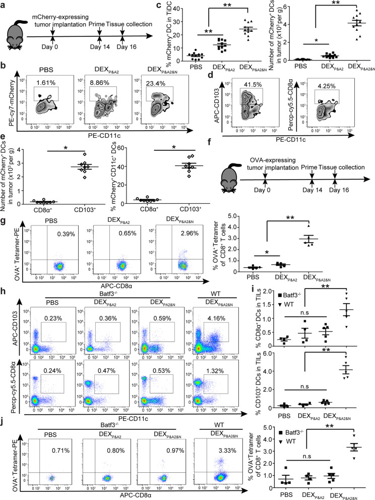
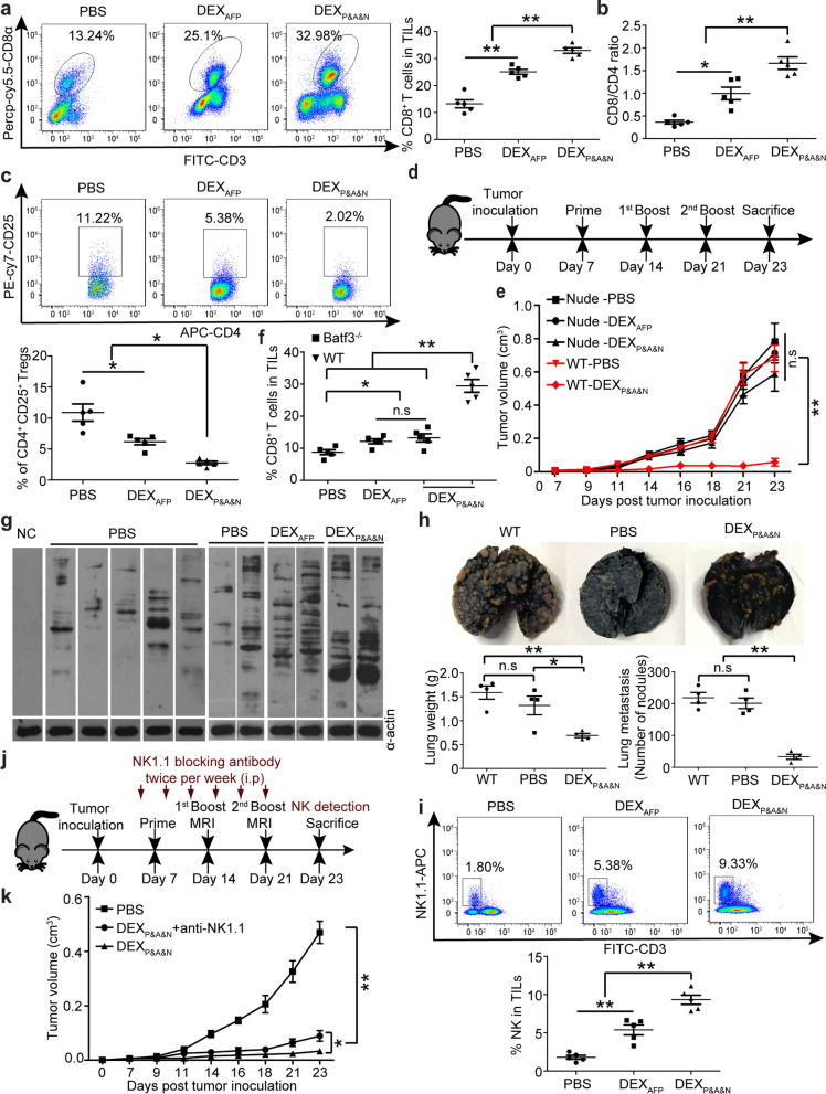
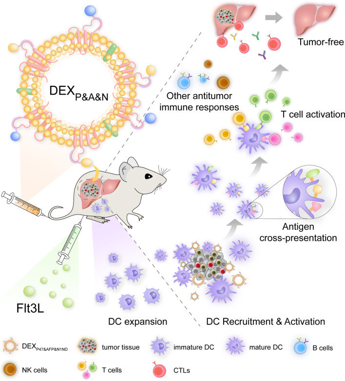
Methods: Here, we demonstrate a general strategy for biological nanovaccines that trigger tailored tumor-specific immune responses for hepatocellular carcinoma (HCC). Dendritic cell (DC)-derived exosomes (DEX) are painted with a HCC-targeting peptide (P47-P), an α-fetoprotein epitope (AFP212-A2) and a functional domain of high mobility group nucleosome-binding protein 1 (N1ND-N), an immunoadjuvant for DC recruitment and activation, via an exosomal anchor peptide to form a "trigger" DEX vaccine (DEXP&A2&N).

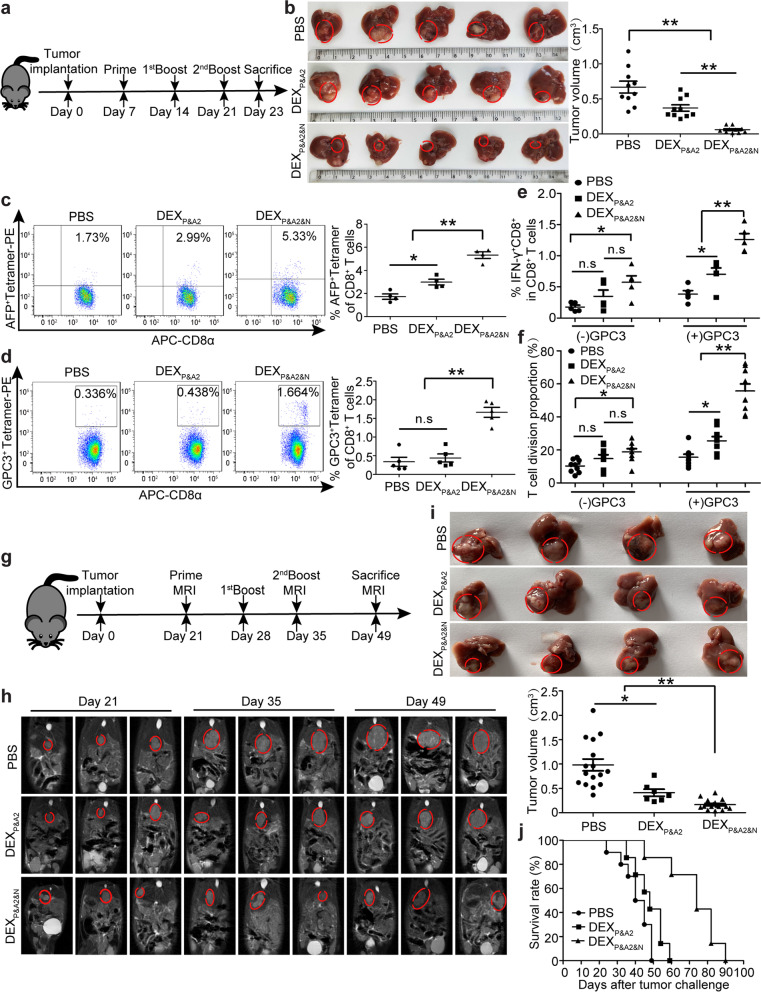
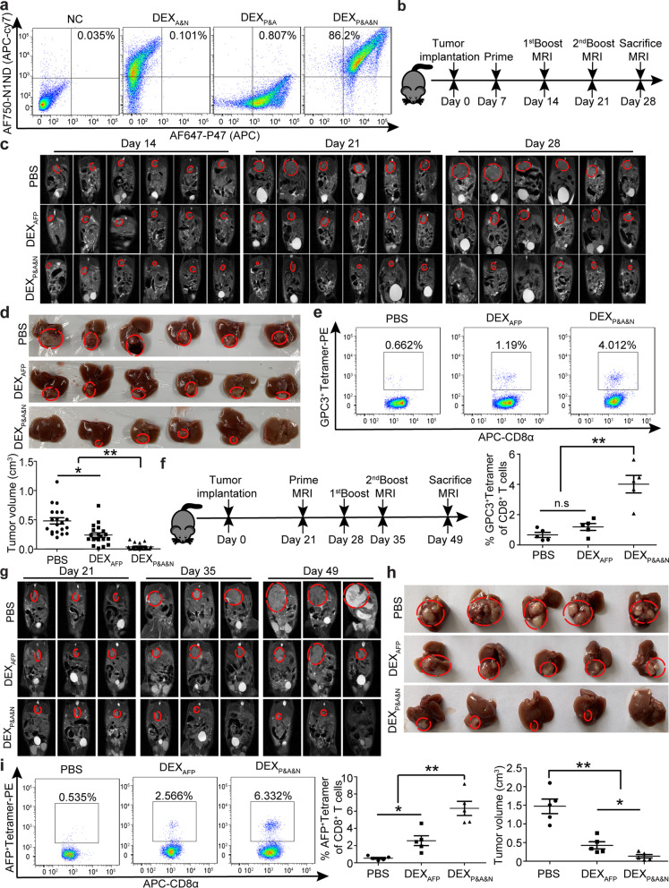
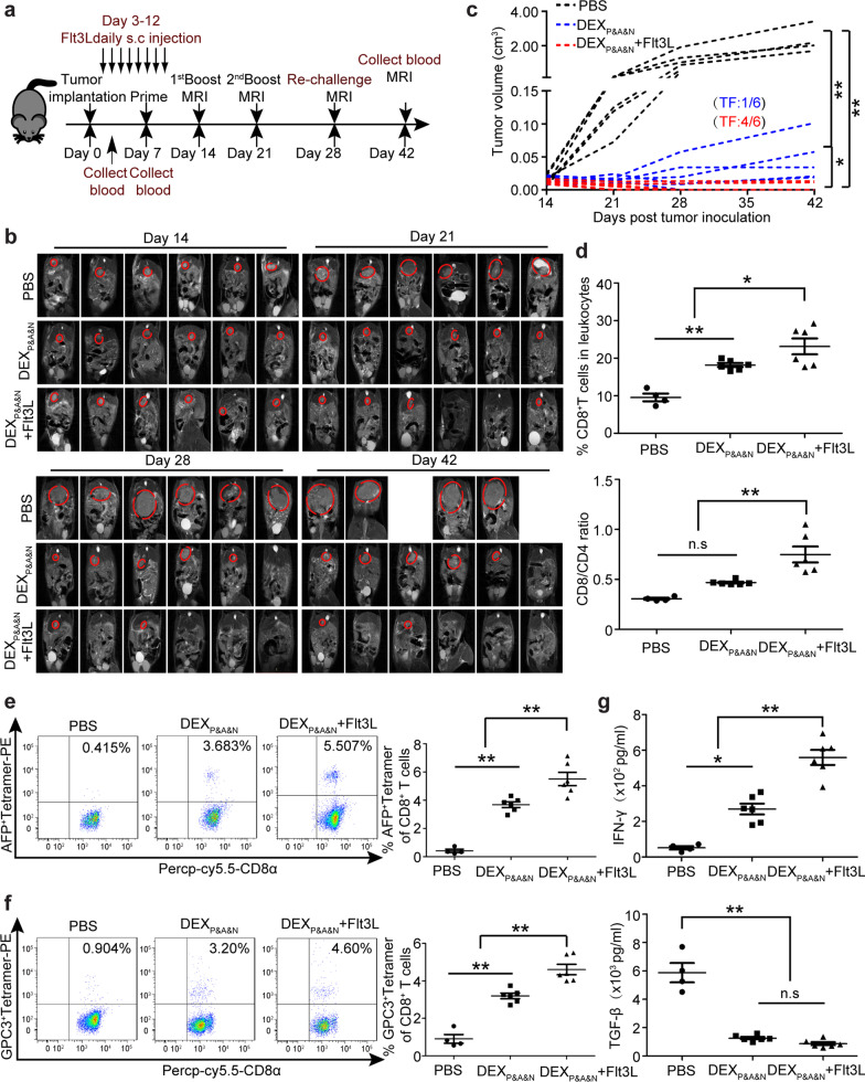
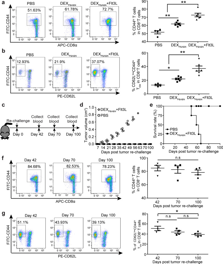
Results: DEXP&A2&N specifically promoted recruitment, accumulation and activation of DCs in mice with orthotopic HCC tumor, resulting in enhanced cross-presentation of tumor neoantigens and de novo T cell response. DEXP&A2&N elicited significant tumor retardation and tumor-specific immune responses in HCC mice with large tumor burdens. Importantly, tumor eradication was achieved in orthotopic HCC mice when antigenic AFP peptide was replaced with the full-length AFP (A) to form DEXP&A&N. Supplementation of Fms-related tyrosine kinase 3 ligand greatly augmented the antitumor immunity of DEXP&A&N by increasing immunological memory against tumor re-challenge in orthotopic HCC mice. Depletion of T cells, cross-presenting DCs and other innate immune cells abrogated the functionality of DEXP&A&N.

Conclusions: These findings demonstrate the capacity of universal DEX vaccines to induce tumor-specific immune responses by triggering an immune response tailored to the tumors of each individual, thus presenting a generalizable approach for personalized immunotherapy of HCC, by extension of other tumors, without the need to identify tumor antigens.

Keywords: Adaptive and innate immunity; Exosome; Hepatocellular carcinoma; Personalized immunotherapy.


Conflict of interest statement

H.Y., X.G. and B.Z. hold a patent (ZL 20151052565.7). The authors declare no competing financial interests.

Figures

Fig. 1
Fig. 1
Evaluation of DEXP&A2&N’s tumor-targeting, DC-recruiting and DC-activating ability in orthotopic mCherry-expressing HCC mice. a Schematic illustration for designer DEX vaccine-DEXP&A2&N. P-P47; A2-AFP212; N-N1ND. DEX refers to DC-derived exosomes. b Flow cytometric analysis to assess the simultaneous binding efficiency of three moieties on DEX (n = 4). Diagram for dosing regimen (c) and tissue distribution and quantitative analysis of labeled DEXP&A2&N (d) in day-14 orthotopic HCC mice bearing mCherry-expressing tumors. DiR-labeled DEXA2&N (n = 9), DEXP&A2&N (n = 9) (80 μg/mouse) or PBS (n = 4) were injected into day-14 orthotopic HCC mice bearing mCherry-expressing tumors intravenously, and tissues were harvested 2 h after injection (one-way ANOVA post hoc Student–Newman–Keuls test was used except for liver and tumor in which one-way ANOVA on ranks was used). mLN-mesenteric lymph node; iLN-inguinal lymph node; Tu-tumor. Flow cytometric and quantitative analysis of CD11c+ DCs in tumor-infiltrating lymphocytes (TILs) from HCC mice bearing mCherry-expressing tumors (n = 13; one-way ANOVA post hoc Student–Newman–Keuls test). Flow cytometric (f) and quantitative analysis (g) of CD103+CD11c+ (one-way ANOVA on ranks) and CD8α+ CD11c+ DCs (one-way ANOVA post hoc Student–Newman–Keuls test) in TILs from HCC mice (n = 9). h Flow cytometric and quantitative analysis of surface protein markers on CD11c+ DCs from tumors of orthotopic HCC mice treated with DEXP&A2 or DEXP&A2&N (n = 5; one-way ANOVA post hoc Student–Newman–Keuls test). i Flow cytometric and quantitative analysis of surface protein markers on CD103+CD11c+ and CD8α+CD11c+ DCs from tumors of orthotopic HCC mice treated with DEXP&A2 or DEXP&A2&N (n = 5; one-way ANOVA post hoc Student–Newman–Keuls test). *p < 0.05, **p < 0.001; n.s, not significant
Fig. 2
Fig. 2
Investigation of DEXP&A2&N promoting DC uptake and cross-presentation of tumor neoantigens in orthotopic HCC mice. a Diagram for dosing regimen of DEXP&A2&N. DEXP&A2 or DEXP&A2&N (80 μg/mouse) were injected into day-14 orthotopic HCC mice bearing mCherry-expressing tumors intravenously, and tissues were harvested 2 days after injection. Flow cytometric (b) and quantitative analysis (c) of mCherry+CD11c+ DCs from tumor of orthotopic HCC mice treated with DEXP&A2 or DEXP&A2&N (n = 11) (One-way ANOVA on ranks was used for the number and one-way ANOVA post hoc Student–Newman–Keuls test was applied for the percent analysis). TiDC: tumor-infiltrating DC. Flow cytometric (d) and quantitative analysis (e) of mCherry+CD103+CD11c+ or CD8α+CD11c+ DCs from tumor of orthotopic HCC mice treated with DEXP&A2&N (n = 8; Mann–Whitney rank-sum test). f Diagram for dosing regimen of DEXP&A2&N. DEXP&A2 or DEXP&A2&N (80 μg/mouse) were injected into day-14 orthotopic HCC mice bearing OVA-expressing tumors intravenously, and tissues were harvested 2 days after injection. g Flow cytometric and quantitative analysis of OVA+ tetramer T cells from splenocytes of orthotopic HCC mice treated with DEXP&A2 or DEXP&A2&N (n = 5; one-way ANOVA on ranks). Flow cytometric (h) and quantitative analysis (i) of CD103+CD11c+ or CD8α+CD11c+ DCs from TILs of orthotopic Batf3−/− HCC mice treated with PBS or DEXP&A2 (n = 4) or DEXP&A2&N (n = 5) and orthotopic wild-type (WT) HCC mice treated with DEXP&A2&N (n = 5) (one-way ANOVA on ranks). j Flow cytometric and quantitative analysis of OVA+ tetramer T cells from tumor of orthotopic Batf3−/− HCC mice treated with PBS or DEXP&A2 (n = 4) or DEXP&A2&N (n = 5) and orthotopic wild-type (WT) HCC mice treated with DEXP&A2&N (n = 5) (one-way ANOVA post hoc Student–Newman–Keuls test). *p < 0.05, **p < 0.001; n.s, not significant
Fig. 3
Fig. 3
Antitumor effects of DEXP&A2&N in orthotopic HCC mice. a Diagram for dosing regimen of DEXP&A2&N. DEXP&A2 or DEXP&A2&N (80 μg/mouse) were injected into day-7 orthotopic HCC mice intravenously three times weekly, and tissues were harvested 2 days after last injection. b Measurement of tumor volume in day-7 orthotopic HCC mice treated with DEXP&A2 or DEXP&A2&N (n = 10; one-way ANOVA on ranks). c Flow cytometric and quantitative analysis of AFP+ tetramer T cells from splenocytes of orthotopic HCC mice treated with DEXP&A2 or DEXP&A2&N (n = 4; one-way ANOVA post hoc Student–Newman–Keuls test). d Flow cytometric and quantitative analysis of GPC3+ tetramer T cells from splenocytes of orthotopic HCC mice treated with DEXP&A2 or DEXP&A2&N (n = 5; one-way ANOVA post hoc Student–Newman–Keuls test). Flow cytometric and quantitative analysis of IFN-γ+CD8+ T cells (e) (n = 5; one-way ANOVA post hoc Student–Newman–Keuls test) and T cell division (f) (n = 9; one-way ANOVA on ranks) in splenocytes of orthotopic HCC mice treated with DEXP&A2 or DEXP&A2&N, followed by in vitro stimulation of GPC3 epitopes. g Diagram for dosing regimen of DEXP&A2&N. DEXP&A2 or DEXP&A2&N (120 μg/mouse) were injected into day-21 orthotopic HCC mice bearing large established tumors intravenously three times weekly, and tissues were harvested 2 weeks after last injection. h MRI monitoring of tumor growth in day-21 orthotopic HCC mice bearing large established tumors at different time points. i Assessment of tumor size in orthotopic HCC mice bearing large established tumors treated with PBS (n = 16), DEXP&A2 (n = 7) or DEXP&A2&N (n = 16) at 28 days after initial treatment (one-way ANOVA on ranks). j Survival rate of orthotopic HCC mice treated with PBS (n = 9), DEXP&A2 or DEXP&A2&N (n = 8). *p < 0.05, **p < 0.001; n.s, not significant
Fig. 4
Fig. 4
Antitumor immunity of DEXP&A&N in orthotopic HCC mice. a Flow cytometric analysis to assess the double loading efficiency of two moieties on DEXAFP. DEXP&A&N refer to DEXP47&AFP&N1ND. b Diagram for dosing regimen of DEXP&A&N. DEXAFP or DEXP&A&N (80 μg/mouse) were injected into day-7 orthotopic HCC mice intravenously three times weekly, and tissues were harvested 7 days after last injection. c Dynamic monitoring of tumor growth in orthotopic HCC mice with MRI at different time points. d Measurement of tumor size in orthotopic HCC mice at 21 days after priming (n = 20; one-way ANOVA on ranks). e Flow cytometric and quantitative analysis of GPC3+ tetramer T cells from tumor of orthotopic HCC mice treated with DEXAFP or DEXP&A&N (n = 5; one-way ANOVA post hoc Student–Newman–Keuls test). f Diagram for dosing regimen of DEXP&A&N. DEXAFP or DEXP&A&N (120 μg/mouse) were injected into day-21 orthotopic HCC mice bearing large established tumors intravenously three times weekly, and tissues were harvested 2 weeks after last injection. g Dynamic monitoring of tumor growth in orthotopic HCC mice bearing large established tumors treated with DEXAFP or DEXP&A&N with MRI at different time points. h Measurement of tumor size in orthotopic HCC mice at 28 days after priming (n = 5; one-way ANOVA post hoc Student–Newman–Keuls test). i Flow cytometric and quantitative analysis of AFP+ tetramer T cells from tumor of orthotopic HCC mice treated with DEXAFP or DEXP&A&N (n = 5; one-way ANOVA post hoc Student–Newman–Keuls test). *p < 0.05, **p < 0.001; n.s, not significant
Fig. 5
Fig. 5
DEXP&A&N evoke potent antitumor immunity with efficacy dependent on innate and adaptive immune responses. Flow cytometric and quantitative analysis of tumor-infiltrating CD8+ (a) and ratio of tumor-infiltrating CD8+ to CD4+ (b) and flow cytometric and quantitative analysis of tumor CD4+CD25+ (c) T cells of DEXP&A&N -treated orthotopic HCC mice bearing large established tumors (n = 5). Diagram for dosing regimen of DEXP&A&N. DEXAFP or DEXP&A&N (80 μg) were injected into day-7 thymus-deficient nude mice bearing subcutaneous HCC tumors intravenously three times weekly, and tissues were harvested 2 days after last injection. e Measurement of tumor size in nude mice bearing subcutaneous HCC tumors at different time points (n = 5). One-way ANOVA on ranks test was used for day 7 and 21; one-way ANOVA post hoc Student–Newman–Keuls test was used for day 9, 11, 14, 16, 18 and 23. f Flow cytometric and quantitative analysis of tumor-infiltrating CD8+ T cells of DEXP&A&N-treated orthotopic Batf3−/− HCC mice (n = 5; one-way ANOVA on ranks). g Western blot to examine Hepa1-6 tumor cell lysates with serum from DEXP&A&N- or DEXAFP-treated mice. α-actin was used as a loading control. h Representative images and quantification of tumor nodules in lungs 21 days after serum re-infusion and tumor challenge (n = 4). i Flow cytometric and quantitative analysis of tumor-infiltrating NK cells of DEXP&A&N-treated orthotopic HCC mice bearing large established tumors (n = 5). j Diagram for NK depletion experiments. k Measurement of tumor volume in DEXP&A&N and anti-NK1.1-treated ectopic HCC mice at different time points (n = 5). One-way ANOVA post hoc Student–Newman–Keuls test was used for day 7, 9, 11, 14, 21 and 23; one-way ANOVA on ranks test was applied for day 16 and 18; and Two-tailed t test was applied for DEXP&A&N with or without anti-NK1.1 antibody on day 23. *p < 0.05, **p < 0.001; n.s means not significant. Note: One-way ANOVA post hoc Student–Newman–Keuls test was used for Fig. 5a-c and 5 h, 5i
Fig. 6
Fig. 6
Flt3L augments DEXP&A&N’s antitumor potency in orthotopic HCC mice. a Diagram for dosing regimen of DEXP&A&N and Flt3L in orthotopic HCC mice. Flt3L was administered subcutaneously to day-3 orthotopic HCC mice at the dose of 800 μg /kg/day for 8 days consecutively and DEXP&A&N (80 μg/mouse) were intravenously administered on day 7 after tumor implantation three times weekly. b Dynamic monitoring of tumor growth in orthotopic HCC mice treated with DEXP&A&N or DEXP&A&N and Flt3L with MRI at different time points. c Measurement of tumor volume for each individual mouse treated with PBS, DEXP&A&N or DEXP&A&N and Flt3L (n = 6). One-way ANOVA on ranks test was used for day 21, 28 and 42; one-way ANOVA post hoc Student–Newman–Keuls test was used for day 14. TF means tumor-free. d Quantitative analysis of CD8+ T cells and ratio of CD8+ to CD4+ T cells in blood of orthotopic HCC mice treated with PBS (n = 4), DEXP&A&N or DEXP&A&N and Flt3L (n = 6) at day 42 after tumor implantation (One-way ANOVA on ranks). Flow cytometric and quantitative analysis of AFP+ tetramer (e) and GPC3+ tetramer (f) T cells in blood of orthotopic HCC mice treated with PBS (n = 4), DEXP&A&N or DEXP&A&N and Flt3L (n = 6) at day 42 after tumor implantation (one-way ANOVA post hoc Student–Newman–Keuls test). g Assessment of IFN-γ and TGF-β in blood of orthotopic HCC mice treated with PBS (n = 4), DEXP&A&N or DEXP&A&N and Flt3L (n = 6) at day 42 after tumor implantation (one-way ANOVA post hoc Student–Newman–Keuls test). *p < 0.05, **p < 0.001; n.s, not significant
Fig. 7
Fig. 7
Long-term protective memory immune responses elicited by DEXP&A&N and Flt3L against tumor re-challenge. Flow cytometric and quantitative analysis of CD44hi CD8+ (a) and CD62LlowCD44hiCD8+ (b) T cells in blood of orthotopic HCC mice treated with PBS (n = 4), DEXP&A&N, DEXP&A&N and Flt3L (n = 6) at day 42 after tumor implantation (one-way ANOVA post hoc Student–Newman–Keuls test). c Diagram for tumor re-challenge in DEXP&A&N and Flt3L-treated tumor-free mice. d Measurement of tumor volume in DEXP&A&N and Flt3L-treated tumor-free mice after re-challenge (n = 4). Mann–Whitney rank-sum test was used for day 7; one-way ANOVA on ranks test was used for day 14, 21, 28, 35 and 42; one-way ANOVA post hoc Student–Newman–Keuls test was used for day 49. e Survival rate of DEXP&A&N and Flt3L-treated tumor-free mice after re-challenge (n = 4). Flow cytometric and quantitative analysis of CD44hiCD8+ (f) and CD62LlowCD44hi CD8+ (g) T cells in blood of DEXP&A&N and Flt3L-treated tumor-free mice at different time points after tumor re-challenge (n = 4; one-way ANOVA post hoc Student–Newman–Keuls test). *p < 0.05, **p < 0.001; n.s, not significant
Fig. 8
Fig. 8
Graphical abstract for illustrating the mechanism underlying the antitumor immunogenicity of universal DEX vaccines

References

    1. Zhu RX, Seto WK, Lai CL, Yuen MF. Epidemiology of hepatocellular carcinoma in the Asia-Pacific Region. Gut Liver. 2016;10:332–339. - PMC - PubMed
    1. Xu W, et al. Immunotherapy for hepatocellular carcinoma: recent advances and future perspectives. Ther Adv Med Oncol. 2019;11:1758835919862692. - PMC - PubMed
    1. Blass E, Ott PA. Advances in the development of personalized neoantigen-based therapeutic cancer vaccines. Nat Rev Clin Oncol. 2021;18:215–229. - PMC - PubMed
    1. Ott PA, et al. A phase Ib trial of personalized neoantigen therapy plus anti-PD-1 in patients with advanced melanoma, non-small cell lung cancer, or bladder cancer. Cell. 2020;183:347–362.e324. - PubMed
    1. Loffler MW, et al. Multi-omics discovery of exome-derived neoantigens in hepatocellular carcinoma. Genome Med. 2019;11:28. - PMC - PubMed

Publication types