. 2022 Apr 29;22(1):174.
doi: 10.1186/s12935-022-02587-x.

FGFR1 is a potential therapeutic target in neuroblastoma

Affiliations

FGFR1 is a potential therapeutic target in neuroblastoma

Flora Cimmino et al. Cancer Cell Int. .

Abstract

Background: FGFR1 regulates cell-cell adhesion and extracellular matrix architecture and acts as oncogene in several cancers. Potential cancer driver mutations of FGFR1 occur in neuroblastoma (NB), a neural crest-derived pediatric tumor arising in sympathetic nervous system, but so far they have not been studied experimentally. We investigated the driver-oncogene role of FGFR1 and the implication of N546K mutation in therapy-resistance in NB cells.

Methods: Public datasets were used to predict the correlation of FGFR1 expression with NB clinical outcomes. Whole genome sequencing data of 19 paired diagnostic and relapse NB samples were used to find somatic mutations. In NB cell lines, silencing by short hairpin RNA and transient overexpression of FGFR1 were performed to evaluate the effect of the identified mutation by cell growth, invasion and cologenicity assays. HEK293, SHSY5Y and SKNBE2 were selected to investigate subcellular wild-type and mutated protein localization. FGFR1 inhibitor (AZD4547), alone or in combination with PI3K inhibitor (GDC0941), was used to rescue malignant phenotypes induced by overexpression of FGFR1 wild-type and mutated protein.

Results: High FGFR1 expression correlated with low relapse-free survival in two independent NB gene expression datasets. In addition, we found the somatic mutation N546K, the most recurrent point mutation of FGFR1 in all cancers and already reported in NB, in one out of 19 matched primary and recurrent tumors. Loss of FGFR1 function attenuated invasion and cologenicity in NB cells, whereas FGFR1 overexpression enhanced oncogenicity. The overexpression of FGFR1N546K protein showed a higher nuclear localization compared to wild-type protein and increased cellular invasion and cologenicity. Moreover, N546K mutation caused the failure in response to treatment with FGFR1 inhibitor by activation of ERK, STAT3 and AKT pathways. The combination of FGFR1 and PI3K pathway inhibitors was effective in reducing the invasive and colonigenic ability of cells overexpressing FGFR1 mutated protein.

Conclusions: FGFR1 is an actionable driver oncogene in NB and a promising therapy may consist in targeting FGFR1 mutations in patients with therapy-resistant NB.

Keywords: FGFR1; Metastasis; Mutation; Neuroblastoma; Targeted therapy.


Conflict of interest statement

Authors declare no competing interests.

Figures

Fig. 1
Fig. 1
Association of FGFR1 expression with clinical outcomes in patients with NB. A The association of FGFR1 expression with clinical outcomes was evaluated in the following datasets: Seeger, Asgharzadeh TARGET and Versteeg. (n = number of patients). B FGFR1 expression analysis in datasets of primary and relapsed tumors. FGFR1 expression levels in two embryonic cells (ES), one neuronal crest cells (NC), one metastatic xenograft tumors (X) and four primary NB (T) datasets. In B and C the number of samples is reported in brackets. p = p-value
Fig. 2
Fig. 2
FGFR1 silencing impairs cell growth, cell invasion and clonogenicity in NB cells. A FGFR1 silencing efficiency was evaluated by western blotting and RT-PCR in SHSY5Y and SKNBE2 transduced by lentiviral vectors encoding shFGFR1#A and shFGFR1#B. FGFR1 levels folded on shCTR mRNA levels are reported. B Cell viability in shFGFR1#A and shFGFR1#B cells is shown as fold change compared to shCTR. C Invading cells and D colony number in FGFR1 silenced and control cells are reported. Vehicle = DMSO. p = p-value
Fig. 3
Fig. 3
FGFR1wt and FGFR1N546K proteins localization. A FGFR1 localization in HEK293 overexspressing FGFR1wt or FGFR1N546K protein was analyzed by Image Stream flow Citometry. The representative figures of single cells at 60X magnification are shown above, whereas the values of mean intensity, standard deviation, cells counted and p-value are reported in the table. B Immunostaining of HEK293, SHSY5Y and SKNBE2 transfected with FGFR1wt or FGFR1N546K analyzed by confocal microscopy. C FGFR1 protein levels in both cytosol and nucleus protein fractions from HEK293, SHSY5Y and SKNBE2 transfected cells was evaluated by western blotting. GAPDH, Lamin β and PARP1 protein levels were used as loading controls
Fig. 4
Fig. 4
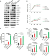
FGFR1wt and FGFR1N546K protein overexpression in NB cells. SH5YSY and SKNBE2 cells were transiently transfected with pCMV6-empty vector, pCMV6-FGFR1wt and pCMV6-FGFR1N546K. A Total protein extracts were analyzed by western blotting to evaluate the levels of phosphorylated and total FGFR1, STAT3, ERK, and-AKT. The t-GFP and β-Actin protein levels were used as transfection control and loading control, respectively. B Cell viability in FGFR1wt and FGFR1N546K overexpressing cells is shown as fold change compared to the control (pCMV6). C Invading cells, D colony number and area were analyzed. mm2 = square millimetres. p = p-value
Fig. 5
Fig. 5
Targeting of FGFR1 signaling by combination treatment with AZD4547 and GDC0941. SH5YSY and SKNBE2 cells were transiently transfected with pCMV6-FGFR1wt, pCMV6-FGFR1N546K and pCMV6 empty vector. A, B Total protein extracts were analyzed by western blotting to evaluate the levels of phosphorylated and total FGFR1, STAT3, ERK and AKT. The β-Actin protein levels were used as loading control. C The ability of cells treated with single AZD4547 0.1 µM and with combination of AZD4547 0.1 µM and GDC0941 1 µM to invade and migrate through a matrigel-coated membrane support was evaluated. The number of invading FGFR1wt or FGFR1N546K overexpressing cells are shown in percentage respect to untreated cells (100% vehicle). D The ability of cells to form neuropheres after treatment with AZD4547 0.1 µM alone and in combination with GDC0941 1 µM was evaluated. The colony number of FGFR1wt or FGFR1N546K overexpressing cells are shown in percentage respect to untreated cells (100% vehicle). Vehicle = DMSO; p = p-value

Similar articles

Cited by

References

    1. Matthay KK, Maris JM, Schleiermacher G, Nakagawara A, Mackall CL, Diller L, et al. Neuroblastoma. Nat Rev Dis Prim. 2016;2:16078. - PubMed
    1. Capasso M, Diskin SJ. Genetics and genomics of neuroblastoma. Cancer Treat Res. 2010;155:65–84. - PubMed
    1. Lasorsa VA, Cimmino F, Ognibene M, Mazzocco K, Erminio G, Morini M, et al. 19p loss is significantly enriched in older age neuroblastoma patients and correlates with poor prognosis. NPJ Genom Med. 2020;5:18. - PMC - PubMed
    1. McDaniel LD, Conkrite KL, Chang X, Capasso M, Vaksman Z, Oldridge DA, et al. Common variants upstream of MLF1 at 3q25 and within CPZ at 4p16 associated with neuroblastoma. PLoS Genet. 2017;13(5):e1006787. - PMC - PubMed
    1. Avitabile M, Succoio M, Testori A, Cardinale A, Vaksman Z, Lasorsa VA, et al. Neural crest-derived tumor neuroblastoma and melanoma share 1p13.2 as susceptibility locus that shows a long-range interaction with the SLC16A1 gene. Carcinogenesis. 2019;41(3):284–295. - PMC - PubMed

LinkOut - more resources