

Alveolar ridge preservation with guided bone regeneration or socket seal technique. A randomised, single-blind controlled clinical trial
- PMID: 35488477
- PMCID: PMC9541021
- DOI: 10.1111/clr.13933
Alveolar ridge preservation with guided bone regeneration or socket seal technique. A randomised, single-blind controlled clinical trial
Abstract
Objectives: To compare radiographic bone changes, following alveolar ridge preservation (ARP) using Guided Bone Regeneration (GBR), a Socket Seal (SS) technique or unassisted socket healing (Control).
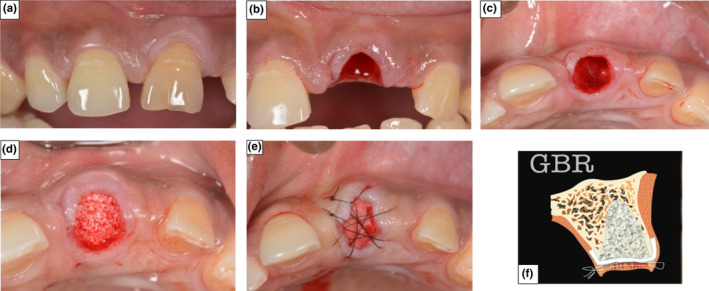
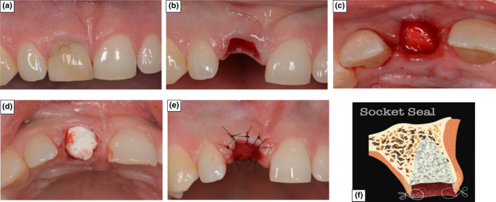
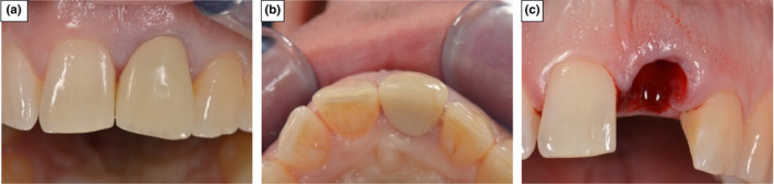
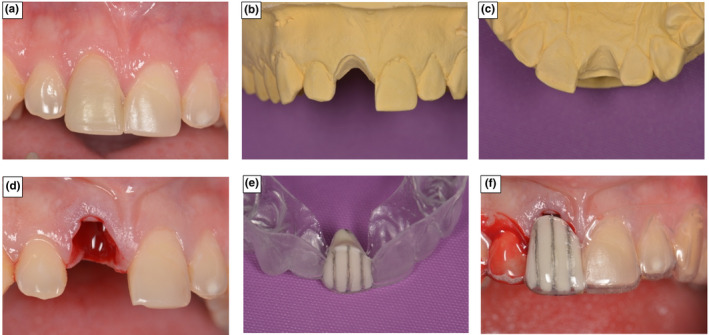
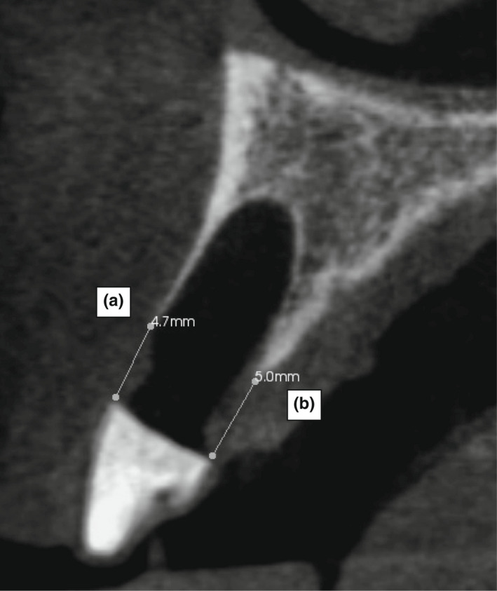
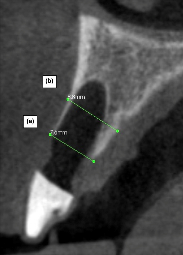
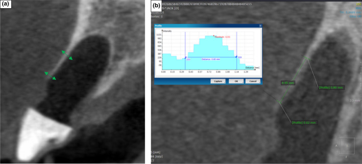
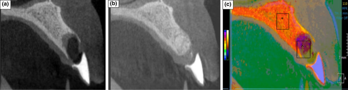
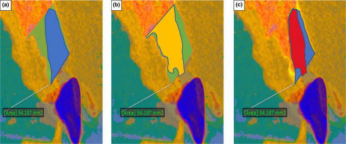
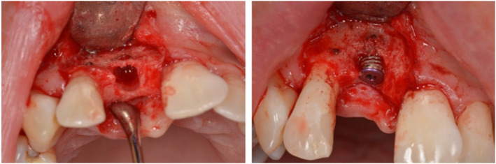
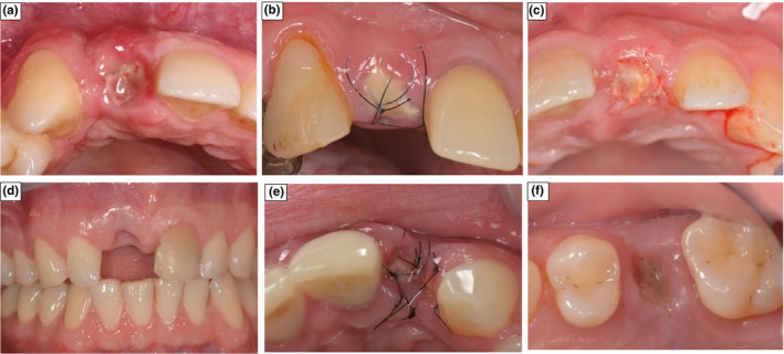
Material and methods: Patients requiring a single rooted tooth extraction in the anterior maxilla, were randomly allocated into: GBR, SS and Control groups (n= 14/). Cone Beam Computed Tomography (CBCT) images were recorded post-extraction and at 4 months, the mid-buccal and mid-palatal alveolar ridge heights (BARH/PARH) were measured. The alveolar ridge width, cross-sectional socket and alveolar-process area changes, implant placement feasibility, requirement for bone augmentation and post-surgical complications were also recorded.
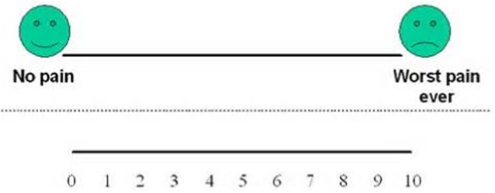
Results: BARH and PARH was found to increase with the SS (0.65 mm ± 1.1/0.65 mm ± 1.42) techniques, stabilise with GBR (0.07 mm ± 0.83/0.86 mm ±1.37) and decrease in the Control (-0.52 mm ± 0.8/-0.43 mm ± 0.83). Statistically significance was found when comparing the GBR and SS BARH (p = .04/.005) and GBR PARH (p = .02) against the Control. GBR recorded the smallest reduction in alveolar ridge width (-2.17 mm ± 0.84), when compared to the Control (-2.3 mm ± 1.11) (p = .89). A mid-socket cross-sectional area reduction of 4% (-2.27 mm2 ± 11.89), 1% (-0.88 mm2 ± 15.48) and 13% (-6.93 mm2 ± 8.22) was found with GBR, SS and Control groups (GBR vs. Control p = .01). The equivalent alveolar process area reduction was 8% (-7.36 mm2 ± 10.45), 6% (-7 mm2 ± 18.97) and 11% (-11.32 mm2 ± 10.92). All groups supported implant placement, with bone dehiscence noted in 57% (n = 4), 64%(n = 7) and 85%(n = 12) of GBR, SS and Control cases (GBR vs. Control p = .03). GBR had a higher risk of swelling and mucosal colour change, with SS associated with graft sequestration and matrix breakdown.
Conclusion: GBR ARP was found to be more effective at reducing radiographic bone dimensional changes following tooth extraction.
Keywords: alveolar bone dimensions; alveolar ridge preservation; bone healing complications and visual analogue pain scores; cone beam computerised tomography; guided bone regeneration; optical scanning; randomised controlled trial; socket seal.
© 2022 The Authors. Clinical Oral Implants Research published by John Wiley & Sons Ltd.
Conflict of interest statement
The authors declare that there is nothing to disclose.
Figures
References
-
- Aimetti, M. , Romano, F. , Griga, F. B. , & Godio, L. (2009). Clinical and histologic healing of human extraction sockets filled with calcium sulfate. International Journal of Oral and Maxillofacial Implants, 24, 902–909. - PubMed
-
- Al‐Hezaimi, K. , Levi, P. , Rudy, R. , Al‐Jandan, B. , & Al‐Rasheed, A. (2011). An extraction socket classification developed using analysis of bone type and blood supply to the buccal bone in monkeys. International Journal of Periodontics and Restorative Dentistry, 31, 421. - PubMed
-
- Araujo, M. G. , & Lindhe, J. (2005). Dimensional ridge alterations following tooth extraction. An experimental study in the dog. Journal of Clinical Periodontology, 32, 212–218. - PubMed
-
- Araujo, M. G. , Silva, C. O. , Misawa, M. , & Sukekava, F. (2015). Alveolar socket healing: What can we learn? Periodontology 2000, 68, 122–134. - PubMed
Publication types
MeSH terms
Grants and funding
LinkOut - more resources
Full Text Sources
Miscellaneous
