. 2022 Jun;143(6):663-685.
doi: 10.1007/s00401-022-02425-4. Epub 2022 Apr 30.

Unique seeding profiles and prion-like propagation of synucleinopathies are highly dependent on the host in human α-synuclein transgenic mice

Affiliations

Unique seeding profiles and prion-like propagation of synucleinopathies are highly dependent on the host in human α-synuclein transgenic mice

Grace M Lloyd et al. Acta Neuropathol. 2022 Jun.

Abstract

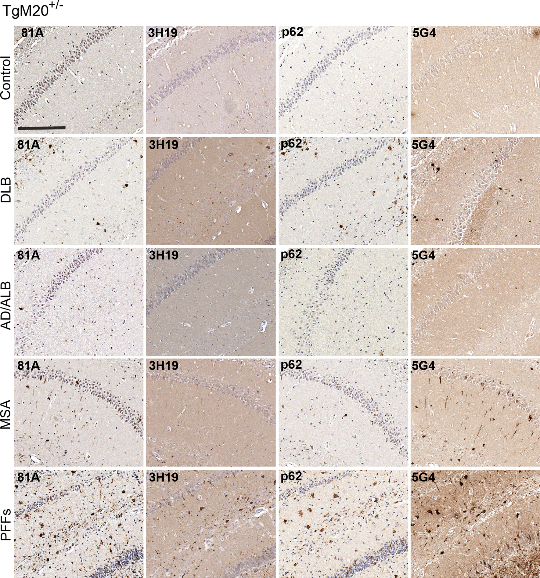
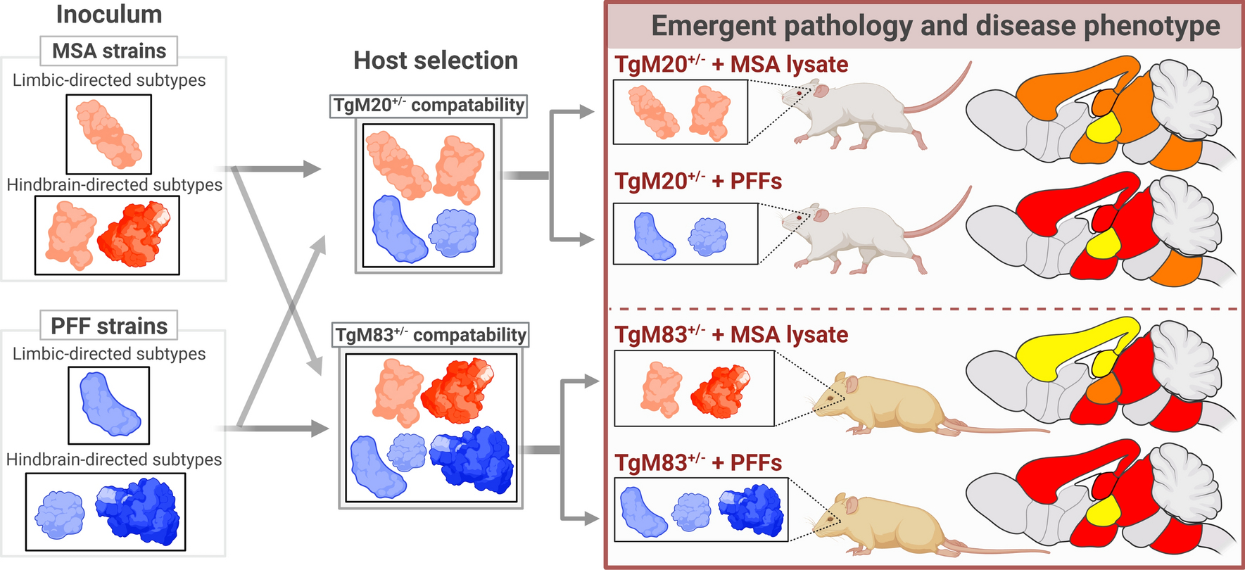
α-synuclein (αSyn) is an intrinsically disordered protein which can undergo structural transformations, resulting in the formation of stable, insoluble fibrils. αSyn amyloid-type nucleation can be induced by misfolded 'seeds' serving as a conformational template, tantamount to the prion-like mechanism. Accumulation of αSyn inclusions is a key feature of dementia with Lewy bodies (DLB) and multiple system atrophy (MSA), and are found as additional pathology in Alzheimer's disease (AD) such as AD with amygdala predominant Lewy bodies (AD/ALB). While these disorders accumulate the same pathological protein, they exhibit heterogeneity in clinical and histological features; however, the mechanism(s) underlying this variability remains elusive. Accruing data from human autopsy studies, animal inoculation modeling, and in vitro characterization experiments, have lent credence to the hypothesis that conformational polymorphism of the αSyn amyloid-type fibril structure results in distinct "strains" with categorical infectivity traits. Herein, we directly compare the seeding abilities and outcome of human brain lysates from these diseases, as well as recombinant preformed human αSyn fibrils by the intracerebral inoculation of transgenic mice overexpressing either human wild-type αSyn or human αSyn with the familial A53T mutation. Our study has revealed that the initiating inoculum heavily dictates the phenotypic and pathological course of disease. Interestingly, we have also established relevant host-dependent distinctions between propagation profiles, including burden and spread of inclusion pathology throughout the neuroaxis, as well as severity of neurological symptoms. These findings provide compelling evidence supporting the hypothesis that diverse prion-type conformers may explain the variability seen in synucleinopathies.

Keywords: Alzheimer’s disease with amygdala predominant Lewy bodies; Amyloid; Dementia with Lewy bodies; Multiple system atrophy; Prion; Strains; Synucleinopathy; α-synuclein.


Conflict of interest statement

Conflict of interest We declare no conflict of interest in this manuscript.

Figures

Fig. 1
Fig. 1
Study design and characterization of brain tissues used for inoculations a) Schematic of experimental design and types of brain inoculations in hemizygous TgM83+/− and TgM20+/− mice. b) Immunoblots with anti-αSyn antibody 3H11 of high-salt (HS) soluble and Triton X-100 insoluble fractions derived from human brain homogenates with either DLB, AD/ALB, MSA or control amygdala or white matter cerebellum as described in Materials and Methods. The relative mobility of molecular mass markers is indicated on the left of the blots. c) Representative images of αSyn immunostaining in tissue sections from the amygdala or cerebellum white matter from control, DLB, AD/ALB and MSA patients, (as indicated at the top right), with αSyn antibody, 3H11. Sections were counterstained with hematoxylin. Scale bar = 150μm; insert = 10μm. AD/ALB Alzheimer’s Disease with amygdala predominant Lewy bodies; AMY amygdala; CB cerebellum; Ct control; DLB Dementia with Lewy bodies; MSA multiple system atrophy; PFFs preformed fibrils.
Fig. 2
Fig. 2
Pathological outcome and histological pathology in TgM83+/− mice induced by inoculation a) Kaplan-Meier paralysis curves of TgM83+/− mice inoculated with brain extract from the amygdala of control, DLB or AD/ALB brains or the cerebellum of control or MSA brains, or WT human αSyn PFFs. b) Representative immunohistochemistry images of pathological αSyn deposition in TgM83+/− mice using antibodies specific for αSyn phosphorylated on Ser129 (81A), for αSyn (3H19), and p62/sequestrasome-1. Regions from the hippocampus are shown for DLB, AD/ALB and control amygdala lysates and PFF injected mice, while regions from the midbrain are depicted for MSA and control CB lysates injected mice to accurately represent relative pathology. Scale bars = 200μm. AD/ALB Alzheimer’s Disease with amygdala predominant Lewy bodies; AMY amygdala; CB cerebellum; DLB Dementia with Lewy bodies; MSA multiple system atrophy; PFFs preformed fibrils.
Fig. 3
Fig. 3
Characterization of histological αSyn pathology in TgM20+/− Mice after inoculation Representative IHC images of pathological αSyn deposition in TgM20+/− mice using antibodies specific for αSyn phosphorylated on Ser129 (81A), for αSyn (3H19), p62/sequestrasome-1 and for aggregated αSyn (5G4). Regions from the hippocampus are shown. Scale bars = 200μm. AD/ALB Alzheimer’s Disease with amygdala predominant Lewy bodies; DLB Dementia with Lewy bodies; MSA multiple system atrophy; PFFs preformed fibrils.
Fig. 4
Fig. 4
Elucidation of regional distribution of pathological αSyn spread in TgM83+/− mice after inoculation a) Diagram depicting distribution of αSyn pathological deposition (red dots) using an antibody specific for αSyn phosphorylated at pSer129. Column on the right indicates the number of animals that did not exhibit any detectible pathology following inoculation (no pathology in controls) over total number of animals within the corresponding cohort. b) Quantitative analysis of percentage of area positive for phosphorylated αSyn deposition, normalized to maximum measured pathology (data represents the mean +/− SEM) within indicated brain regions from TgM83+/− mice following inoculation with brain lysate from amygdala of control, DLB or AD/ALB brains or the cerebellum of control or MSA brains, or WT human αSyn PFFs. c) Representative IHC images of the distinct pSer129 αSyn deposition illustrating the regional differences between cohorts. Scale bar = 25μm. AD/ALB Alzheimer’s Disease with amygdala predominant Lewy bodies; CTX cortex; DLB Dementia with Lewy bodies; ENTI entorhinal cortex; HPF hippocampal formation; HY hypothalamus; MB midbrain; MSA multiple system atrophy; MY medulla; PFFs preformed fibrils; PAG periaqueductal gray; PIR piriform cortex; SUBv ventral subiculum; TH thalamus.
Fig. 5
Fig. 5
Elucidation of regional distribution of pathological αSyn spread in TgM20+/− mice after inoculation a) Diagram depicting distribution of αSyn deposition (red dots) using antibody specific for αSyn phosphorylated at pSer129. Column on the right indicates the number of animals that did not exhibit any detectible pathology following inoculation (no pathology in controls) over total number of animals within the corresponding cohort. *10 mice were harvested at endpoint; however, one sample was lost during tissue processing. b) Quantitative analysis of percentage of area positive for phosphorylated αSyn deposition, normalized to maximum measured pathology (data represents the mean +/− SEM) within indicated brain regions from TgM20+/− mice following inoculation with brain lysate from amygdala of control, DLB or AD/ALB brains or the cerebellum of control or MSA brains, or WT human αSyn PFFs. c) Representative IHC images of the distinct pSyn deposition illustrating the regional differences between cohorts. Scale bar = 25μm. AD/ALB Alzheimer’s Disease with amygdala predominant Lewy bodies; CTX cortex; DLB Dementia with Lewy bodies; ENTI entorhinal cortex; HPF hippocampal formation; HY hypothalamus; MB midbrain; MSA multiple system atrophy; MY medulla; PFFs preformed fibrils; PAG periaqueductal gray; PIR piriform cortex; SUBv ventral subiculum; TH thalamus.
Fig. 6
Fig. 6
Investigation of brain astrocytic activation in Tg mice with low or no αSyn-positive inclusions Representative images showing anti-GFAP immuno-staining counterstained with hematoxylin from a) TgM83+/− mice and b) TgM20+/− mice inoculated with amygdala lysate from control, DLB or AD/ALB brains. Scatter dot plots compare percent of area with GFAP reactivity within transgenic mice between different inoculums (a-b), (two-way ANOVA followed by Tukey’s multiple comparisons test; data represents the mean +/− SEM), within indicated brain regions. Scale bars = 500μm. AD/ALB Alzheimer’s Disease with amygdala predominant Lewy bodies; CTX cortex; DLB Dementia with Lewy bodies; ENTI entorhinal cortex; HPF hippocampal formation; HY hypothalamus; MB midbrain; MY medulla; PAG periaqueductal gray; PIR piriform cortex; SUBv ventral subiculum; TH thalamus.
Fig. 7
Fig. 7
Characterization of astrocytic activation in Tg mice with extensive αSyn pathology Representative images showing anti-GFAP immuno-staining counterstained with hematoxylin from a) TgM83+/− mice and b) TgM20+/− mice inoculated with PFFs or brain lysate from the cerebellum of control or MSA brains. Scatterplots compare percent of area with GFAP reactivity within transgenic mice between different inoculums (a-b), (two-way ANOVA followed by Tukey’s multiple comparisons test; data represents the mean +/− SEM), within indicated brain regions. Scale bars = 500μm. CTX cortex; ENTI entorhinal cortex; HPF hippocampal formation; HY hypothalamus; MB midbrain; MSA multiple system atrophy; MY medulla; PFFs preformed fibrils; PAG periaqueductal gray; PIR piriform cortex; SUBv ventral subiculum; TH thalamus.
Fig. 8
Fig. 8
Inoculation with MSA-derived αSyn results in distinct, host-dependent morphological profiles a) Representative images of different morphologies from MSA-inoculated TgM83+/− and TgM20+/− mice, shows region-specific morphological profiles of αSyn pSer129 positive inclusions that are discernibly different between hosts. b) Heat map showing the percentage of animals within each cohort that were found to have each morphological type, demonstrates that TgM83+/− mice show greater consistency than TgM20+/− mice. c) Graph summarizing the fraction of total morphologies found within each cohort and region, shows that morphological variation diminishes in the caudal regions of TgM20+/− mice. Scale bar = 15μm. BS brainstem; Cb cerebellum; CH cerebrum; Sp spine.
Fig. 9
Fig. 9
Inoculation with PFFs results in manifold morphologies a) Representative images of distinct morphologies from PFF-inoculated TgM83+/− and TgM20+/− mice, shows region-specific morphological profiles of pSer129 positive inclusions, that are discernibly different between hosts. b) Heat map showing the percentage of animals within each cohort that were found to have each morphological type, demonstrates that TgM83+/− mice show greater consistency than TgM20+/− mice. c) Graph summarizing the fraction of total morphologies found within each cohort and region, shows that morphological variation diminishes in the caudal regions of TgM20+/− mice. Scale bar = 15μm. BS brainstem; Cb cerebellum; CH cerebrum; Sp spine.
Fig. 10
Fig. 10
Theoretical diagram outlining potential interactions between conformation proteoforms and αSyn transgenic mice after inoculation. Nucleating factors in inoculum may contain a limited allotment of conforms, represented by different colors and shapes, of which, only a subset would be able to transmit within the host environment. Intrinsic misfolding properties of the host protein relative to the interactions with the nucleating seeds are important factors in prion-like transmission capability and kinetics, but other factors, such as differences in cellular and regional expression, post-translational modifications, and altered glial activity (e.g. neuroinflammation), may guide the preferential amplification of specific αSyn polymorphs. Once formed, these structures may maintain characteristics that restrict pathogenicity, leading to the formation of strains. In this diagrammed example, ‘clouds’ of αSyn prion-like strains that are preferentially compatible with either the limbic or hindbrain regions in mouse (depicted in the boxes on the left) may undergo further selectivity dictated by mouse host permissivity. Host, region, and cell-specific mechanisms, such as protease expression and type, lysosomal activity, molecular chaperones or homology of amino acid sequence with host protein may be involved in determining the ultimate emergence of pathology. Color in brain indicates generalized level of pathology: red = high, orange. = moderate, yellow = low/none. Gray indicates this region was not quantified. Created with Biorender.com.

Similar articles

Cited by

References

    1. Aguzzi A, Rajendran L (2009) The Transcellular Spread of Cytosolic Amyloids, Prions, and Prionoids. Neuron 64:783–790 - PubMed
    1. Ayers JI, Brooks MM, Rutherford NJ, Howard JK, Sorrentino ZA, Riffe CJ, Giasson BI (2017) Robust Central Nervous System Pathology in Transgenic Mice following Peripheral Injection of α-Synuclein Fibrils. J Virol 91:e02095–16. doi: 10.1128/jvi.02095-16 - DOI - PMC - PubMed
    1. Ayers JI, Lee J, Monteiro O, Woerman AL, Lazar AA, Condello C, Paras NA, Prusiner SB (2022) Different α-synuclein prion strains cause dementia with Lewy bodies and multiple system atrophy. Proc Natl Acad Sci U S A 119:e2113489119. doi: 10.1073/pnas.2113489119 - DOI - PMC - PubMed
    1. Bartz JC (2016) Prion strain diversity. Cold Spring Harb Perspect Med 6:a024349. doi: 10.1101/cshperspect.a024349 - DOI - PMC - PubMed
    1. Bartz JC (2021) Environmental and host factors that contribute to prion strain evolution. Acta Neuropathol 142:5–16. doi: 10.1007/s00401-021-02310-6 - DOI - PMC - PubMed

Publication types