Review
. 2022 May 20;50(9):4813-4839.
doi: 10.1093/nar/gkac290.

DNA binding by polycomb-group proteins: searching for the link to CpG islands

Affiliations
Review

DNA binding by polycomb-group proteins: searching for the link to CpG islands

Brady M Owen et al. Nucleic Acids Res. .

Abstract

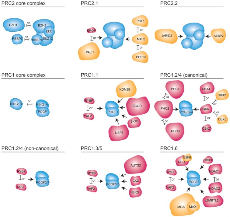
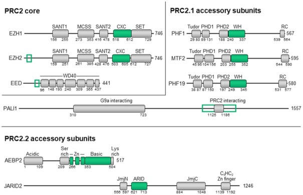
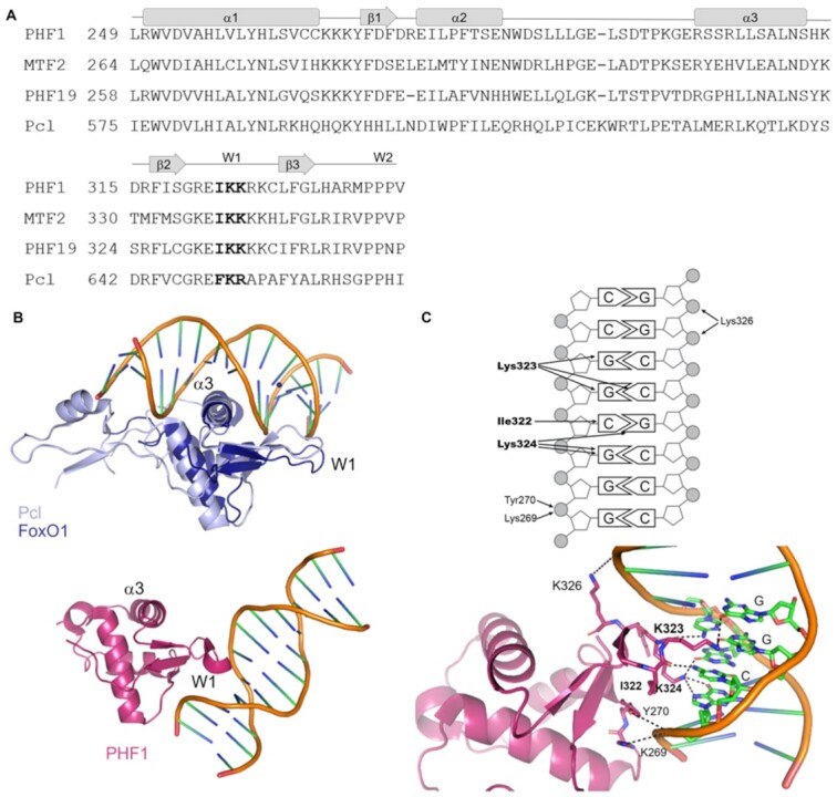
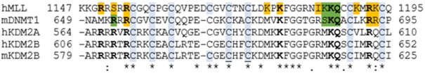
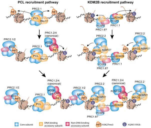
Polycomb group proteins predominantly exist in polycomb repressive complexes (PRCs) that cooperate to maintain the repressed state of thousands of cell-type-specific genes. Targeting PRCs to the correct sites in chromatin is essential for their function. However, the mechanisms by which PRCs are recruited to their target genes in mammals are multifactorial and complex. Here we review DNA binding by polycomb group proteins. There is strong evidence that the DNA-binding subunits of PRCs and their DNA-binding activities are required for chromatin binding and CpG targeting in cells. In vitro, CpG-specific binding was observed for truncated proteins externally to the context of their PRCs. Yet, the mere DNA sequence cannot fully explain the subset of CpG islands that are targeted by PRCs in any given cell type. At this time we find very little structural and biophysical evidence to support a model where sequence-specific DNA-binding activity is required or sufficient for the targeting of CpG-dinucleotide sequences by polycomb group proteins while they are within the context of their respective PRCs, either PRC1 or PRC2. We discuss the current knowledge and open questions on how the DNA-binding activities of polycomb group proteins facilitate the targeting of PRCs to chromatin.


Figures

Figure 1.
Figure 1.
Components of PRC1 and PRC2 complexes in mammals. PcG proteins form two predominant families of complexes; PRC1 and PRC2. Complexes within each family share common core proteins (shown in blue) and interact with accessory subunits (in magenta or yellow) which modulate their enzymatic activity and are required for chromatin binding and correct targeting. Known DNA-binding accessory subunits which may contribute to sequence selective recruitment of PcG proteins are highlighted in yellow. Arrows indicate binding to the core complex, but not necessarily the sites of protein-protein interactions since these are not always known. Mutually exclusive interactions are indicated by ‘or’.
Figure 2.
Figure 2.
Proposed DNA-binding regions of PRC2 proteins. PRC2 subunits are presented only if DNA-interacting regions were previously identified in them. DNA-binding domains are shown in green. DNA-binding regions outside of defined domains in EZH2 and EED are outlined in green.
Figure 3.
Figure 3.
ARID domain mechanism of DNA binding. (A) Hydrogen bonds and salt bridges between the ARID domain of DRI and DNA are shown with black arrows. DRI Thr351 and Ser352 are shown in bold and make direct contacts with DNA bases which may confer AT selectivity. Contacts were mapped based on the structure of the ARID domains from DRI in a complex with DNA (PDB ID: 1KQQ (118)). (B) The structure of the ARID domain of DRI (dark blue, PDB ID: 1KQQ (118)) with DNA was superimposed over the structure of the ARID domain from JARID2 that was solved without DNA (light blue, PDB ID: 2RQ5 (121)). (C) Close contacts between Thr351 and Ser352 (green) of DRI and the DNA bases are indicated in dashed black lines. The equivalent residues in JARID2 are labelled in light blue. (D) Sequence alignment of JARID2, DRI and MRF2 ARID domains with DRI DNA contacts from (A) highlighted. In all panels, amino acids in DRI from the loop between H5 and H6 contact bases in the major groove and are labelled in green. Additional interactions are mediated by amino acids in the loop between H2 and H3 (magenta), a pocket between H4 and H5 (orange) and the end of H8 (red).
Figure 4.
Figure 4.
Mechanism of DNA binding by PCL proteins. (A) Sequence alignment of WH domains. Amino acids in the conserved Ile-Lys-Lys motif are shown in bold. (B) Top structure: A DNA-binding mode of Drosophila Pcl (light blue, PDB ID: 5OQD) proposed by Choi et al. (34) based on alignment to the DNA bound structure of the FoxO1 WH domain (dark blue) (PDB ID: 3CO6 (228)). Bottom structure: The DNA-bound structure of PHF1 (magenta), which forms non-canonical interactions with the DNA (PDB ID: 5XFQ (31)). (C) Contacts between PHF1 and the CpG dinucleotide. Contacts as reported in Li et al. (31) are represented by arrows (top) and dashed lines (bottom). Amino acids which contact the DNA bases, and hence are proposed to contribute to sequence selective binding (31) are in bold.
Figure 5.
Figure 5.
The ZF-CxxC domain of KDM2B bind CpG dinucleotides. Sequence alignment of ZF-CxxC domains. Residues highlighted in green were reported to contact the bases of the CpG dinucleotide (PDB: 2KKF (152), PDB: 3PT6 (153)). Residues in gold were reported to contact the DNA backbone. DNA-contacting residues which are conserved in human KDM2B are in bold. Conserved cysteine residues which coordinate zinc ions are highlighted in blue and two of these cysteines which mutating them disrupt KDM2B function in mice (36) are underlined.
Figure 6.
Figure 6.
Model of DNA-dependent recruitment of PcG proteins. PRC2.1 is seeded on target DNA by the PCL proteins (top left). PRC1.1 is seeded on DNA by KDM2B (top right) and PRC1.6 may also be recruited at some sites by MGA-MAX or E2F6. Once either the H3K27me3 or the H2AK119ub marks were nucleated, they can recruit the canonical PRC1 (middle left) or the PRC2.2 (middle right), respectively. After both the H2AK119Ub and H3K27me3 repressive marks were established, positive feedback loops involving PRC1, PRC2 and their respective chromatin modifications then lead to the maintenance of the repressive marks and possibly contributed to the recruitment of other PcG proteins (bottom). Orange arrows indicate enhancement of enzymatic activity by accessory subunits (in yellow) or the H3K27me3 mark (in orange). Transparent shapes and question marks indicate uncertainty regarding the precise role taken by the indicated protein in the presented pathway. For simplicity, histone tails are only shown for one copy of H3 and H2A for each nucleosome.

Similar articles

Cited by

References

    1. Barbour H., Daou S., Hendzel M., Affar E.B.. Polycomb group-mediated histone H2A monoubiquitination in epigenome regulation and nuclear processes. Nat. Commun. 2020; 11:5947. - PMC - PubMed
    1. Schuettengruber B., Bourbon H.-M., Di Croce L., Cavalli G.. Genome regulation by polycomb and trithorax: 70 years and counting. Cell. 2017; 171:34–57. - PubMed
    1. Laugesen A., Højfeldt J.W., Helin K.. Molecular mechanisms directing PRC2 recruitment and H3K27 methylation. Mol. Cell. 2019; 74:8–18. - PMC - PubMed
    1. Lewis E.B. A gene complex controlling segmentation in Drosophila. Nature. 1978; 276:565–570. - PubMed
    1. Voncken J.W., Roelen B.A.J., Roefs M., de Vries S., Verhoeven E., Marino S., Deschamps J., van Lohuizen M.. Rnf2 (Ring1b) deficiency causes gastrulation arrest and cell cycle inhibition. Proc. Natl. Acad. Sci. USA. 2003; 100:2468–2473. - PMC - PubMed