. 2022 Aug;163(2):481-494.
doi: 10.1053/j.gastro.2022.04.028. Epub 2022 Apr 27.

β-Catenin Sustains and Is Required for YES-associated Protein Oncogenic Activity in Cholangiocarcinoma

Affiliations

β-Catenin Sustains and Is Required for YES-associated Protein Oncogenic Activity in Cholangiocarcinoma

Yi Zhang et al. Gastroenterology. 2022 Aug.

Abstract

Background & aims: YES-associated protein (YAP) aberrant activation is implicated in intrahepatic cholangiocarcinoma (iCCA). Transcriptional enhanced associate domain (TEAD)-mediated transcriptional regulation is the primary signaling event downstream of YAP. The role of Wnt/β-Catenin signaling in cholangiocarcinogenesis remains undetermined. Here, we investigated the possible molecular interplay between YAP and β-Catenin cascades in iCCA.

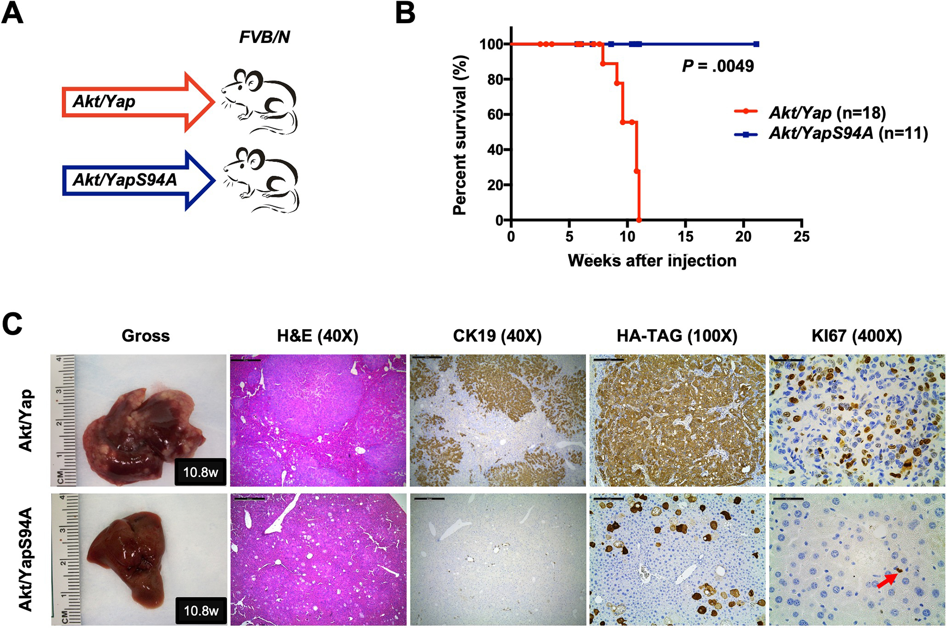
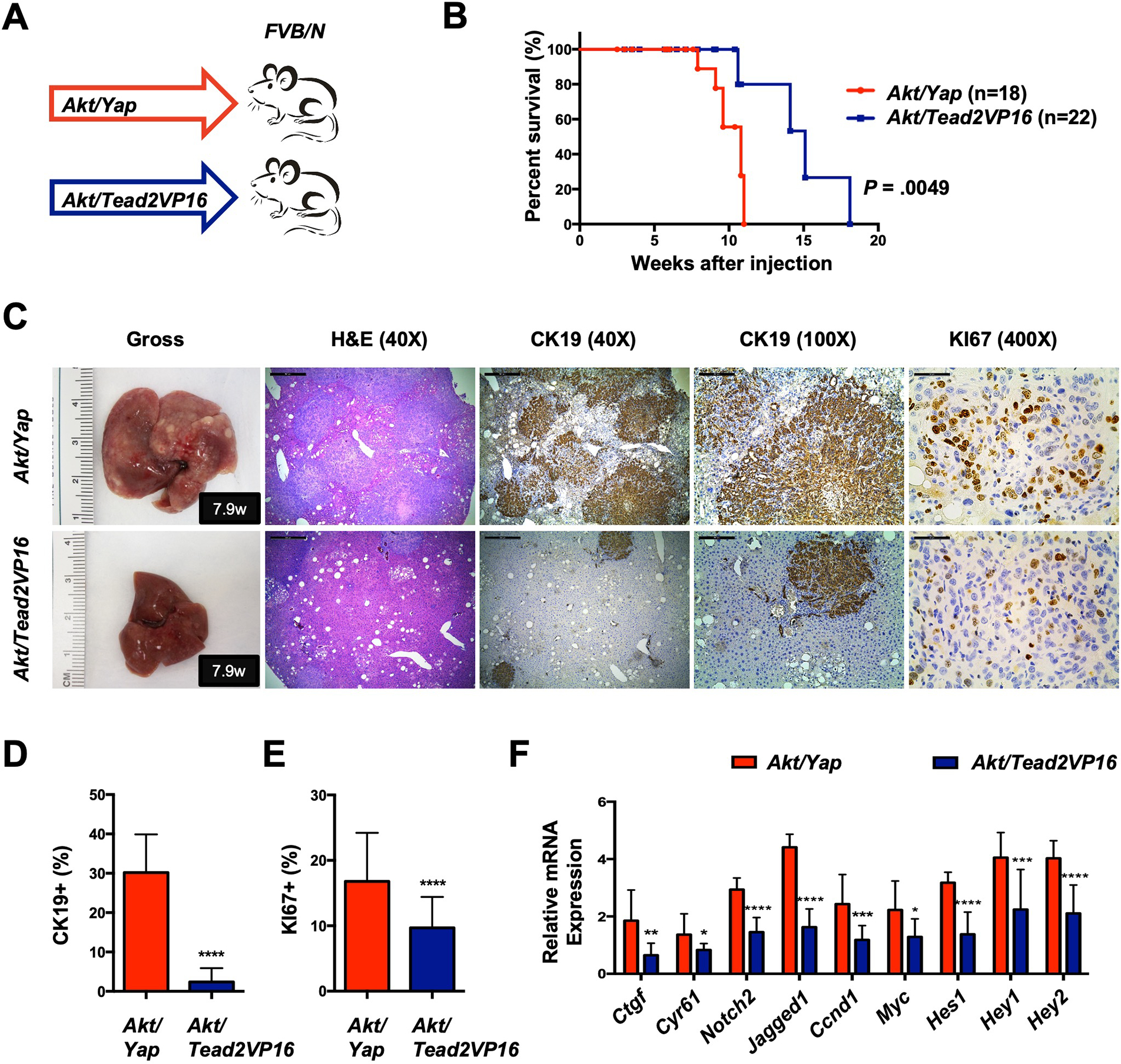
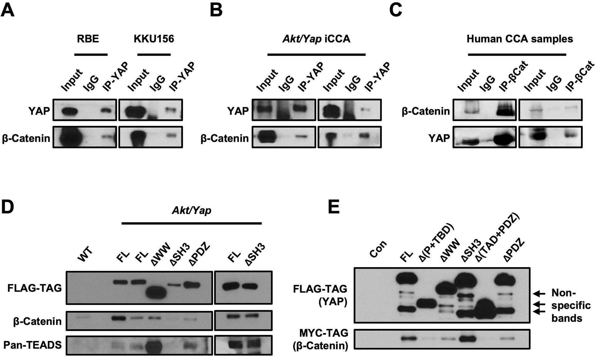
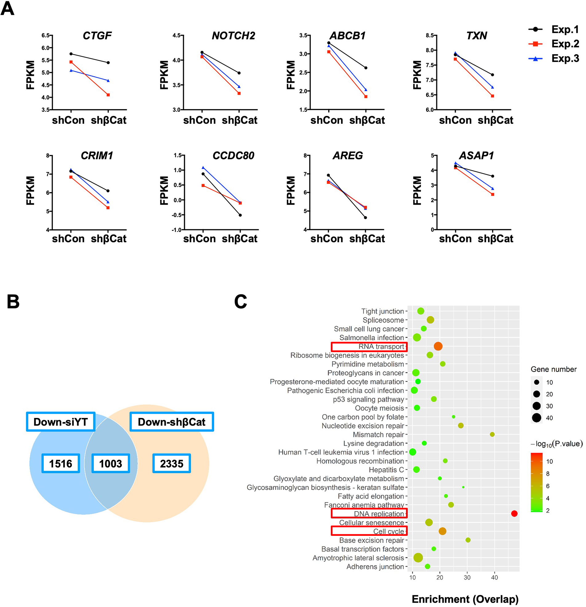
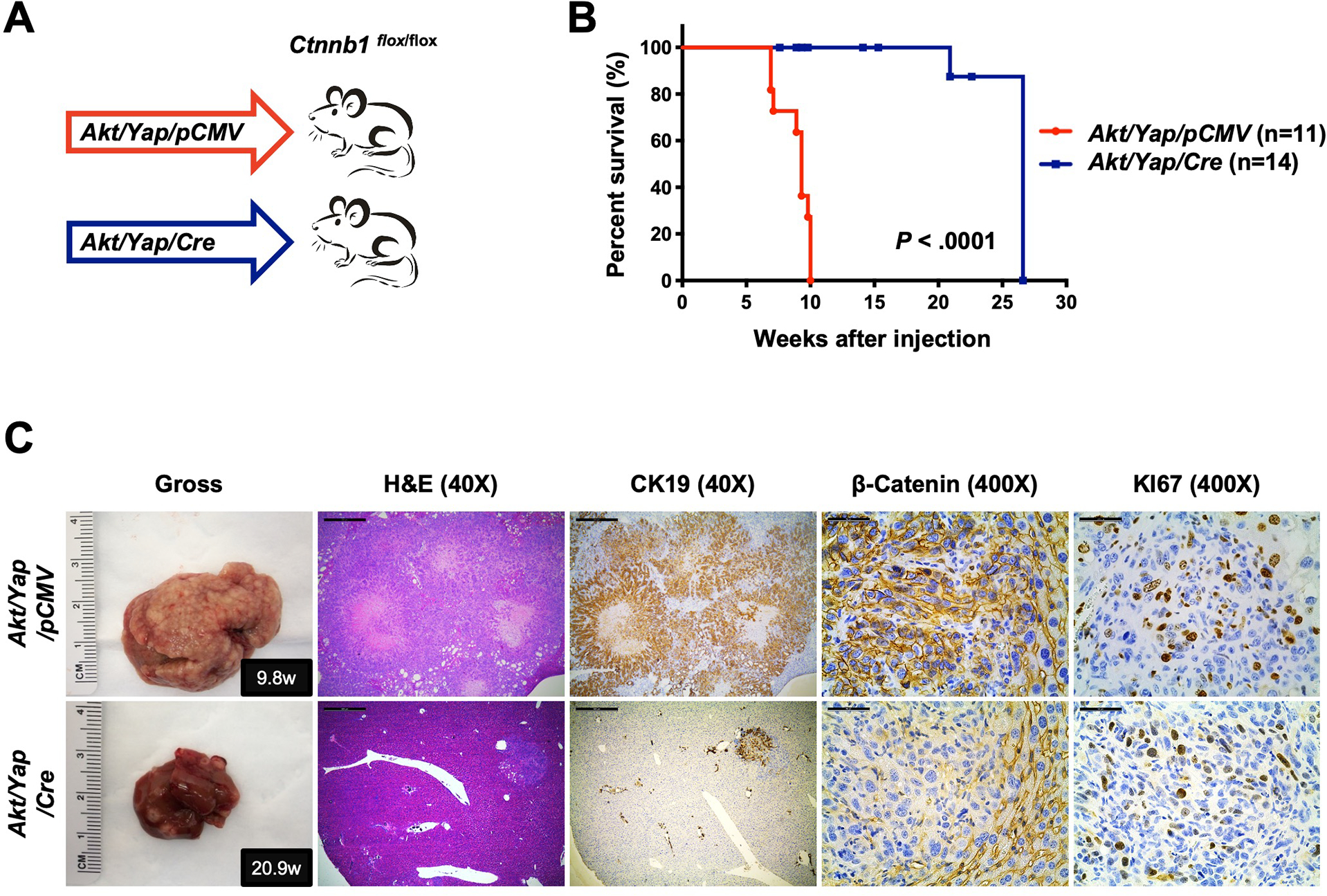
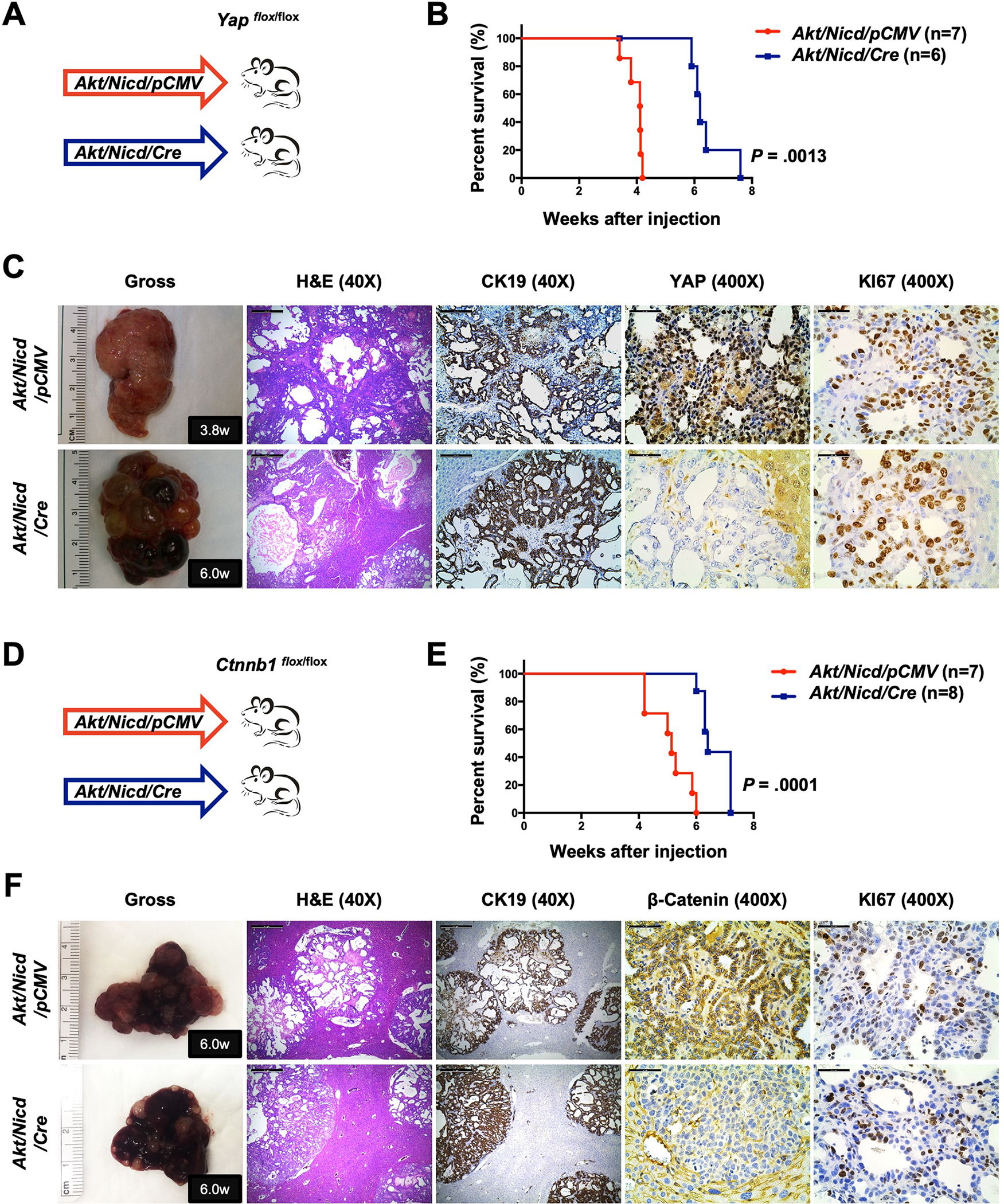
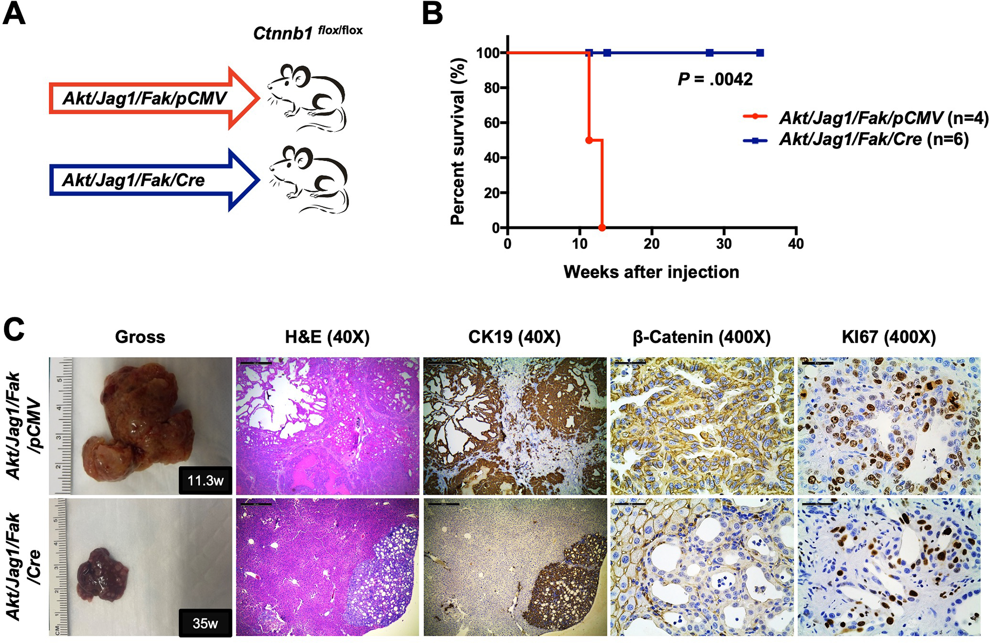
Methods: Activated AKT (Myr-Akt) was coexpressed with YAP (YapS127A) or Tead2VP16 via hydrodynamic tail vein injection into mouse livers. Tumor growth was monitored, and liver tissues were collected and analyzed using histopathologic and molecular analysis. YAP, β-Catenin, and TEAD interaction in iCCAs was investigated through coimmunoprecipitation. Conditional Ctnnb1 knockout mice were used to determine β-Catenin function in murine iCCA models. RNA sequencing was performed to analyze the genes regulated by YAP and/or β-Catenin. Immunostaining of total and nonphosphorylated/activated β-Catenin staining was performed in mouse and human iCCAs.

Results: We discovered that TEAD factors are required for YAP-dependent iCCA development. However, transcriptional activation of TEADs did not fully recapitulate YAP's activities in promoting cholangiocarcinogenesis. Notably, β-Catenin physically interacted with YAP in human and mouse iCCA. Ctnnb1 ablation strongly suppressed human iCCA cell growth and Yap-dependent cholangiocarcinogenesis. Furthermore, RNA-sequencing analysis revealed that YAP/ transcriptional coactivator with PDZ-binding motif (TAZ) regulate a set of genes significantly overlapping with those controlled by β-Catenin. Importantly, activated/nonphosphorylated β-Catenin was detected in more than 80% of human iCCAs.

Conclusion: YAP induces cholangiocarcinogenesis via TEAD-dependent transcriptional activation and interaction with β-Catenin. β-Catenin binds to YAP in iCCA and is required for YAP full transcriptional activity, revealing the functional crosstalk between YAP and β-Catenin pathways in cholangiocarcinogenesis.

Keywords: Hippo/YAP; Intrahepatic Cholangiocarcinoma; TEADs; β-Catenin.


Conflict of interest statement

Disclosures

The authors declare no potential conflicts of interest.

Figures

Figure 1.
Figure 1.. TEADs is required for Yap-driven iCCA formation in mice.
(A) Study design. FVB/N mice were subjected to HTVi of either Akt/Yap (n=18) or Akt/YapS94A (n = 11) plasmids. (B) Mouse survival curves. (C) Representative gross images, H&E, and immunohistochemistry for CK19, HA-TAG and KI67 of liver sections from Akt/Yap (M10.8w.p.i) and Akt/YapS94A (F10.8w.p.i) mice. The red arrow indicates KI67 positive stained nuclei. Scale bar: 500 μm (40x), 100 μm (100x), and 50 μm (400x). Abbreviations: H&E, hematoxylin and eosin staining; w.p.i, weeks post-injection.
Figure 2.
Figure 2.. TEAD mediated transcriptional activation is not sufficient to induce Yap-dependent cholangiocarcinogenesis.
(A) Study design. FVB/N mice were subjected to HTVi of either Akt/Yap (n=18) or Akt/Tead2VP16 (n = 22) plasmids. Akt/Yap, Akt/YapS94A (Fig.1B), and Akt/ead2VP16 mice were generated in parallel. (B) Mouse survival curves. (C) Representative gross images, H&E, and immunohistochemistry for CK19 and KI67 of liver sections from Akt/Yap (M7.9w.p.i) and Akt/Tead2VP16 (M7.9w.p.i) mice. (D) Analysis of CK19-positive areas in Akt/Yap and Akt/Tead2VP16 mouse liver tissues at 2.5–11 w.p.i. (E) Quantification of the KI67 positive staining in liver sections from the depicted mice. (F) mRNA expression of Yap and Notch targets. Data were analyzed by the Mann-Whitney test. Statistical significance: *P < .05, **P < .01, ***P < .001, ****P < .0001. Scale bar: 500 μm (40x), 100 μm (100x) and 50 μm (400x). Abbreviations: H&E, hematoxylin and eosin staining; w.p.i, weeks post-injection.
Figure 3.
Figure 3.. Yap physically interacts with β-Catenin in iCCA.
Co-immunoprecipitation assays to detect β-Catenin and Yap interaction. (A, B) Lysates from human CCA cells transfected with the pT3-EF1αH-FLAG-YapS127A plasmid (A) or Akt/Yap iCCA tumors (B) were subjected to immunoprecipitation with an anti-Yap antibody. (C) Lysates from human CCA samples were subjected to immunoprecipitation with an anti-β-Catenin antibody. (D, E) Lysates from Akt/Yap (FL), Akt/YapΔWW, Akt/YapΔSH3, Akt/YapΔPDZ tumors (D), or 293FT cells transfected with truncated Flag-Yap and Myc-β-Catenin plasmids (E) were subjected to immunoprecipitation with anti-FLAG beads. FVB/N mouse livers (WT) (D) and untreated 293FT cells (E) were used as the controls.
Figure 4.
Figure 4.. Genomic profiling reveals the crosstalk between YAP and β-Catenin signaling cascade in iCCA.
(A) Fragments per kilobase of transcript per million mapped reads (FPKM) results of YAP target genes expression. (B) The overlapping downregulated genes in the shβCat and siYT RBE cell lines. (C) KEGG analysis of downregulated genes in both shβCat and siYT RBE cell lines. P-value ranked the genes. Red inserts indicate overlapping pathways related to tumorigenesis.
Figure 5.
Figure 5.. Deletion of β-Catenin significantly inhibits Akt/Yap iCCA development in mice.
(A) Study design. Ctnnb1flox/flox mice were subjected to HTVi of either Akt/Yap/pCMV (control, n = 11) or Akt/Yap/Cre (n = 14) plasmids. (B) Mouse survival curves. (C) Representative gross images, H&E, and immunohistochemistry for CK19, β-Catenin, and KI67 of liver sections from Akt/Yap/pCMV (M9.8w.p.i) and Akt/Yap/Cre (M20.9w.p.i) mice. Data were analyzed using the Mann-Whitney test. Scale bar: 500 μm (40x) and 50 μm (400x). Abbreviations: H&E, hematoxylin and eosin staining; w.p.i, weeks post-injection.
Figure 6.
Figure 6.. Deletion of β-Catenin mildly delays Akt/Nicd driven iCCA.
(A) Study design. Yapflox/flox conditional knockout mice were subjected to HTVi of either Akt/Nicd/pCMV (control, n = 7) or Akt/Nicd/Cre (n = 6) plasmids. (B) Mouse survival curves. (C) Representative gross images, H&E, and immunohistochemistry for CK19, YAP, and KI67 of liver sections from Yapflox/flox Akt/Nicd/pCMV (M3.8w.p.i) and Yapflox/flox Akt/Nicd/Cre (M6.0w.p.i) mice. (D) Study design. Ctnnb1 flox/flox mice were subjected to HTVi of either Akt/Nicd/pCMV (control, n = 7) or Akt/Nicd/Cre (n = 8) plasmids. (E) Mouse survival curves. (F) Representative gross images, H&E, and immunohistochemistry for CK19, β-Catenin, and KI67 of liver sections from Ctnnb1flox/flox Akt/Nicd/pCMV (M6.0w.p.i) and Ctnnb1flox/flox Akt/Nicd/Cre (M6.0w.p.i) mice. Scale bar: 500 μm (40x) and 50 μm (400x). Abbreviations: H&E, hematoxylin and eosin staining; w.p.i, weeks post-injection.
Figure 7.
Figure 7.. Deletion of β-Catenin strongly suppresses Akt/Jag1/Fak-induced iCCA.
(A) Study design. Ctnnb1flox/flox mice were subjected to HTVi of either Akt/Jag1/Fak/pCMV (control, n = 4) or Akt/Jag1/Fak/Cre (n = 6) plasmids. (B) Mouse survival curves. (C) Representative gross images, H&E, and immunohistochemistry for CK19, β-Catenin, and KI67 of liver sections from Akt/Jag1/Fak/pCMV (F11.3w.p.i) and Akt/Jag1/Fak/Cre (F35w.p.i) mice. Scale bar: 500 μm (40x) and 50 μm (400x). Abbreviations: H&E, hematoxylin and eosin staining; w.p.i, weeks post-injection.

Comment in

References

    1. Banales JM, Marin JJG, Lamarca A, et al. Cholangiocarcinoma 2020: the next horizon in mechanisms and management. Nat Rev Gastroenterol Hepatol 2020;17:557–588. - PMC - PubMed
    1. Siegel RL, Miller KD, Jemal A. Cancer statistics, 2018. CA Cancer J Clin 2018;68:7–30. - PubMed
    1. Cheng R, Du Q, Ye J, et al. Prognostic value of site-specific metastases for patients with advanced intrahepatic cholangiocarcinoma: A SEER database analysis. Medicine (Baltimore) 2019;98:e18191. - PMC - PubMed
    1. Nguyen-Lefebvre AT, Selzner N, Wrana JL, et al. The hippo pathway: A master regulator of liver metabolism, regeneration, and disease. FASEB J 2021;35:e21570. - PubMed
    1. Sugihara T, Isomoto H, Gores G, et al. YAP and the Hippo pathway in cholangiocarcinoma. J Gastroenterol 2019;54:485–491. - PMC - PubMed

Publication types

MeSH terms